Guillain-Barré syndrome: signs, diagnosis, treatment - Online diagnostics. Guillain-Barre syndrome: symptoms, causes, diagnosis, treatment Guillain-Barre disease prognosis and complications
Guillain-Barré syndrome (acute inflammatory demyelinating polyradiculoneuropathy) (G61.0) is an acutely developing autoimmune inflammatory disease of the peripheral nervous system, characterized by acute demyelination of the roots of the spinal and cranial nerves, clinically manifested by paresthesias of the limbs, muscle weakness and/or flaccid paralysis.
Prevalence of the disease: 1-1.9 per 100 thousand people. The onset of the disease is observed at the age of 30-50 years.
The causes of the disease are unknown, so the syndrome is otherwise called idiopathic polyneuropathy. Immune-mediated factors play a role in the development of the disease. 2 weeks before the onset of symptoms of the disease, most patients notice symptoms of a respiratory or gastrointestinal infection.
Symptoms of Guillain-Barré syndrome
Symptoms of the disease appear acutely. Most patients experience pain (up to 80%) and paresthesia (up to 20%). Weakness in the legs, then in the arms, and muscles of the torso increases over several days (90%). Muscle weakness develops quickly but stops progressing within 4 weeks of onset. Numbness, pain in the feet, hands, and sometimes around the mouth have been present since the onset of the disease (70%). Weakness in the facial muscles, difficulty swallowing and breathing appear after 1-2 weeks. 30% of patients may experience dysfunction of the sphincters.
An objective examination reveals symmetrical flaccid predominantly distal tetraparesis (lower paraparesis), up to tetraplegia; paresthesia, hyperesthesia of the “socks”, “gloves” type; pain on palpation along the nerve trunks (up to 100%). In 30% of cases, symptoms of tension can be detected (Lasega, Neri). Characterized by a sharp depression or loss of deep reflexes. In 60-80% of cases, bulbar disorders and paresis of facial muscles are observed. Typically, damage to the sympathetic nervous system is manifested by dysautonomous disorders (profuse sweating, hypertension, postural hypotension, etc.). The development of respiratory failure (paresis of the diaphragm and respiratory muscles) and heart rhythm disturbances can be life-threatening (30%).
Diagnostics
- Study of cerebrospinal fluid (protein-cell dissociation, from the 2nd week - a moderate increase in protein content).
- Serological blood tests for infections.
- ENMG (primary demyelinating lesion).
- Blood pressure monitoring, ECG, respiratory function testing.
Differential diagnosis:
- Other polyneuropathies (with diphtheria, porphyria).
- Transverse myelitis.
- Acute cerebrovascular accidents in the vertebrobasilar region.
Treatment of Guillain-Barré syndrome
Treatment is prescribed only after confirmation of the diagnosis by a medical specialist. Maintenance of vital functions (ventilation), plasmapheresis, and pulse therapy with class G immunoglobulins are required.
Essential drugs
There are contraindications. Specialist consultation is required.


- (human immunoglobulin class G). Dosage regimen: administered intravenously at a dose of 0.4 g/kg once a day for 5 days.
- (non-steroidal anti-inflammatory drug). Dosage regimen: IM - 100 mg 1-2 times a day; after pain relief, it is prescribed orally in a daily dose of 300 mg in 2-3 doses, a maintenance dose of 150-200 mg/day.
- (anticonvulsant). Dosage regimen: orally, starting with 0.1 g 2 times a day, then the dose is increased by 0.1 g per day to 0.6-0.8 g (in 3-4 doses). After the pain disappears, the dose is gradually reduced to 0.1-0.2 g per day.
- (sedative, hypnotic, antihistamine). Dosage regimen: 1-5 ml of 1% solution intramuscularly. Orally 0.025-0.05 g 1-3 times a day. The course of treatment is 10-15 days.
- Prozerin (inhibitor of acetylcholinesterase and pseudocholinesterase). Dosage regimen: orally for adults, 10-15 mg 2-3 times a day; subcutaneously - 1-2 mg 1-2 times a day.
Definition. Guillain-Barré syndrome (GBS) is a severe autoimmune disease of the peripheral nervous system that is the most common cause of acute flaccid tetraparesis.
Epidemiology. According to global epidemiological studies, GBS occurs in 1 - 2 cases per 100,000 population per year, regardless of gender and age. The incidence of GBS in individual cities and regions of the Russian Federation corresponds to global data and varies from 0.34 to 1.9 per 100,000, with an average of 1.8 per 100,000 population per year.
Etiology. The leading role in the pathogenesis of the development of GBS is given to autoimmune mechanisms, while the peculiarity of this disease is a self-limiting, monophasic course with extremely rare relapses (up to 3 - 5%).
GBS usually develops 1 to 3 weeks after an infectious disease (ARVI, influenza, sinusitis, bronchitis, pneumonia, tonsillitis, measles, mumps, diarrhea, etc.). Epstein-Barr virus, Mycoplasma pneumoniae, Campylobacter jejuni and cytomegalovirus are considered the main triggers of the autoimmune process in GBS. It is assumed that the antigenic similarity of the shell of the infectious agent with individual structural elements of peripheral nerves (sheath, axon) causes the production of specific autoantibodies and the formation of circulating immune complexes that attack peripheral nerves in a “molecular mimicry” manner.
Less commonly, GBS occurs after vaccination (against influenza, hepatitis, rabies, etc.), surgical interventions (hernia repair, appendectomy, artificial termination of pregnancy, etc.), stressful situations, hypothermia, or against the background of complete health.
Classification. There are several forms of GBS, differing in the characteristics of the pathological process, the primary point of application of autoimmune aggression (nerve sheath or axonal core), prognosis of recovery, and clinical manifestations.
Most often (70 - 80%) all over the world, including in Russia, acute inflammatory demyelinating polyneuropathy (AIDP) is diagnosed as part of GBS, in which autoantibodies attack the myelin sheath of the nerve. In the second most common place (5 - 10%) are axonal forms - acute motor and motor-sensory axonal neuropathies (OMAN and OMSAN), characterized by primary damage to the axons of peripheral nerves and differing from each other in involvement (OMSAN) or intactness (OMAN) sensitive fibers. Other forms of GBS (Miller Fisher syndrome, pharyngo-cervico-brachial, acute pandysautonomia, paraparetic, sensory, Bickerstaff brainstem encephalitis [SEB]) are diagnosed extremely rarely (1 - 3%).
reference Information. Bickerstaff brainstem encephalitis (BTE) is clinically characterized by a combination of depression of consciousness, ophthalmoplegia, ataxia and hyperreflexia. Today, the autoimmune mechanism for the development of EBS is beyond doubt: the condition is associated with diarrhea caused by Campylobacter jejuni in 23% of cases, or is often associated with infection with cytomegalovirus or Mycoplasma pneumoniae. Anti-GQ1b IgG antibodies are detected in 66–68% of patients with EBS.
Diagnostic difficulties arise in the presence of so-called overlap syndromes, when the same patient simultaneously exhibits clinical, biochemical, serological and instrumental signs characteristic of 2 diseases or syndromes. The foreign literature presents clinical cases of crossover syndromes of GBS and EBS. The addition of flaccid tetraparesis to the symptoms of EBS indicates a possible parallel damage to peripheral nerves due to the development of overlap syndrome with GBS, which aggravates the course of EBS.
It turned out that up to 60% of cases of EBS are associated with the development of GBS and, as a rule, with its axonal forms. Despite the rarity of overlapping autoimmune neurological syndromes and the subtle differences in their constituent pathological conditions, their existence should always be remembered.
read also the post: Bickerstaff encephalitis(to the website)
GBS is also classified according to the severity of the condition depending on the clinical manifestations: [ 1 ] the mild form is characterized by the absence or minimal paresis, which does not cause significant difficulties when walking and self-care; [ 2 ] with moderate severity, walking impairment occurs, limiting the patient’s movement or requiring outside help or support; [ 3 ] in severe forms of the disease, the patient is bedridden and requires constant care, dysphagia is often observed; [ 4 ] in extremely severe forms, patients require artificial pulmonary ventilation (ALV) due to weakness of the respiratory muscles.
Clinic. The disease is characterized by a rapid (up to 4 weeks) increase in muscle weakness with initial involvement of the lower extremities and an “ascending pattern” of spread from distal to proximal muscle groups. Patients complain of increasing weakness in the legs and difficulty walking. As the disease progresses, the hands and often facial muscles are involved in the pathological process. In some cases, symptoms begin with damage to the cranial nerves or proximal muscle groups, and may predominantly affect the upper extremities. In every fourth or fifth case, the musculature of the trunk is involved in the pathological process, accompanied by weakness of the respiratory muscles (intercostal muscles, diaphragm), as a result of which every third patient with severe tetraparesis requires artificial pulmonary ventilation (ALV). With GBS, bulbar syndrome is often observed, primarily manifested by difficulties in swallowing and aspiration of fluid.
Muscle weakness is accompanied by sensory disorders - pain hyposthesia of the polyneuritic type and loss of deep sensitivity, as well as tendon areflexia. A fairly common symptom of GBS is pain. There are forms of the disease in which motor deficits are observed in isolation. Disorders of pelvic functions are not typical for GBS and can be observed in bedridden patients, mainly in the form of urinary retention.
Often there are signs of autonomic dysfunction in the form of changes in blood pressure (hypertension, hypotension), tachycardia, heart rhythm disturbances, hypersalivation, hyperhidrosis, paralytic ileus, which is an extreme manifestation of dynamic intestinal obstruction.
Diagnostics. The diagnosis of GBS is based on international criteria adopted by the World Health Organization in 1993. Signs required for diagnosis: [ 1 ] progressive muscle weakness in the legs and/or arms; [ 2 ] absence or extinction of tendon reflexes in the first days of the disease.
Signs supporting the diagnosis: [ 1 ] relative symmetry of the lesion; [ 2 ] symptoms progress over a period of no more than 4 weeks; [ 3 ] disturbance of sensitivity of the polyneuritic type; involvement of cranial nerves (most often - damage to the facial nerve); [ 5 ] recovery usually begins 2 to 4 weeks after the disease stops growing, but can sometimes be delayed for several months; [ 6 ] autonomic disorders: tachycardia, arrhythmias, postural hypotension, hypertension, vasomotor symptoms; [ 7 ] absence of fever at the onset of the disease (some patients experience fever at the onset of the disease due to intercurrent infections); fever does not exclude GBS, but raises the question of the possibility of another disease; [ 8 ] increased protein in the cerebrospinal fluid with normal cytosis - protein-cell dissociation (observed from the second week of the disease); [ 9 ] electroneuromyographic (ENMG) signs of demyelination and/or axonal damage to peripheral nerves.
Signs, questionable in diagnosis: [ 1 ] pronounced persistent asymmetry of motor disorders; [ 2 ] conduction level of sensory disturbances, pyramidal and cerebral symptoms; [ 3 ] persistent violations of pelvic functions; [ 4 ] more than 50 mononuclear leukocytes in the cerebrospinal fluid;[ 5 ] the presence of polymorphonuclear leukocytes in the cerebrospinal fluid.
These criteria apply to ARDP, axonal, paraparetic and pharyngo-cervico-brachial forms. Miller Fisher syndrome and acute pandysautonomia are clinically significantly different from other forms of GBS, so it is difficult to apply generally accepted diagnostic criteria for this disease. The diagnosis in these cases is established primarily on the basis of anamnestic data and the clinical picture of the disease.
Characteristics of Miller Fisher syndrome: [ 1 2 ] rapidly developing ataxia, tendon areflexia, ophthalmoplegia; [ 3 ] moderate weakness in the limbs may occur; [ 4 ] pain sensitivity is usually preserved; disturbances of deep sensitivity may be observed; [ 5 ] full recovery within 1 - 3 months; [ 6 ] with ENMG, the amplitude of sensory potentials is reduced or absent; The H-reflex is not evoked.
Characteristics of acute pandysautonomia: [ 1 ] the occurrence of neurological symptoms 1 - 2 weeks after a viral or bacterial infection; [ 2 ] the presence of an isolated lesion of the autonomic nervous system; [ 3 ] the cardiovascular system is often affected (postural hypotension, arterial hypertension, tachycardia, cardiac arrhythmias); [ 4 ] blurred vision, dry eye, anhidrosis; [ 5 ] dysfunction of the gastrointestinal tract (paralytic ileus); [ 6 ] difficulty urinating, acute urinary retention; [ 7 ] increased sweating, bluish coloration of the skin of the hands and feet, coldness of the extremities; [ 8 ] stunning, confusion due to hyponatremia associated with overproduction of antidiuretic hormone; Convulsions may occur when plasma sodium levels are less than 120 mmol/l; [ 9 ] recovery occurs gradually and often not completely.
read also the article: Acute pandisautonomia(to the website)
Neurophysiological criteria for diagnosis. Electroneuromyography (ENMG) is the only instrumental diagnostic method that allows you to confirm lesions of the peripheral nervous system and the diagnosis of GBS, respectively, as well as clarify the nature of pathological changes (demyelinating or axonal) and their prevalence. The protocol and scope of ENMG studies in patients with GBS depends on the clinical manifestations of the disease:
[1 ] for predominantly distal paresis, long nerves on the arms and legs are examined: at least 4 motor and 4 sensory (motor and sensory portions of the median and ulnar nerves; peroneal, tibial, superficial peroneal and sural nerves on one side);
[2 ] the main ENMG parameters are assessed: motor responses (distal latency, amplitude, shape and duration), the presence of excitation conduction blocks and response dispersion is assessed; the speed of excitation propagation along motor fibers in the distal and proximal areas is analyzed; sensory responses (amplitude) and speed of excitation along sensory fibers in the distal sections; late ENMG phenomena (F-waves): the latency, shape and amplitude of responses, the magnitude of chronodispersion, the percentage of dropouts are analyzed;
[3 ] in the presence of proximal paresis, additional examination of two short nerves (axillary, musculocutaneous, femoral, etc.) with assessment of motor response parameters (latency, amplitude, shape) is mandatory.
Neurophysiological criteria for the classification of GBS (R. Hadden, D. Cornblath, R. Hughes et al., 1998):
[1 ] group with primary demyelinating lesion: the presence of at least one of the following signs is necessary in at least 2 nerves or two signs in one nerve if all other nerves are non-excitable and the amplitude of the M-response at the distal point is 10% and more than the lower limit of the norm: the velocity of propagation of excitation (SPT) is less than 90% of the lower limit of the norm, or less than 85% when the amplitude of the M-response at the distal point is less than 50% of the lower limit of the norm; the distal latency of the M-response exceeds the upper limit of the norm by more than 10%, or by more than 20% if the amplitude of the M-response at the distal point is below the lower limit of the norm; presence of dispersion or excitation block; F-wave latency exceeds the upper limit of normal by more than 20%;
[2 ] group with primary axonal lesion: there are no signs of demyelination listed above in any nerve (excluding any one sign in 1 nerve if the amplitude of the M-response at the distal point is more than 10% below the lower limit of normal), and in at least two nerves the amplitude of the M-response at the distal point is more than 80% below the lower limit of normal;
[3 ] group with non-excitable nerves: the M-response cannot be recorded in any of the examined nerves or is present in only one nerve with an amplitude at the distal point more than 10% below the lower limit of normal;
[4 ] indeterminate group: changes identified during stimulation ENMG do not meet the criteria of any of the above groups.
Thus, to make a diagnosis of GBS, it is necessary to clearly determine the history of the development of the disease, together with an assessment of the neurological status, and compare it with the diagnostic criteria for GBS (WHO; 1993). It is advisable to perform a lumbar puncture with a study of the cerebrospinal fluid, as well as confirm the neural level of the lesion and clarify the form of the disease according to the ENMG examination.
Additionally, the following diagnostic tests may be recommended to confirm the diagnosis and clarify the characteristics of GBS in a particular case: [ 1 ] blood test for autoantibodies to gangliosides, with mandatory testing of GM1, GD1a, and GQ1b if the patient has oculomotor disorders; [ 2 ] blood test for IgA antibodies to Campylobacter jejuni; [ 3 ] study of the content of biomarkers of neurofilament heavy chains, tau protein and gliofibrillary acidic protein in blood serum.
Differential diagnosis. Based on the characteristics of the clinical picture of the disease, GBS should first of all be differentiated from conditions that can lead to the development of acute peripheral tetraparesis.

The data in the presented table reflects how labor-intensive the differential diagnosis of GBS is in certain cases. However, the differential diagnostic search is greatly simplified when using a unique algorithm developed by researchers from the Federal State Budgetary Institution "NTsN" RAMS, with the help of which the percentage of erroneous diagnoses in patients with acute flaccid tetraparesis syndrome is sharply reduced, and the economic costs associated with the use of the entire arsenal of diagnostic methods kept to a minimum.

note: OVT - acute flaccid tetraparesis; EMG - electromyography; PNP - polyneuropathy; GBS - Guillain-Barre syndrome; LP - lumbar puncture; BHAK - biochemical blood test; RF - rheumatic factor; CRP - C-reactive protein; CPK - creatinine phosphokinase; MRI - magnetic resonance imaging (at least 1 Tesla); CT - computed tomography.
Pathogenetic (specific) therapy for GBS. Specific methods of treating GBS include program plasmapheresis and a course of intravenous immunotherapy with immunoglobulin G preparations. The effectiveness of both methods is equivalent, and the choice of one or another type of therapy depends on its availability, and is also determined by the presence of indications and contraindications. The goal of pathogenetic therapy is, first of all, to stop the influence of autoimmune mechanisms leading to the development of polyneuropathy, which will stop the further increase in neurological symptoms, accelerate the onset of the recovery period, and also reduce the severity of residual deficits.
Indications for specific therapy for GBS: [ 1 ] increase in neurological symptoms (up to 4 weeks of illness); [ 2 ] repeated increase in neurological disorders after temporary improvement (with or without treatment); [ 3 ] spontaneous stabilization of the condition or regression of neurological deficit in patients with severe and extremely severe forms of GBS (a course of specific therapy can accelerate the rate of recovery and reduce the severity of the consequences).
High-volume programmed plasmapheresis:
[1 ] Mechanism of action: mechanical removal of autoantibodies and circulating immune complexes involved in peripheral nerve damage.
[2 ] Contraindications: anemia, thrombocytopenia, hypofibrinogenemia, erosive and ulcerative lesions of the gastrointestinal tract, exacerbation of hemorrhoids, menses, coagulopathy, as well as any other reasons that may contribute to the development of hemorrhagic complications.
[3 ] Mode: from 3 to 5 sessions of plasmapheresis are carried out with the mandatory removal of at least 35 - 50 ml/kg of the patient’s plasma in one procedure. During a two-week course, plasma should be removed in an amount of at least 140-160 (up to 250) ml/kg of the patient’s weight. The intervals between sessions should be short (usually every other day), but it is always necessary to assess the state of the hemostatic system after each procedure.
[4 ] Methodology: plasmapheresis operations for GBS should be performed on continuous separators. A prerequisite for determining the effectiveness of this type of treatment is the simultaneous removal of a significant volume of plasma. The recommended blood sampling speed is 30-60 ml/min, the rotation speed of the separator centrifuge is up to 7500 rpm. Heparin is used as an anticoagulant at a dose of 50–350 U/kg. An alternative is the membrane (filtration) method of plasmapheresis using plasma filters or cascade plasma filtration.
[5 ] Replacement media: crystalloid solutions (isotonic sodium chloride solution and other saline solutions, glucose-potassium mixture), colloidal plasma substitutes (solutions of hydroxyethyl starch (HES)), as well as donor albumin (5%, 10% or 20% solution), sometimes in combination with fresh frozen plasma from a donor (in case of antithrombin III deficiency). Albumin is recommended to be administered at the end of plasmapheresis operations, in volumes constituting at least 30 - 35% of the total amount of replacement media. Vascular access is carried out by puncture and catheterization of two peripheral veins or a central vein (subclavian or jugular) with the installation of a two-channel catheter. In the case of using peripheral access, a cuff is placed on the patient’s shoulder area on the blood sampling side, in which a pressure of 40 to 70 mm Hg is maintained during blood sampling. Premedication is used extremely rarely in patients with GBS and includes an analgesic, an antihistamine and a tranquilizer (midazolam). In case of unstable hemodynamics, drug correction (dopamine, dobutamine) can be used, which is carried out in parallel with rehydration and hemodilution. Hemodilution is carried out in cases of hypovolemia with hemoconstriction (hematocrit more than 45%, hemoglobin more than 140 g/l). Intravenous infusion of low molecular weight colloids and crystalloids in a ratio of 1:3 is carried out at a rate of up to 20 ml/kg of patient weight. In patients with hypovolemia without hemoconstriction and dehydration, infusion preparation before plasmapheresis is carried out by administering colloidal solutions (albumin, HES, gelatinol).
[6 ] Complications: may be associated with the operation of filters or separators (hemolysis of red blood cells, destruction of platelets, overheating of the blood, inadequate flow of anticoagulant and/or replacement media into the blood supply systems); and/or caused by the procedure itself (possible transfer of hepatitis viruses, HIV, cytomegalovirus, etc. through donor plasma, allergic reactions to administered solutions and drugs, hemorrhagic syndrome, fluid imbalance, activation of coagulation, complement system, fibrinolytic cascade and platelet aggregation). Prevention of complications of plasmapheresis is carried out during preparation, performance of plasmapheresis sessions and subsequent management of the patient, and is aimed at preventing serious complications. A carefully collected medical history and preoperative examination, including endoscopy, will minimize the risk of hemorrhagic complications. Before starting therapy, adequate hydration of the patient is necessary. The following indicators are monitored and corrected during the entire plasmapheresis operation and after it: plasma electrolytes, hematocrit, blood clotting time using the Sukharev method (during the operation, the clotting time must be at least 25 minutes, after the operation three measurements are taken at intervals of 4 hours, an additional 5 thousand units of heparin are injected subcutaneously with a clotting time of less than 5 minutes). It is recommended to adhere to the policy of refusing to replace with donor plasma, except in cases associated with severe hypovolemia and the need to correct the hemostatic system. Before blood sampling begins, the patient is first given 250 to 500 ml of isotonic sodium solution or 6% HES solution.
Intravenous immunotherapy:
[1 ] For the treatment of GBS, only intravenous human immunoglobulin preparations containing at least 95% immunoglobulins G are used. 5% or 10% ready-to-use solutions are preferable.
[2 ] Mechanism of action: class G immunoglobulins block the production of autoantibodies, reduce the production of pro-inflammatory cytokines, reduce the formation of damaging circulating immune complexes, etc. Immunoglobulin G is also a first-line drug in the treatment of GBS in children.
[3 ] Contraindications: low IgA level during immunological testing, the presence of an anaphylactic reaction to previous administration of human immunoglobulin preparations.
[4 ]Regimen: the course of treatment consists of administering the drug at a dose of 0.4 g/kg of patient’s body weight per day every day for 5 days (2 g/kg of body weight per course).
[5 ] Methodology: if the drug was stored in the refrigerator, before administration it must be warmed to room temperature to avoid pyrogenic reactions. The rate of administration is determined depending on the drug chosen. Usually in the first 15 minutes it should not exceed 1.4 ml/kg/hour, then - 1.9 - 2.5 ml/kg/hour; for some drugs the maximum possible rate of administration can reach 5 ml/kg/hour . An infusion pump is used to ensure the required rate of administration.
[6 ] Vascular access: if peripheral access is preserved, there is no need to install a central venous catheter.
[7 ] Complications: adverse reactions occur no more often than in 10% of cases. These include headache, muscle pain, chest discomfort, fever, nausea, and vomiting. Reducing the rate of drug infusion will usually reduce these reactions. For the purpose of prevention, before starting the IV infusion, paracetamol and Reopoliglucin (or Infucol HES) can be administered. Serious complications include: increased risk of thromboembolism (prevented by low rates of drug administration and the use of prophylactic doses of direct anticoagulants); urticaria, petechiae, migraine. Hemolysis and renal tubular necrosis are extremely rare.
Nonspecific methods of therapy for GBS. Nonspecific treatments for GBS include the following: [ 1 ] qualified care for immobilized patients and patients on mechanical ventilation (prevention of bedsores, hypostatic pneumonia, contractures, etc.); [ 2 ] prevention and timely adequate correction of secondary infectious complications; [ 3 ] drug and non-drug prevention of deep vein thrombosis and pulmonary embolism; [ 4 ] control and correction of swallowing and breathing disorders (tube feeding, mechanical ventilation), as well as hemodynamic disorders; [ 5 ] monitoring the state of the bladder and gastrointestinal tract functions; [ 6 ] correction of pain syndrome (pregabalin, gabapentin, carbamazepine, non-steroidal anti-inflammatory drugs, tramadol); [ 7 ] psychological support.
note! The complex of restorative therapy (for GBS) deserves special attention, which is determined individually taking into account the stage and severity of the disease, the presence of indications and contraindications. Patients with severe forms of GBS are shown: [ 1 ] with immobility - passive gymnastics, and [ 2 ] in the future - exercise therapy (a prerequisite is the duration and continuity of classes), massage of the limbs, verticalization for hemodynamic training, electrical stimulation, for developing contractures - paraffin therapy, etc. Once the patient reaches the ability to stand, holding the torso in an upright position, it is possible to join exercises on simulators for training walking (Lokomat and others). To speed up the recovery of limb function, exercises on simulators with biofeedback are recommended (Armeo, Pablo, Amadeo, RT-300 and others)
Unacceptable: [1 ] prescription of glucocorticosteroid drugs: [ !!! ] it has been proven that this type of immunosuppressive therapy for GBS is absolutely ineffective; the use of corticosteroids in the acute period of the disease causes erosive and ulcerative damage to the gastrointestinal mucosa, which makes plasmapheresis impossible; and long-term oral administration of corticosteroids in patients with GBS contributes to the persistence of persistent residual effects and the development of side effects; [ 2 ]carrying out software plasmapheresis operations using a discrete method; [ 3 ] the use of IVIG preparations containing less than 95% of class G immunoglobulins or with an unspecified composition of immunoglobulins in the treatment of GBS; [ 4 ] in the case of severe forms of GBS, non-compliance with international and domestic recommendations on the volume of pathogenetic therapy: removal of plasma less than 140 ml/kg of body weight or administration of IVIG less than 2 g/kg per course.
Forecast. With the correct therapeutic tactics for managing patients with GBS and timely pathogenetic therapy, the recovery prognosis is favorable - most patients return to their previous lifestyle and professional activities. It should be noted that axonal forms of GBS are characterized by slower and worse recovery, therefore this category of patients requires special attention - the early start of pathogenetic therapy to the fullest extent possible with the implementation of all recommendations on the method and mode of its implementation.
Unfavorable prognostic factors are also a high rate of increase in neurological disorders (immobility of the patient in the first week of the disease), age over 60 years, the presence of previous diarrhea, registration of low amplitudes of motor responses during an ENMG examination (less than 10% of the lower limit of normal) and some others . However, in the case of adequate pathogenetic therapy, the vast majority of patients after AIDP are able to move independently by a month, and after axonal forms by six months, from the onset of the disease. However, in 5 - 10% of patients who have suffered, as a rule, axonal forms of GBS, persistent gross neurological deficit persists, completely changing their lifestyle and requiring constant assistance.
additional literature:
article “Guillain-Barré syndrome” D.E. Kutepov, N.I. Litvinov, Clinical Hospital No. 1 of the Administration of the President of the Russian Federation, Moscow, Russia (Kazan Medical Journal, 2015, volume 96, no. 6) [read];
article “Guillain-Baré syndrome: clinical features, diagnosis, prognosis” by I.V. Damulin, Department of Nervous Diseases, First Moscow State Medical University. THEM. Sechenov (Neurological Journal, No. 6, 2013) [read];
article “Features of the course of GBS in Russia: analysis of 186 cases” by N.A. Suponeva, E.G. Mochalova, D.A. Grishina, M.A. Piradov; Federal State Budgetary Institution "Scientific Center of Neurology" RAMS, Moscow; Moscow State University named after M.V. Lomonosov (magazine “Neuromuscular Diseases” No. 1, 2014) [read];
presentation “Differential diagnostic aspects of diseases accompanied by AFP syndrome” L.I. Yasinskaya, Candidate of Medical Sciences, Associate Professor, Belarusian State Medical University, Department of Nervous and Neurosurgical Diseases (2014) [read];
abstract for the academic degree of Doctor of Medical Sciences “Guillain-Barré syndrome: epidemiology, differential diagnosis, pathomorphosis, risk factors” Suponeva N.A., Moscow, 2013 [read]
Clinical guidelines for the diagnosis and treatment of Guillain-Barré syndrome(All-Russian Society of Neurologists, 2014) [download]
© Laesus De Liro
Piradov M.A. 2000
Rehabilitation is possible
This disease has at least eight different names - Landry syndrome (named after the French neurologist who first described it in 1859), Guillain-Barre-Strol syndrome (scientists who made a significant contribution to the study of the disease), acute polyradiculoneuritis, etc. Today According to the International Classification of Diseases, it is officially called Guillain-Barré syndrome (GBS) or acute post-infectious polyneuropathy. In neurology, GBS is considered a unique disease. And not so much because of its relative rarity (occurs in 2 people per 100 thousand population), but because of the possibility of complete rehabilitation of the patient, although sometimes the severity of GBS damage is comparable to the most severe diseases. The deputy director for science of the Research Institute of Neurology of the Russian Academy of Medical Sciences, head of the neuroreanimation department, Professor Mikhail PIRADOV, tells in more detail. Guillain-Barré syndrome is the most common cause of acute peripheral tetraparesis and paralysis. Neurological symptoms develop very quickly, while not only motor but also sensory functions (primarily joint-muscular sensitivity) are disrupted, and sometimes very severely, and tendon reflexes are reduced until they completely disappear. GBS is not characterized by pelvic disorders, but in a third of patients, the respiratory and swallowing muscles are seriously affected. In severe cases, the doctor sees a person lying motionless in bed, who cannot breathe, swallow or even open his eyes at all. But if an electroencephalogram is taken from a patient, it will be the same as that of a healthy person, and as a person he is not intellectually changed one iota. In 70 percent cases of GBS occurs a few days after the onset of flu-like phenomena: moderate fever, muscle pain, runny nose - all that is usually called acute respiratory infections. About 15 percent Of cases, the syndrome appears after profuse diarrhea, in 5 percent. - after surgical procedures, be it abortion, hernia repair, appendectomy or more complex operations. Sometimes the disease develops after various types of vaccinations. GBS occurs in any region of the globe, at any time of the year, and is equally common in both sexes. The average age in most observations is about 40 years. At the same time, two small age peaks stand out: at 20-25 years and over 60 years. In classic cases, the diagnosis of GBS is simple and includes two mandatory signs: increasing muscle weakness in at least two limbs and a significant decrease in tendon reflexes up to complete loss. Additional diagnostic criteria are a decrease in the speed of nerve impulses through the muscles with the formation of a conduction block and protein-cell dissociation in the cerebrospinal fluid. Guillain-Barré syndrome is based on autoimmune mechanisms, where the role of triggering factor is assigned to certain viruses and bacteria. However, there is still no definitive opinion on the nature of the antigen or antigens that cause the development of cascade immune reactions. In the last five years, it has been established that the name GBS includes a whole spectrum of polyneuropathies: acute inflammatory demyelinating polyneuropathy (occurs in 75-80 percent of cases); acute motor neuropathy and, as its variant, acute motor-sensory axonal neuropathy (15-20 percent); Fisher syndrome (3 percent). Most autoimmune diseases are irreversible. But with GBS, the picture is completely different, unique: this disease is self-limiting. If a seriously ill patient is given only artificial ventilation for several months, the affected nerves are restored. And almost as completely as when using the main modern methods of treating GBS - plasmapheresis or intravenous therapy with class G immunoglobulins. The question may arise: why treat the patient with expensive methods? But imagine what it means to be on a ventilator for 3-6 months and be bedridden? Timely use of plasmapheresis and class G immunoglobulins can reduce the time spent on mechanical ventilation to several weeks or even days, and fundamentally change the course and outcome of the disease. It is no secret that today in the country many patients with severe forms of GBS die. This is largely due to the fact that many hospitals are not equipped with high-quality breathing equipment or do not have qualified personnel to provide long-term artificial ventilation. Patients die due to common infections and bedsores. In addition, it is not always possible to perform plasmapheresis operations with the replacement of large volumes of plasma (up to 200 ml of plasma/kg per course of treatment, consisting of 4-5 operations). It is completely unacceptable to treat such patients in a rural or small district hospital - they must be hospitalized in larger hospitals equipped with the necessary facilities and equipment. A characteristic mistake in many cases remains the treatment of patients with GBS with hormonal drugs: special studies of more than one thousand patients have shown that hormones do not affect the rate of restoration of impaired functions, but, on the contrary, carry many complications. However, hormones continue to be used unreasonably even in a number of clinics in the largest Russian cities. Abroad, this can simply lead to deprivation of a doctor’s license. If we talk about the financial side of the matter, of course, today for most patients, treatment with imported class G immunoglobulins, widely used in the West, is simply unaffordable, but, fortunately, a course of program plasmapheresis in our country is much cheaper. And the therapeutic effect of these two treatment methods is the same: approximately 85-90 percent. cases, a person with Guillain-Barré syndrome, despite severe damage to the peripheral nervous system, recovers completely, and only 10-15 percent. patients experience residual effects. Of course, in terms of prevalence, Guillain-Barré syndrome is incomparable with stroke, traumatic brain injury or epilepsy. But with a stroke, at best, 20 percent is recovered. people, and timely treatment of Guillain-Barre syndrome with no less severity of damage gives a significantly greater effect. And if every year in Moscow alone about 200 people suffer from GBS, fully restoring the health of 180 is no small feat. In my practice, there was a case when the disease struck an 18-year-old guy, a candidate for master of sports in athletics: he could not breathe, swallow, or move on his own. A year later, this man fulfilled the standard of a master of sports. And there are many such examples - young women, after proper treatment of GBS, give birth to children, the vast majority of patients return to a full life.
RCHR (Republican Center for Health Development of the Ministry of Health of the Republic of Kazakhstan)
Version: Clinical protocols of the Ministry of Health of the Republic of Kazakhstan - 2016
Guillain-Barre syndrome (G61.0)
Neurology
general information
Short description
Approved
Joint Commission on Healthcare Quality
Ministry of Health and Social Development of the Republic of Kazakhstan
dated November 29, 2016
Protocol No. 16
Guillain-Barre syndrome(Guillain-Barrésyndrome) (GBS) is an acute, rapidly progressive autoimmune lesion of the peripheral nervous system, manifested in the form of paresthesia of the limbs, muscle weakness and/or flaccid paralysis (monophasic immune-mediated neuropathy).
Synonyms of Guillain-Barré syndrome: acute inflammatory demyelinating polyneuropathy, acute idiopathic polyneuropathy, infectious polyneuritis (polyneuropathy), acute polyradiculitis, Guillain-Barré-Strohlsyndrome, Landry-Guillain-Barré syndrome, Landry-Guillain-Barré-Strohlsyndrome, Landry's syndrome, Landry's ascending paralysis, French polio, etc.
A feature of this disease is a self-limiting, monophasic course with extremely rare relapses.
Correlation of ICD-10 and ICD-9 codes
| CodeICD-10 | ICD-9 code | ||
|
G61.0 |
Guillain-Barré syndrome |
357.0 |
Guillain-Barré syndrome |
Date of protocol development/revision: 2016
Protocol users: GPs, therapists, resuscitators, neurologists (adults, children).
Level of evidence scale:
| A | A high-quality meta-analysis, systematic review of RCTs, or large RCTs with a very low probability (++) of bias, the results of which can be generalized to an appropriate population. |
| IN | High-quality (++) systematic review of cohort or case-control studies or High-quality (++) cohort or case-control studies with very low risk of bias or RCTs with low (+) risk of bias, the results of which can be generalized to relevant population. |
| WITH |
Cohort or case-control study or controlled trial without randomization with low risk of bias (+). Results that can be generalized to the relevant population or RCTs with very low or low risk of bias (++ or +) whose results cannot be directly generalized to the relevant population. |
| D | Case series or uncontrolled study or expert opinion. |
Classification
Classification
GBS is classified as both a neuroinfection and a post-infectious condition. There are several forms of GBS, differing in the characteristics of the pathological process, the primary point of application of autoimmune aggression (nerve sheath or axonal core), prognosis of recovery, and clinical manifestations.
According to modern concepts, there are at least 8 varieties (clinical variants/subtypes) of Guillain-Barré syndrome:
1) acute inflammatory demyelinating polyneuropathy (classic form of Guillain-Barré syndrome);
2) acute motor-sensory axonal neuropathy (AMSAN);
3) acute motor axonal neuropathy (AMAN);
4) Miller-Fisher syndrome (MFS);
5) acute panautonomic neuropathy (acute panautonomic Guillain-Barre syndrome, acute pandysautonomia);
6) Bickerstaff brainstem encephalitis;
7) pharyngo-cervico-brachial variant;
8) acute cranial polyneuropathy.
There are also options for combining Miller-Fisher syndrome with other forms of Guillain-Barre syndrome (MFS/GBS overlap syndrome).
GBS is also classified according to the severity of the condition depending on the clinical manifestations:
· mild form is characterized by the absence or minimal paresis that does not cause significant difficulties when walking and self-care;
· in moderate cases, a walking disorder occurs that limits the patient’s movement or requires outside help or support;
· in severe cases of the disease, the patient is bedridden and requires constant care, dysphagia is often observed;
· in extremely severe cases, patients require artificial ventilation (ALV) due to weakness of the respiratory muscles.
Neurophysiological criteria for the classification of GBS(R.
Hadden,
D.
Cornblath,
R.
Hughesetal., 1998).
Group with primary demyelinating lesions:
the presence of at least one of the following signs is necessary in at least 2 nerves or two signs in one nerve if all other nerves are non-excitable and the amplitude of the M-response at the distal point is 10% or more of the lower limit of normal:
· the velocity of propagation of excitation (SPT) is less than 90% of the lower limit of normal, or less than 85% when the amplitude of the M-response at the distal point is less than 50% of the lower limit of normal;
· the distal latency of the M-response exceeds the upper limit of the norm by more than 10%, or by more than 20% if the amplitude of the M-response at the distal point is below the lower limit of the norm;
· presence of dispersion or excitation block;
· F-wave latency exceeds the upper limit of normal by more than 20%.
Group with primary axonal lesion:
There are no signs of demyelination listed above in any nerve (excluding any one sign in 1 nerve if the amplitude of the M-response at the distal point is more than 10% below the lower limit of normal), and in at least two nerves the amplitude of the M-response at the distal point is more than than 80% below the lower limit of normal.
Group with non-excitable nerves:
· The M-response cannot be recorded in any of the examined nerves or is present in only one nerve with an amplitude at the distal point more than 10% below the lower limit of normal.
Unspecified group:
· changes identified during stimulation ENMG do not meet the criteria of any of the above groups.
Diagnostics (outpatient clinic)
OUTPATIENT DIAGNOSTICS
Diagnostic criteria:
Complaints:
· Increasing muscle weakness in the arms and/or legs;
Numbness and decreased sensitivity;
· increased sensitivity (tactile, temperature, etc.) in the hands and feet;
· pain in the back, shoulder and pelvic girdle;
· impaired swallowing, both solid food and liquids;
· violation of respiratory functions, up to the absence of spontaneous breathing, due to weakening of the respiratory muscles, weakening of the voice and cough;
· heart rate disorder, in some it can be very rapid, in others it can be slow;
· paralysis of facial muscles;
· increased sweating;
fluctuations in blood pressure;
· uncontrolled emission of urine may occur;
loss of tendon reflexes;
· shaky and uncertain gait, poor coordination of movements;
· changes in the volume of the abdomen, this happens because it is difficult for a person to breathe using the diaphragm, and he is forced to use the abdominal cavity;
· decreased visual acuity - double vision and strabismus most often occur.
Symptoms are common to both adults, children and newborns.
Anamnesis: GBS usually develops 1-3 weeks after an infectious disease (ARVI, influenza, sinusitis, bronchitis, pneumonia, tonsillitis, measles, mumps, diarrhea, etc.).
Neurological symptoms appear suddenly; Most patients experience pain and paresthesia.
When collecting anamnesis, it is important to clarify the following aspects.
Presence of provoking factors. In approximately 80% of cases, the development of Guillain-Barré syndrome is preceded by one or another disease or condition by 1-3 weeks.
· infections of the gastrointestinal tract, upper respiratory tract, can develop after an intestinal infection caused by Campylobacter jejuni, after infections caused by herpes viruses (cytomegalovirus, Epstein-Barr virus, varicella-zoster virus), Haemophilus influenzae, mycoplasmas, measles, mumps, Lyme borreliosis, etc. In addition, with HIV infection, the development of Guillain-Barré syndrome is possible.
· vaccination (rabies, tetanus, influenza, etc.);
· surgical interventions or injuries of any localization;
· taking certain medications (thrombolytic drugs, isotretinoin, etc.) or contact with toxic substances;
· sometimes Guillain-Barré syndrome develops against the background of autoimmune (systemic lupus erythematosus) and tumor (lymphogranulomatosis and other lymphomas) diseases.
There is a certain pattern in the increase in symptoms, based on which 3 stages of the disease are distinguished:
· progression (1-4 weeks) - the appearance and intensification of neurological disorders;
· plateau (10-14 days) - stabilization of the clinical picture;
· reverse development (from several weeks to 2 years) - restoration of normal functioning of the body.
Physical examination includes:
· general somatic status: general condition and its severity, body temperature, measurement of the patient’s weight, examination of the skin, breathing, pulse, blood pressure, condition of internal organs (lungs, heart, liver, kidneys, etc.).
· neurological status:
A neurological examination is aimed at identifying and assessing the severity of the main symptoms of Guillain-Barré syndrome - sensory, motor and autonomic disorders.
· assessment of limb muscle strength;
· study of reflexes - Guillain-Barré syndrome is characterized by areflexia (that is, the absence of most reflexes);
· sensitivity assessment - the presence of skin areas with a feeling of numbness or tingling;
· assessment of pelvic organ function - short-term urinary incontinence is possible;
· assessment of cerebellar function - the presence of unsteadiness in the Romberg position (standing with arms outstretched in front of you and eyes closed), lack of coordination of movements;
· assessment of eye movements - with Guillain-Barré syndrome there may be a complete lack of ability to move the eyes;
· Carrying out autonomic tests - to assess damage to the nerves innervating the heart;
· the heart's reaction to sudden rising from a lying position and physical activity is assessed;
· assessment of swallowing function.
Assessment of the severity of motor deficit in children over 3 years of age is carried out using the North American scale:
Stage 0 of Guillain-Barre syndrome is normal;
Stage 1 - minimal motor impairment;
Stage II - ability to walk 5m without support or support;
Stage III - ability to walk 5m with support or support;
Stage IV - inability to walk 5m with support or support (confined to a bed or wheelchair);
Stage V of Guillain-Barre syndrome - the need for artificial ventilation;
Stage VI - death.
In clinical practice, to assess the severity of movement disorders, a limb muscle strength scale is used (A. Szobor, 1976).
0 points - there is no movement in the muscle.
1 point - minimal movements in the muscle, but the patient cannot support the weight of the limb.
2 points - the patient maintains the weight of the limb, but the resistance provided to the researcher is minimal.
3 points - the patient resists efforts to change the position of the limb, but this is insignificant.
4 points - the patient resists efforts well to change the position of the limb, but there is some decrease in strength.
5 points - muscle strength corresponds to the age and constitutional norm of the subject.
Clinical variants of AIDP
| Option | Main clinical symptoms |
| With a typical clinical picture | |
| Acute inflammatory demyelinating polyradiculoneuropathy (typical GBS) (>85%) | Weakness in the limbs with relatively mild sensory disturbances (possibly isolated motor disturbances). |
| Acute motor axonal polyneuropathy (>5%) | Weakness in the limbs with no changes in sensitivity. Deep reflexes may be preserved. Rapid restoration of functions. Mostly occurs in children. |
| Acute motor-sensory axonal polyneuropathy (>1%) | Weakness and sensory disturbances in the limbs. Rapid development of severe motor deficits with slow and incomplete recovery. Mostly occurs in adults. |
| With an atypical clinical picture | |
| Miller-Fisher syndrome (>3%) | A combination of ataxia, predominantly of the cerebellar type, with areflexia, ophthalmopolegia, and sometimes mild weakness in the limbs. Sensitivity is usually preserved. |
Laboratory research:
· OAC - to exclude inflammatory disease of internal organs, accompanied by polyneuropathic syndrome;
· blood sugar test (to exclude diabetic polyneuropathy);
· biochemical blood tests - creatine, urea, AST, ALT, bilirubin (to exclude metabolic polyneuropathy);
· blood tests for gas composition and electrolyte concentrations - biochemical blood tests help to exclude metabolic polyneuropathies;
· PCR of blood for hepatitis viruses - to exclude polyneuropathic syndrome in hepatitis
· blood test for HIV infection - to exclude polyneuropathy associated with HIV infection;
· PCR blood test for viral infections (cytomegalovirus, Epstein-Barr virus, Borreliaburgdorferi, Campylobacter jejuni, etc.) - if an infectious etiology of GBS is suspected.
Instrumental studies:
· X-ray of the chest organs - to exclude inflammatory lung disease or associated pulmonary complications when the respiratory muscles are weakened;
· ECG - to identify or exclude vegetative heart rhythm disturbances in the GBS clinic;
· Ultrasound of the abdominal organs - diseases of the internal organs (liver, kidneys, etc.) may be accompanied by polyneuropathy, similar to GBS;
· MRI of the brain *-necessary for differential diagnosis with CNS pathology (acute cerebrovascular accident, encephalitis);
· MRI of the spinal cord* - to exclude lesions (myelitis) at the level of the cervical thickening of the spinal cord (C4 - Th2);
· Electroneuromyography** (ENMG) - may be normal during the first week of the disease; if muscles are damaged, a denervation type of ENMG curve is detected, pulse conduction is slow, signs of damage to myelin or axons. Needle electromyography is characterized by the presence of signs of the ongoing denervation-reinnervation process in polyneuropathy. Most often, distal muscles of the upper and lower extremities are examined (for example, tibialis anterior, extensor digitorum communis), and, if necessary, proximal muscles (for example, quadriceps femoris).
*N.B.!
Absolute contraindications to MRI are: a metal foreign body in the orbit; intracranial aneurysms clipped with ferromagnetic material; electronic devices in the body (pacemaker); hematopoietic anemia (for contrast).
Relative contraindications to MRI are:
· severe claustrophobia;
· metal prostheses, clips located in non-scanned organs;
· intracranial aneurysms clipped with non-ferromagnetic material.
** N.B.! ENMG is the only instrumental diagnostic method that allows you to confirm lesions of the peripheral nervous system and the diagnosis of GBS, respectively, as well as clarify the nature of pathological changes (demyelinating or axonal) and their prevalence.
The protocol and scope of ENMG studies in patients with GBS depends on the clinical manifestations of the disease:
- for predominantly distal paresis, long nerves on the arms and legs are examined: at least 4 motor and 4 sensory (motor and sensory portions of the median and ulnar nerves; peroneal, tibial, superficial peroneal and sural nerves on one side). The main ENMG parameters are assessed:
· motor responses (distal latency, amplitude, shape and duration), the presence of excitation conduction blocks and response dispersion is assessed; the speed of excitation propagation along motor fibers in the distal and proximal areas is analyzed;
· sensory responses (amplitude) and speed of excitation along sensory fibers in the distal sections;
· late ENMG phenomena (F-waves): the latency, shape and amplitude of responses, the magnitude of chronodispersion, and the percentage of dropouts are analyzed.
- in the presence of proximal paresis, additional examination of two short nerves (axillary, musculocutaneous, femoral, etc.) with assessment of motor response parameters (latency, amplitude, shape) is mandatory.
It must be remembered that the first signs of the denervation process appear no earlier than 2-3 weeks after the onset of the disease, and signs of the reinnervation process - no earlier than 4-6 weeks.
Diagnostic criteria for classic GBS by Asbury A.K. and Cornblath D.R.
based on clinical and laboratory data:
· the presence of progressive motor weakness involving more than one limb in the pathological process;
areflexia or severe hyporeflexia;
· cerebrospinal fluid analysis - the presence in 1 μl of cerebrospinal fluid of no more than 50 monocytes and/or 2 granulocytes 2+.
The system for diagnosing GBS, the criteria of which are formulated by the National Institute of Neurological and Communicative Disorders and Stroke (USA):
Mandatory criteria:
progressive motor weakness in more than one limb;
· the severity of paresis varies from minimal weakness in the legs to tetraplegia;
· inhibition of reflexes of varying degrees.
Auxiliary criteria for diagnosing the syndrome:
1. weakness increases within 4 weeks from the onset of the disease;
2. relative symmetry of the lesion;
3. mild sensory impairment;
4. involvement of cranial nerves in the pathological process;
5. recovery;
6. symptoms of autonomic dysfunction;
7. the usual absence of a febrile period at the onset of the disease;
8. increased protein levels in the cerebrospinal fluid (CSF) 1 week after the onset of symptoms of the disease, provided that the number of mononuclear leukocytes usually does not exceed 10 cells per 1 mm3;
9. disruption of the conductive function of nerves during the course of the disease in approximately 80% of cases;
10. absence of established causes of damage to peripheral nerves, such as the influence of hexacarbon, porphyria, diphtheria, other toxic and infectious diseases that mimic GBS.
Signs that absolutely exclude the diagnosis of GBS:
· asymmetry of paresis;
· exclusively sensory disorders;
· persistent pelvic disorders;
· pronounced pelvic disorders;
· recent diphtheria;
· presence of psychopathological symptoms - hallucinations, delusions;
· proven poisoning with salts of heavy metals and others.
Diagnostic algorithm:
Diagnostics (hospital)
DIAGNOSTICS AT THE INPATIENT LEVEL
Diagnostic criteria at the hospital level: see outpatient level.
Complaints and anamnesis: see outpatient level.
Physical examination: see outpatient level.
* N.B.! The criteria that are given in paragraph 9, subparagraph 1 are characteristic of GBS, axonal, paraparetic and pharyngo-cervico-brachial forms, and forms such as Miller Fisher syndrome and acute pandysautonomia are clinically significantly different from other forms of GBS, therefore the generally accepted criteria for diagnosing this disease difficult to apply for them. The diagnosis in these cases is established primarily on the basis of anamnestic data and the clinical picture of the disease.
Characteristics of Miller Fisher syndrome.
· stunning, confusion due to hyponatremia associated with overproduction of antidiuretic hormone. Convulsions may occur when plasma sodium levels are less than 120 mmol/L.
Characteristics of acute pandysautonomia.
· the appearance of neurological symptoms 1-2 weeks after a viral or bacterial infection;
· presence of isolated damage to the autonomic nervous system;
· the cardiovascular system is often affected (postural hypotension, arterial hypertension, tachycardia, cardiac arrhythmias);
blurred vision, dry eyes, anhidrosis;
· dysfunction of the gastrointestinal tract (paralytic ileus);
Difficulty urinating, acute urinary retention;
· increased sweating, bluish coloration of the skin of the hands and feet, coldness of the extremities;
· stunning, confusion due to hyponatremia associated with overproduction of antidiuretic hormone. Convulsions may occur when plasma sodium levels are less than 120 mmol/l;
· recovery occurs gradually and often not completely.
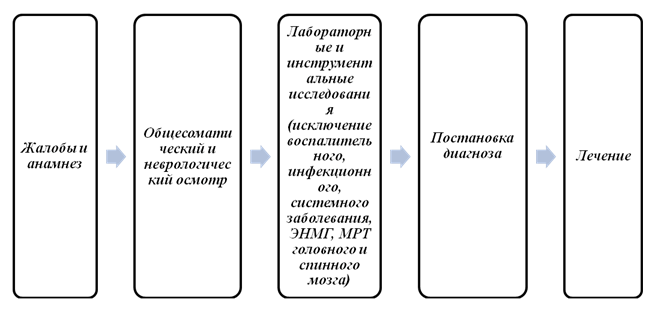
To make a diagnosis of Guillain-Barré syndrome, it is necessary to clearly determine the history of the development of the disease, together with an assessment of the neurological status, and compare it with the diagnostic criteria for GBS (WHO; 1993). It is advisable to perform a lumbar puncture with a study of the cerebrospinal fluid, as well as confirm the neural level of the lesion and clarify the form of the disease according to the ENMG examination.
Diagnostic algorithm:
GBS should first of all be differentiated from conditions that can lead to the development of acute peripheral tetraparesis. Differential diagnostic search is greatly simplified when using a unique algorithm developed by researchers from the Federal State Budgetary Institution “NTS” of the Russian Academy of Medical Sciences.
Differential diagnostic algorithm for acute flaccid tetraparesis (AFT)

Note: OVT-acute flaccid tetraparesis; EMG electromyography; PSP polyneuropathy; GBS - Guillain-Barre syndrome; LP - lumbar puncture; BHAK - biochemical blood test; RF - rheumatic factor; CRP - C-reactive protein; CPK - creatinine phosphokinase; MRI - magnetic resonance imaging (at least 1 Tesla); CT - computed tomography.
Laboratory research: see outpatient level (for those examinations that were listed additionally).
List of basic laboratory tests:
· blood for immunoglobulins - when planning specific therapy with class G immunoglobulins, it is necessary to determine Ig fractions in the blood, low concentrations of IgA are usually associated with its hereditary deficiency, in such cases there is a high risk of developing anaphylactic shock (immunoglobulin therapy is contraindicated);
· studies of cerebrospinal fluid (cytosis, protein concentration). When analyzing cerebrospinal fluid, the following three indicators are usually considered among the diagnostic criteria confirming GBS:
presence of high protein content,
increased albumin fraction,
· absence of concomitant increase in cytosis.
Additionally, the following diagnostic tests may be recommended to confirm the diagnosis and clarify the characteristics of GBS in a particular case:
· blood test for autoantibodies to gangliosides, with mandatory testing of GM1, GD1a, and GQ1b if the patient has oculomotor disorders;
· blood test for IgA antibodies to Campylobacter jejuni;
· study of the content of biomarkers of neurofilament heavy chains, tau protein and gliofibrillary acidic protein in blood serum.
Instrumental studies: see outpatient level.
In severe cases of the disease (rapid progression, bulbar disorders), 24-hour monitoring of blood pressure, ECG, pulse oximetry and examination of pulmonary function (spirometry, peak flowmetry), monitoring of pulmonary function (determination of vital capacity of the lungs) should be carried out (in an intensive care unit). ) for timely identification of indications for transferring the patient to mechanical ventilation.
Differential diagnosis
GBS must be differentiated from other diseases manifested by acute peripheral paresis, primarily from poliomyelitis (especially in young children) and other polyneuropathies (diphtheria, porphyria). In addition, lesions of the spinal cord and brain stem (transverse myelitis, stroke in the vertebrobasilar system) and diseases with impaired neuromuscular transmission (myasthenia gravis, botulism) may have a similar clinical picture.
|
Diagnosis |
Rationale for differential diagnosis |
Surveys |
Exclusion criteria diagnosis |
| Poliomyelitis (especially in young children) | Acute peripheral paresis |
· ENMG; · needle EMG; · consultation with a therapist; · consultation infectious disease specialist |
· epidemiological anamnesis; · presence of fever at the onset of the disease; · symptoms from the gastrointestinal tract; asymmetry of the lesion; · absence of objective sensitivity disorders; high cytotosis in the cerebrospinal fluid; · the diagnosis of poliomyelitis is confirmed using virological or serological studies. |
|
Other polyneuropathies (inflammatory: chronic inflammatory polyneuropathy with acute onset, Sjögren's disease, Churg-Strauss disease, cryoglobulinemic vasculitis; Infectious: HIV-associated, Lyme disease; Toxic: diphtheria, porphyria, medicinal, acute alcohol, heavy metal poisoning Dysmetabolic: polyneuropathy of critical conditions, with renal, liver failure, acute hyperglycemic polyneuropathy) |
Acute peripheral paresis |
· ENMG; · needle EMG; · Consultant therapist; · infectious disease specialist; · biochemical blood and urine tests |
· signs of the current denervation-reinnervation process; · in favor of porphyria is the combination of predominantly motor polyneuropathy with severe abdominal pain, intestinal paresis, arterial hypertension, tachycardia, severe mental changes (from depression to delirium), sleep disturbance, and epileptic seizures. Porphyria has a change in the color of urine, which in the light takes on a reddish tint, and then a rich reddish-brown color |
|
Transverse myelitis. Lesion at the level of the cervical thickening of the spinal cord (C4 - Th2) post-infectious (M.pneumoniae, Schistosoma), post-vaccination, viral (enteroviruses, herpes), myelitis associated with HIV, with demyelinating diseases of the central nervous system, with systemic diseases (systemic red lupus, Sjögren's disease, acute necrotizing vasculitis) |
Acute peripheral paresis |
MRI of the spinal cord and brain; · ENMG; · cons. therapist; · infectious disease consultant. |
· segmental border of sensory impairment; · persistent pelvic disorders; · lack of involvement of facial and respiratory muscles in severe tetraparesis. |
|
Acute violation of spinal circulation, in the vertebero-basilar region. (spinal cord vascular thrombosis, vascular malformation, aneurysm, compression, trauma, spinal cord neoplasm) |
Acute peripheral paresis |
MRI of the brain and spinal cord; · ENMG; · cons. therapist; · cons. neurosurgeon. |
Acute development (usually within a few minutes); · in most cases, depression of consciousness (coma); · The diagnosis is finally confirmed using MRI of the brain/spinal cord. |
| Myasthenia gravis | Acute peripheral paresis | · ENMG. |
· variability of symptoms; · absence of sensory disorders; characteristic changes in tendon reflexes; · the diagnosis is confirmed using EMG (detection of the decrement phenomenon); · positive pharmacological test with proserin. |
| Botulism | Acute peripheral paresis |
· ENMG; · infectious disease consultant. |
· relevant epidemiological data, · descending type of spread of paresis, · safety in some cases of tendon reflexes, absence of sensory disorders, no changes in quora. |
Treatment abroad
Get treatment in Korea, Israel, Germany, USA
Get advice on medical tourism
Treatment
Drugs (active ingredients) used in treatment
Treatment (outpatient clinic)
OUTPATIENT TREATMENT
Treatment tactics:
Suspicion of Guillain-Barré syndrome, even with minimal severity of symptoms, is the basis for emergency hospitalization, and symptomatic treatment is carried out at the outpatient stage, and when a diagnosis is made, they are sent to the hospital, and the patient and his relatives must be warned about the possible rapid deterioration of the condition.
Non-drugtreatment: No.
Drug treatment:
Symptomatic therapy:
· if blood pressure increases, nifedipine can be prescribed, 10-20 mg sublingually;
To reduce tachycardia, propranalol is used, at an initial dose of 20 mg 3 times a day; then the dose is gradually increased to 80-120 mg in 2-3 doses, under the control of blood pressure, heart rate, ECG;
· for bradycardia - atropine, adults: intravenous bolus under ECG and blood pressure control - 0.5-1 mg, if necessary, repeat the administration after 3-5 minutes; maximum dose 0.04 mg/kg (3 mg). Children - 10 mcg/kg;
To reduce pain, analgesics and non-steroidal anti-inflammatory drugs are administered:
· ketorolac, orally in a single dose of 10 mg or repeatedly, depending on the severity of the pain syndrome, 10 mg up to 4 times a day. The maximum daily dose should not exceed 40 mg, or no more than 60 mg is administered intramuscularly in 1 administration; usually 30 mg every 6 hours.
Diclofenac, intramuscularly. Single dose - 75 mg, maximum daily dose - 150 mg (with a break between doses of at least 30 minutes).
ibuprofen, 1-2 tablets 3-4 times a day; if necessary - 1 tablet every 4 hours. Do not take more often than every 4 hours. The maximum daily dose for adults should not exceed 1200 mg (no more than 6 tablets in 24 hours).
Algorithm of action in emergency situations: symptomatic therapy measures.
Other types of treatment: No.
· consultation with an infectious disease specialist - establishing or ruling out an infectious disease (infectious mononucleosis, Lyme disease, HIV, etc.);
· consultation with a therapist - establishing or excluding a therapeutic disease (inflammatory disease of internal organs: lungs, kidneys, liver, etc.);
· consultation with an endocrinologist, nephrologist, rheumatologist - if necessary, to exclude somatic pathology.
Preventive actions:
· There is no specific prevention of the disease; doctors can recommend treating all infectious diseases at the very beginning of their development, this will reduce the negative impact of pathogens on the nervous system.
Patient monitoring:
· assessment of the patient’s general condition with a description of the condition of the skin; patient's weight;
· hemodynamic indicators: number of respiratory movements, A/D, heart rate, Pulse;
· assessment of neurological status.
· etiopathogenetic treatment is not carried out at this stage, and therefore there are no indicators.
Treatment (ambulance)
DIAGNOSIS AND TREATMENT AT THE EMERGENCY CARE STAGE
Diagnostic measures:
GBS often has an acute course and is potentially life-threatening, because the lesion, starting from the legs, progresses, spreading to the bulbar and other cranial nerves, and therefore the following measures are necessary:
Swallowing assessment- in case of bulbar palsy, swallowing impairment, to prevent aspiration
· nasogastric tube.
Breathing assessment- possible development of progressive respiratory failure, and not only of the obstructive type due to bulbar paralysis, but also with damage to the phrenic nerve (characterized by a paradoxical type of breathing - when inhaling, the anterior abdominal wall collapses) and intercostal nerves.
· tracheal intubation (for further transfer of the patient to mechanical ventilation).
Heart function assessment:
· ECG - decrease and even inversion of the S-T segment, increase in the Q-T interval, cardiac arrest is possible.
During transportation, it is important to take care to maintain airway patency, carefully monitor blood pressure and heart rhythm (tachycardia, orthostatic hypotension, arrhythmia, etc.).
Drug treatment:
· syndromic therapy according to the emergency medical care protocol.
Treatment (inpatient)
INPATIENT TREATMENT
Treatment tactics: The main goal of treatment is: restoration of vital functions, elimination of symptoms of an autoimmune disease using specific techniques, rehabilitation period of the patient, prevention of complications. The first thing that needs to be done is to place the patient in a hospital, and, if necessary, connect him to a ventilator, install a catheter in case of difficulty urinating, install a nasogastric tube in case of difficulty swallowing.
Non-drug treatment:
In severe cases with severe paresis, proper care is of particular importance to prevent complications associated with prolonged immobility of the patient (infections, bedsores, pulmonary embolism). It is necessary to periodically (at least once every 2 hours) change the patient’s position, skin care, control over the functions of the bladder and intestines, passive exercises, and prevention of aspiration. In cases of persistent bradycardia and the risk of developing asystole, installation of a temporary pacemaker may be required.
Drug treatment:
Specific therapy for Guillain-Barré syndrome, aimed at stopping the autoimmune process, currently uses pulse therapy with class G immunoglobulins and plasmapheresis (see paragraph - other types of treatment). The effectiveness of each method is relatively the same, so their simultaneous use is considered inappropriate.
Immunoglobulin class G, like plasmapheresis, reduces the duration of stay on mechanical ventilation; it is administered intravenously daily for 5 days at a dose of 0.4 g/kg. Possible side effects: nausea, headaches, muscle pain, fever.
Symptomatic therapy for Guillain-Barré syndrome is carried out to correct disturbances in the acid-base and water-electrolyte balance, correct blood pressure levels, and prevent deep vein thrombosis and thromboembolism.
Infusion therapy for the correction of acid-base and water-electrolyte imbalances, severe arterial hypotension.
For persistent severe arterial hypertension, antihypertensive drugs (β-blockers or slow calcium channel blockers) are prescribed (see KP Arterial hypertension).
For severe tachycardia, β-blockers (propranolol) are prescribed; for bradycardia, atropine is prescribed (see below).
With the development of intercurrent infections, antibiotic therapy is necessary (broad-spectrum drugs are used).
To prevent deep vein thrombosis and pulmonary embolism, low molecular weight heparin is prescribed in prophylactic doses twice a day).
For pain of nociceptive origin (muscular, mechanical), NSAIDs are recommended; in the case of neuropathic pain, the drugs of choice are gabapentin, carbamazepine, pregabalin (for adults only!) (see below).
List of essential medicines:.
| Drugs | Single dose | Frequency of administration |
| Immunoglobulin class G | 0.4 g/kg i.v. | . 0.4 g/kg/day for 5 days, 1 time per day, 5 days. |
| gabapentin | 300 mg |
1 day 300 mg 1 time / day, 2 day 300 mg 2 times / day, 3 day 300 mg 3 times / day, then depending on individual tolerability and effectiveness, the dose can be increased by 300 mg/day every 2-3 days to a maximum of 3600 mg/day. |
| carbamazepine | 200 mg | The recommended starting dose is 200-400 mg per day. The dose can be gradually increased until a satisfactory clinical effect is obtained, in some cases it can be 1600 mg per day. After the pain syndrome goes into remission, the dosage can be gradually reduced |
| pregabalin | 150 mg | Treatment begins with a dose of 150 mg per day, divided into two or three doses. Depending on the patient's individual response and tolerability, after 3-7 days the dose can be increased to 300 mg per day, and, if necessary, after another 7 days - to a maximum dose of 600 mg per day. |
List of additional medicines:.
| Drugs | Single dose | Frequency of administration |
| nifedipine | 10 mg | 1-2 times under the tongue |
| Propranalol | 10 mg | 20 mg 3 times a day, then the dose is gradually increased to 80-120 mg in 2-3 doses, under the control of blood pressure, heart rate, ECG |
| Atropine | 0,5-1,0 | adults: intravenous bolus under ECG and blood pressure monitoring - 0.5-1 mg, if necessary, repeat the administration after 3-5 minutes; maximum dose 0.04 mg/kg (3 mg). Children - 10 mcg/kg; |
| Ketorolac | 10 mg | orally once in a dose of 10 mg or repeatedly, depending on the severity of the pain syndrome, 10 mg up to 4 times a day. The maximum daily dose should not exceed 40 mg, or no more than 60 mg is administered intramuscularly in 1 administration; usually 30 mg every 6 hours. Not used in children. |
| Diclofenac | 75 mg | intramuscularly, single dose 75 mg, maximum daily dose - 150 mg (with a break between doses of at least 30 minutes). Children do not apply. |
| Ibuprofen | 0.2 g |
1-2 tablets 3-4 times a day; if necessary - 1 tablet every 4 hours. Do not take more often than every 4 hours. The maximum daily dose for adults should not exceed 1200 mg (no more than 6 tablets in 24 hours). Children: 10-20 mg/kg 3 times a day for 2-3 days. |
Surgical intervention, indicating indications for surgical intervention: Surgical intervention may be necessary for tracheostomy in case of prolonged mechanical ventilation (more than 10 days), as well as gastrostomy for severe and prolonged bulbar disorders.
Other types of treatment:
You should always remember the exceptional importance of a set of rehabilitation measures to prevent complications due to the patient’s immobility and to maintain the functional state of the muscles until a sufficient range of independent movements appears.
The patient needs:
- Physiotherapy
- Massage has a beneficial effect on metabolism, which also accelerates nerve growth and reinnervation
- Physiotherapy to prevent the formation of contractures (electrical stimulation, heat therapy, medicinal electrophoresis, etc.).
- Hyperbaric oxygenation.
Membrane plasmapheresis significantly reduces the severity of paresis and the duration of mechanical ventilation. As a rule, 4-6 sessions are carried out with an interval of one day; the volume of plasma replaced in one session must be at least 40 ml/kg. 0.9% sodium chloride solution or rheopolyglucin are used as replacement media.
You should remember about contraindications to plasmapheresis (infections, bleeding disorders, liver failure), as well as possible complications (electrolyte imbalance, hemolysis, allergic reactions).
Indications for consultation with specialists:
· consultation with an infectious disease specialist if necessary (in the absence of a specialist at the pre-hospital level) - establishment or exclusion of a chronic infection (brucellosis, borreliosis, etc.), as well as in the case of confirmation of an infectious agent for correction of etiological therapy;
· consultation with a therapist if necessary (in the absence of a specialist at the pre-hospital level) - establishment or exclusion of a therapeutic disease (inflammatory disease of internal organs: lungs, kidneys, liver, etc.), correction of hemodynamic parameters, electrolyte balance during therapy;
· consultation with an ICU doctor - treatment of patients with severe forms of Guillain-Barré syndrome is carried out jointly with a doctor in the intensive care unit;
· consultation with a cardiologist - in case of severe cardiovascular disorders (persistent severe arterial hypertension, arrhythmias).
Indications for transfer to the intensive care unit:
· severe and extremely severe degree of neurological disorders;
hemodynamic instability;
· respiratory dysfunction.
Indicators of treatment effectiveness:
· stabilization of the immunological status (quantitative and qualitative composition of IgG in blood and cerebrospinal fluid);
· regression of focal neurological symptoms.
Further management.
After the patient’s health status has normalized, he must be registered with a neurologist. In addition, it will be necessary to undergo preventive examinations in order to identify the preconditions for relapse of the disease at an early stage. Dispensary observation in the clinic at the place of residence.
After the end of the acute period, comprehensive rehabilitation measures are necessary, the plan of which is drawn up individually depending on the severity of residual symptoms (physical therapy, massage, while thermal procedures are contraindicated!).
Patients who have had GBS. should be informed of the need to observe a protective regime for at least 6-12 months after the end of the disease. Physical overload, overheating, hypothermia, excessive insolation, and alcohol intake are unacceptable. You should also refrain from vaccination during this period.
Medical rehabilitation
is carried out in accordance with the Standard for organizing the provision of medical rehabilitation to the population of the Republic of Kazakhstan, approved by order of the Minister of Health of the Republic of Kazakhstan dated December 27, 2013 No. 759.
Palliative care
Depending on the type and severity of complications following the illness, additional treatment may be required, such as:
· immobilized patients are prescribed heparin subcutaneously at a dose of 5,000 units every 12 hours and temporary compression of the calf muscles to prevent deep vein thrombosis;
· massage has a beneficial effect on metabolism, which also accelerates nerve growth and reinnervation;
· kinesiotherapy has been proven to stimulate reinnervation and restore muscle volume;
· physical therapy to improve strength, to prevent the formation of contractures (electrical stimulation, heat therapy, medicinal electrophoresis);
· rehabilitation to develop daily living skills and the use of adaptive products to help with daily living;
· the patient may require orthopedic aids or other auxiliary methods to improve movement;
· psychotherapy;
Hospitalization
Indications for planned hospitalization: No.
Indications for emergency hospitalization:
· patients with GBS are subject to hospitalization in the intensive care unit.
Information
Sources and literature
- Minutes of meetings of the Joint Commission on the Quality of Medical Services of the Ministry of Health and Social Development of the Republic of Kazakhstan, 2016
- 1. Bykova O. V., Boyko A. N., Maslova O. I. Intravenous use of immunoglobulins in neurology (Literature review and own observations) // Nevrol. magazine - 2000, 5. P.32-39. 2. Gekht B. M., Merkulova D. M. Practical aspects of the clinic and treatment of polyneuropathies // Neurol. journal - 1997. - No. 2. - P. 4-9. 3. Piradov M.A., Suponeva N.A. “Guillain-Barré syndrome: diagnosis and treatment. Guide for doctors" - 2011. 4. Suponeva N.A., Piradov M.A. “Intravenous immunotherapy in neurology” -2013. 5. Sladky J. T. Guillain-Barre syndrome in children // J. Child Neurol. 2004. V. 19. P. 191–200. 6. Schmidt B., Toyka K. V., Kiefer R. et al. Inflammatory infiltrates in sural nerve biopsies in Guillain-Barre syndrome and chronic inflammatory demyelinating neuropathy // 1996. V. 19. P. 474–487. 7. Khalili-Shirazi A., Hughes R. A., Brostoff S. W. et al. T cell responses to myelin proteins in Guillin-Barre syndrome // J. Neurol. Sci. 1992. V. 111. P. 200–203. 8. Van Rhijn I., Bleumink-Pluym N. M., Van Putten J. P. et al. Campylobacter DNA is present in circulating myelomonocytic cells of healthy persons and in persons with Guillain-Barre syndrome // J. Infect. Dis. 2002. V. 185. P. 262–265. 9. Cooper J. C., Ben-Smith A., Savage C. O. et al. Unusual T cell receptor phenotype V gene usage of gamma delta T cells in a line derived from peripheral nerve of a patient with Guillain-Barre syndrome // J. Neurol. Neurosurg. Psychiatry. 2000. V. 69. P. 522–524. 10. Ilyas A. A., Chen Z. W., Cook S. D. et al. Immunoglobulin G subclass distribution of autoantibodies to gangliosides in patients with Guillain-Barre syndrome // Res. Commun. Pathol. Pharmacol. 2002. V. 109. P. 115–123. 11. Tsang R. S., Valdivieso-Garcia A. Pathogenesis of Guillain-Barre syndrome // Expert Rev. Anti Infect. Ther. 2003. V. 1. P. 597–608. 12. Kieseier B. C., Kiefer R., Gold R. et al. Advances in understanding and treatment of immune-mediated disorders of the peripheral nervous system // Muscle Nerve. 2004. V. 30. P. 131–156. 13. Adams D., Gibson J. D., Thomas P. K. et al. HLA antigens in Guillain-Barre syndrome // Lancet. 1977. No. 2. P. 504–505. 14. Koga M., Yuki N., Kashiwase K. et al. Guillain-Barre and Fisher’s syndromes subsequent to Campylobacter jejuni enteritis are associated with HLA-54 and Cwl independent of anti-ganglioside antibodies // J. Neuroimmunol. 1998. V. 88. P. 62–66. 15. Magira E. E., Papaijakim M., Nachamkin I. et al. Differential distribution of HLA-DQ beta/DR beta epitopes in the two forms of Guillain-Barre syndrome, acute motor axonal neuropathy and acute inflammatory demyelinating polyneuropatrhy (AIDP); identification of DQ beta epitopes associated with susceptibility to and protection from AIDP // J. Immunol. 2003. V. 170. P. 3074–3080. 16. Geleijns K., Schreuder G. M., Jacobs B. C. et al. HLA class II alleles are not a general susceptibility factor in Guillain-Barre syndrome // Neurology. 2005. V. 64. P. 44–49. 17. Asbury A. K., Cornblath D. R. Assessment of current diagnostic criteria for Guillain-Barre syndrome // Ann. Neurol. 1990. V. 27. S. 21–24.
Information
ABBREVIATIONS USED IN THE PROTOCOL
| CIDP | chronic inflammatory demyelinating polyradiculoneuropathy |
| PNP | polyneuropathy |
| NMSP | hereditary motor-sensory polyneuropathy |
| SGB | Guillain-Barre syndrome |
| HELL | arterial hypertension |
| PNS | peripheral nervous system |
| CNS | central nervous system |
| MRI | Magnetic resonance imaging |
| PCR | polymerase chain reaction |
| CSF | cerebrospinal fluid |
| ESR | erythrocyte sedimentation rate |
| Ig | immunoglobulin |
| Heart rate | heart rate |
| AIDS | acquired immunodeficiency syndrome |
| EMG | electromyography |
| ENMG | electroneuromyography |
| IVIG | normal human immunoglobulin for intravenous administration |
| GK | glucocorticoids |
List of protocol developers with qualification information:
1. Kayshibaeva Gulnaz Smagulovna, Candidate of Medical Sciences, JSC “Kazakh Medical University of Continuing Education”, head of the Department of Neurology, certificate “adult neuropathologist”.
2. Zhumagulova Kulparam Gabibulovna, candidate of medical sciences, certificate “adult neuropathologist of the highest category”, JSC “Kazakh Medical University of Continuing Education”, associate professor of the department of neurology.
3. Tuleutaeva Raikhan Yesenzhanovna, clinical pharmacologist, candidate of medical sciences, professor of RAE, head of the department of pharmacology and evidence-based medicine of Semey State Medical University.
Disclosure of no conflict of interest: No.
List of reviewers:
Dushchanova G.A. - Doctor of Medical Sciences, Professor, Head of the Department of Neurology, Psychiatry and Psychology of the South Kazakhstan State Pharmaceutical Academy.
Indication of the conditions for reviewing the protocol: Review of the protocol 3 years after its publication and from the date of its entry into force or if new methods with a level of evidence are available.
Attached files
Attention!
- By self-medicating, you can cause irreparable harm to your health.
- The information posted on the MedElement website and in the mobile applications "MedElement", "Lekar Pro", "Dariger Pro", "Diseases: Therapist's Guide" cannot and should not replace a face-to-face consultation with a doctor. Be sure to contact a medical facility if you have any illnesses or symptoms that concern you.
- The choice of medications and their dosage must be discussed with a specialist. Only a doctor can prescribe the right medicine and its dosage, taking into account the disease and condition of the patient’s body.
- The MedElement website and mobile applications "MedElement", "Lekar Pro", "Dariger Pro", "Diseases: Therapist's Directory" are exclusively information and reference resources. The information posted on this site should not be used to unauthorizedly change doctor's orders.
- The editors of MedElement are not responsible for any personal injury or property damage resulting from the use of this site.
Guillain-Barré syndrome is an acute form of progressive inflammatory polyneuropathy, which is characterized by muscle weakness and polyneuritic sensitivity disorder. The disease is also called acute idiopathic polyneuritis, Landry's palsy, or inflammatory demyelinating polyradiculoneuropathy. The disease is a representative of autoimmune abnormalities. Typically, pathology has specific signs that make it possible to recognize it at the earliest stages of development and begin adequate treatment in time. It has been proven that more than 80% of patients have a favorable prognosis and are completely cured.

autoimmune demyelination of nerves in Guillain-Barré syndrome
Guillain-Barré syndrome (GBS) is observed in all age groups, but people aged 35-50 years are especially susceptible to it, with equal frequency in both women and men. The incidence per 100,000 people ranges from 0.4 to 4 cases.
Causes of the disease
Scientists from different countries have been studying the syndrome for 100 years, but they still cannot fully figure out the exact reasons that provoke the occurrence of the disease.
 It is believed that the appearance and development of the anomaly occurs due to a disruption of the patient’s immune system. When a person is completely healthy, when foreign cells enter the body, the immune system begins to fight the infection, rejecting all its dangerous elements. The patient is recovering. With GBS, the body begins to confuse “friends and foes”: the patient’s neurons are perceived as foreign and are subject to “attack.” When the nervous system is destroyed, a syndrome occurs.
What causes disturbances in the functioning of the immune system itself is not completely known. Experts include the most common reasons:
- Traumatic brain injuries. A strong blow to the head, any damage to it, as well as swelling, tumors or hemorrhages in the brain can be the main factor in the development of the syndrome. That is why, when a patient consults a specialist, first of all, the doctor must find out about the presence of any traumatic brain injuries.
- Infections. Recent viral infections greatly weaken a person's immune system, thereby increasing the likelihood of developing GBS. The body's defense mechanism perceives neurons as an infection and, with the help of white blood cells, continues to kill them. In this case, the syndrome appears one to three weeks after the infectious disease.
- Allergies. The disease often develops in allergy sufferers, for example, after chemotherapy, vaccination against polio and diphtheria, or severe surgical interventions.
- Genetic predisposition. Most diseases are inherited, and Guillain-Barre syndrome is no exception. If someone in the family has already suffered from a pathology, then most likely it will occur in their descendants. In this case, you need to especially monitor your health: take care of your head and try not to develop infectious diseases.
In childhood, the syndrome develops extremely rarely. The disease may be congenital or acquired. Any anomalies of intrauterine development can lead to the development of the syndrome:
- Preeclampsia;
- Frequent use of medications during pregnancy;
- The presence of autoimmune diseases in the mother;
- Long-term infections during pregnancy;
- Use of drugs, alcohol or smoking.
Acquired reasons include:
- Passive smoking of a child;
- Hormonal imbalances in the body;
- Vaccinations;
- Self-medication;
- Metabolic disease;
- Neurological disorders;
- Development of tumor processes.
Symptoms

The disease can manifest itself in 3 forms:
- Spicy. All signs of the disease appear simultaneously within 1-2 days.
- Sub-acute. The incubation period is 15 to 20 days.
- Sluggish, chronic. This is one of the most dangerous forms, as it is practically untreatable.
The first symptoms of GBS in both children and adults resemble infection with a common viral respiratory infection:
- Aches in bones, joints;
- Hyperthermia;
- Severe weakness;
- Inflammation of the upper respiratory tract;
- Numbness of the limbs;
- Sometimes patients are bothered by various gastrointestinal disorders.
In addition to the general signs, there are also more obvious ones:
- Weakness of the limbs. Due to the destruction of nerve cells, a decrease or complete loss of sensitivity in the muscle area occurs. At first, pain appears only in the lower legs, then discomfort affects the hands and feet. The patient is bothered by tingling and numbness in the fingers. In severe cases, coordination of movements is impaired: it becomes difficult for a person to independently hold a pen or write with the affected limb. It is worth noting that the symptoms of the disease appear symmetrically: 2 arms or legs are simultaneously affected.
- Enlargement of the abdomen, which is noticeable even visually. A protruding abdomen is one of the main indicators of the presence of the disease. This occurs due to the fact that the patient’s breathing changes to the abdominal type due to a weakening of the diaphragm.
- Incontinence. In a person with GBS, the healthy functioning of the bladder is impaired and urine begins to leak involuntarily.
- Difficulty swallowing. The swallowing reflex is impaired due to weakened pharyngeal muscles. In this case, the patient may even choke on saliva. The muscles of the mouth gradually weaken, which leads to discomfort while chewing food.
Guillain-Barré syndrome affects almost all organ systems, so unreasonable hypertension, tachycardia or banal visual impairment may be the first, albeit hidden, sign of the development of pathology.
How dangerous is the course of the syndrome?
Usually the anomaly develops slowly over 2-3 weeks. At first, there is a slight weakness in the joints, which intensifies over time and really begins to cause discomfort to the patient.

Immediately after tingling, in the acute course of the disease, general malaise and weakness in the shoulder and hip regions occur. After a few hours, breathing difficulties appear. In this case, it is imperative to seek help from a hospital. Typically, the patient is immediately connected to an artificial respiration system, and then given the necessary medication and physiotherapeutic treatment.
In the acute form of the disease, the pathology can completely paralyze any limb on the second or third day.
Also, in the absence of timely treatment, the patient faces:
- Decreased immunity;
- Respiratory failure;
- Inactivity of joints;
- Peripheral paralysis;
- Problems of adaptation in society;
- Difficulty in living;
- Disability;
- Death.
Diagnosis of the disease
In order to diagnose a patient with GBS, several aspects need to be clarified:
- When was the last time a person had any viral disease? It has been proven that in 80% of cases the syndrome occurs due to recent infections.
- Is the patient currently taking medications and, if so, what medications. They will also help cause the development of GBS.
- How long ago the patient was vaccinated against any diseases.
- Does the patient suffer from autoimmune or tumor diseases?
- Has the person undergone surgery recently?
- Whether there were serious injuries to any part of the body.

The following studies should also be carried out:
- General blood and urine analysis;
- Blood chemistry;
- Serological and virological examinations;
- Cerebrospinal fluid examination;
- Magnetic resonance imaging;
- Electrocardiography;
- Registration of electrical activity of muscles;
- X-ray or ultrasound of the affected area;
- External respiration examination;
- Study of basic vital signs.
Muscle weakness in several limbs simultaneously and tendon areflexia can be another clear sign of Guillain-Barré syndrome. This also includes various disorders in the pelvic area, polymorphonuclear leukocytes, asymmetries of paresis and sensitivity disorders.
Differential diagnosis
Despite the fact that the symptoms of GBS are similar to those of many other diseases (diphtheria, porphyria, transverse myelitis, botulism and myasthenia), they still need to be distinguished for further correct treatment. When making a differential diagnosis, the following factors should be taken into account:
- If polio is suspected, it is necessary to collect data from an epidemiological study, take into account symptoms of the gastrointestinal tract, identify high cytosis in the cerebrospinal fluid, asymmetry of the lesion and the absence of sensory impairment. The diagnosis can be confirmed using serological or virological analysis.
- Polyneuropathy is characterized by the appearance of psychopathological signs, as well as pain in the pelvic and abdominal areas. Deviation of the main indicators from the norm in the urine also indicates the development of the disease.
- Transverse myelitis accompanies dysfunction of the pelvic organs and the absence of damage to the nerves of the skull.
- Symptoms of the anomaly can be confused with cerebral infarction. But in this case, the pathology affects the body in a few minutes and often leads to coma. MRI will help determine the exact cause of the dysfunction of body systems.
- Botulism is characterized by the absence of sensitivity disorders and any changes in the cerebrospinal fluid.
Treatment

Patients diagnosed with GBS must be hospitalized in a hospital. In approximately 30% of cases, mechanical ventilation is necessary. Pathology therapy is performed at the following levels:
- Reanimation;
- Symptomatic;
- Blood purifying;
- Drug;
- Muscular-restorative;
- Preventive.
Resuscitation therapy
If the anomaly is in acute form, resuscitation treatment is carried out, which is aimed at relieving symptoms:
- The patient is connected to an artificial respiration system;
- A catheter is used to remove urine;
- A tracheal tube and probe are installed if there are problems with swallowing.
Symptomatic therapy
This type of treatment is carried out using various medications:

- Antihypertensive drugs: “Anaprilin”, “Metaprolol”;
- Antibiotic therapy: “Norfloxacin”;
- Drugs that help stabilize heart rate and blood pressure: Propranolol, Anaprilin (for tachycardia), Piracetam (for bradycardia);
- Low molecular weight heparin: “Hemapaxan”, “Certoparin”;
- Pain-relieving drugs – NSAIDs or Gabapentin, Pregabalin;
- Antipyretics, when the temperature rises above 38 degrees: “Ibuklin”, “Next”;
- Laxatives: Bisacodyl, Laxatin.
Plasmapheresis
One of the most effective procedures aimed at treating GBS is hardware blood purification - plasmapheresis. It helps stop the autoimmune process in the body. Indicated for severe and moderate disease. Usually about 4-6 operations are performed with a break of one day. Instead of plasma, a special isotonic solution of sodium or albumin is injected into the blood, through which the blood is cleansed and the functioning of all body systems is normalized.
Surgical treatment of the syndrome
If mechanical ventilation has been carried out for more than 7-10 days, a tracheostomy - an artificial windpipe - should be applied. In severe cases, a gastrostroma may be required - an opening in the stomach, surgically created for feeding the patient.
Non-drug therapy with folk remedies
It is impossible to cure GBS with folk remedies. But it is quite possible to cope with some of its symptoms:
- Fever. It is recommended to drink plenty of fluids and ventilate the room. Tea with lemon, infusions with different berries and dried fruits: cranberries, strawberries, currants, blueberries, raspberries and dried apricots will help lower the temperature. You can brew linden blossom, chamomile, St. John's wort, aspen buds, mint and thyme - leave for half an hour, then drink in small sips.
- Aching bones. Lingonberry tea, a compress of fresh cabbage leaves, horseradish and burdock, baths with pine extract or decoctions of medicinal herbs will help to cope with it.
- Weakness. Doctors recommend breathing fresh air and ventilating the room as often as possible. You should try to eat more proteins. You also need food rich in vitamins and microelements. And sweet strong tea or chocolate will help lift your spirits.
Rehabilitation
Due to the fact that the syndrome affects not only neurons, but also the periosteal muscles, the patient will have to learn to walk again and perform simple movements of the limbs.

To normalize healthy muscle function, you can use traditional treatment, which includes:
- Electrophoresis;
- Trituration;
- Radon baths;
- Massage;
- Baths to relax the body and muscle tone;
- Masks and compresses with paraffin or beeswax;
- Health-improving gymnastics.
During the recovery of the body, you should definitely go on a special therapeutic diet and at the same time take a course of vitamin preparations. Complexes containing calcium, potassium, magnesium and vitamin B will be especially useful.
Patients with GBS should register with a neurologist and undergo regular preventive examinations. It is worth remembering that timely, high-quality therapy can return the patient to a full life.
Prevention of the syndrome
There is no special prevention of pathology. Doctors can only advise against any vaccinations for a year so that the disease does not return again. After this time, vaccination is permitted, but only if it is truly necessary.
You should also stop drinking alcohol, avoid overheating and hypothermia, and reduce physical activity. In this case, relapse of the disease is excluded.
Forecast
Most often, with Guillain-Barré syndrome, the prognosis is favorable. Typically, normal limb function is restored after 7-12 months in 85% of people. The disease becomes chronic in 7-15% of cases. The fatality rate is approximately 5%. The cause of death may be respiratory failure, pneumonia or viral infections. But most often, all this can be prevented by contacting a specialist in time.
Video: lecture on Guillain-Barré syndrome
Video: Guillain-Barre syndrome in the program “Live Healthy”
