What parts does the joint consist of? Syndesmology. Human joints. Elbow joint and its structure
The skeleton is a passive part of the movement apparatus and is a system of levers of movement and support. Consequently, its individual elements must be naturally connected to each other in a movable manner, which would allow the body to move in space. Movable bone joints are primarily characteristic of the limb bones - the thoracic and pelvic bones.
At the same time, part of the skeleton serves as a support and protection for the soft parts of the body and internal organs, so the individual elements of the skeleton must be connected motionlessly. Examples include the bones of the skull and chest cavity. Based on this, we can note a wide variety of types of connections of skeletal bones, depending on the function performed and in connection with the historical development of a particular organism. Thus, all types of bone connections can be divided into two large groups: continuous or synarthrosis (synarthrosis) and intermittent, or diarthrosis (diarthrosis). Science studies the connection of skeletal bones syndesmology(syndesmologia).
Types of continuous bone connectionsThere are five types of continuous bone connections.
1. synsarcosis (synsarcosis) - connection of bones with the help of muscles. For example, the scapula is connected to the torso by the trapezius, rhomboid, serratus ventral, and atlantoacromial muscles. The humerus is connected to the body by the latissimus dorsi, internal and superficial pectoral and brachiocephalic muscles. This connection ensures maximum mobility of the connecting parts.
2. syndesmosis (syndesmosis) - connection of bones using fibrous connective tissue. There are several types of syndesmoses:
· ligaments (ligamentum) - formed by bundles of collagen fibers. In this way, the radius and ulna bones of the forearm, the fibula and tibia of the lower leg are connected. Ligaments are a very strong connection, ranking second after bones in strength. With age, the strength of ligaments increases. However, prolonged absence of physical activity leads to a decrease in the tensile strength of the ligaments;
· membranes (membrana) - formed by flat plates of collagen fibers. For example, the broad pelvic ligament, which connects the sacrum to the pelvic bone, or the membranes of the occipitoatlas joint;
· seams (sutura) - formed by connective tissue and located between the lamellar bones of the skull. There are several types of seams: 1) smooth or flat(sutura plana) - are a fragile connection. They are located between the paired nasal bones, nasal and incisive, nasal and maxillary, 2) gear(sutura serrata) - connection between the frontal and parietal paired bones, 3) scaly(sutura squamosa) - a connection in which the thinned edge of one bone overlaps the thinned edge of another bone. This is how the temporal and parietal bones connect. 4) leafy(sutura foliata) - a connection in which the edges of one bone in the form of leaves protrude far into the recesses of another bone. Such sutures are located between the bones of the brain part of the skull. Scale and leaf joints are the strongest joints;
3. synelastosis (synelastosis) - the connection of bones with the help of elastic fibrous connective tissue capable of stretching and resisting rupture. Synelastosis occurs where the bones move widely apart when moving. In this way, the arches, spinous and transverse processes of the vertebrae are connected. When the spinal column bends, these parts of the vertebrae move significantly away from each other. Elastic fibers are able to form powerful cords, forming the supraspinous and nuchal ligaments, which help connect the head and spinal column to each other.
4. synchondrosis (synchondrosis) - connection of bones using cartilage tissue - hyaline or fibrous. Synchondroses provide significant strength to the connection, allow some of its mobility, and perform a spring function, weakening shocks during movement. Hyaline cartilage has elasticity and strength, but it is brittle. Found in areas of limited mobility, such as connecting the epiphyses and diaphyses of long bones of young animals, or costal cartilages and bony ribs. Fibrous cartilage is elastic and durable. It is located in places with high joint mobility. An example is the intervertebral cartilaginous discs between the heads and fossae of adjacent vertebrae. If, with synchondrosis, there is a gap in the thickness of the cartilage, then this connection is called symphysis. This is how the pelvic bones are connected to each other, forming the pelvic suture - the symphysis.
5. synostosis (synostosis) - connection of bones using bone tissue. There is a complete lack of mobility in it, because they talk about bone fusion. Synostosis occurs between the 4th and 5th bones in the carpus and tarsus, between the bones of the forearm and tibia in ruminants and horses, and between the segments of the sacrum. With age, synostosis spreads in the skeleton; it occurs at the site of syndesmosis or synchondrosis. For example, ossification between the bones of the skull, between the epiphyses and diaphyses of tubular bones, etc. Based on the presence of synostosis, the age of the bones of the skeleton of the torso and skull is determined during forensic and veterinary examination.
Types of intermittent bone connectionsIn phylogenesis, this is the most recent type of bone connection, which appeared only in terrestrial animals. It allows for a large range of motion and is more complexly constructed than a continuous connection. This joint is called diarthrosis (joint). Characterized by the presence of a slit-like cavity between the articulating bones.
Joint structure
Joint - articulatio. In each joint there is a capsule, synovial fluid that fills the articular cavity, and articular cartilage that covers the surface of the connecting bones.
Joint capsule (capsula articularis) - forms a hermetically sealed cavity, the pressure in which is negative, below atmospheric. This promotes a tighter fit of the connecting bones. It consists of two membranes: outer or fibrous and inner or synovial. The thickness of the capsule is not the same in its different parts. Fibrous membrane- membrana fibrosa - serves as a continuation of the periosteum, which passes from one bone to another. Due to the thickening of the fibrous membrane, additional ligaments are formed. Synovial membrane- membrana synovialis - built of loose connective tissue, rich in blood vessels, nerves, folded with villi. Sometimes synovial bursae or protrusions form in the joints, located between the bones and muscle tendons. The joint capsule is rich in lymphatic vessels through which the components of the synovium flow. Any damage to the capsule and contamination of the joint cavity is dangerous to the life of the animal.
Synovia - synovia - viscous yellowish liquid. It is secreted by the synovial membrane of the capsule and performs the following functions: lubricates the articular surfaces of bones and relieves friction between them, serves as a nutrient medium for articular cartilage, and releases metabolic products of articular cartilage into it.
Articular cartilage - cartilago articularis - covers the contacting surfaces of bones. This is hyaline cartilage, smooth, elastic, reduces surface friction between bones. Cartilage is able to weaken the force of shocks during movement.
Some joints have intra-articular cartilage in the form meniscus(tibiofemoral) and disks(temporomandibular). Sometimes found in joints intra-articular ligaments- round (hip) and cruciform (knee). The joint may contain small asymmetrical bones (carpal and tarsal joints). They are connected to each other inside the joint by interosseous ligaments. Extra-articular ligaments- are auxiliary and additional. They are formed by thickening the fibrous layer of the capsule and hold the bones together, directing or limiting movement in the joint. There are lateral lateral and medial ligaments. When an injury or sprain occurs, the bones of the joint are displaced, that is, dislocated.
Rice. 1. Scheme of the structure of simple and complex joints
A, B – simple joint; B – complex joint
1 – pineal gland; 2 – articular cartilage; 3 – fibrous layer of the capsule; 4 – synovial layer of the capsule; 5 – articular cavity; 6 – recession; 7 – muscle; 8 – articular disc.
Types of joints
By structure There are simple and complex joints.
Simple joints- these are joints in which there are no intra-articular inclusions between the two connecting bones. For example, the head of the humerus and the glenoid fossa of the scapula are connected by a simple joint, in the cavity of which there are no inclusions.
Complex joints- these are bone connections in which between the connecting bones there are intra-articular inclusions in the form of discs (temporomandibular joint), menisci (knee joint) or small bones (carpal and tarsal joints).
By nature of movement There are uniaxial, biaxial, multiaxial and combined joints.
Uniaxial joints- movement in them occurs along one axis. Depending on the shape of the articular surface, such joints are block-shaped, helical and rotary. Trochlear joint(ginglym) is formed by part of a block, cylinder or truncated cone on one bone and corresponding depressions on the other. For example, the elbow joint of ungulates. Helical joint- characterized by movement simultaneously in a plane perpendicular to the axis and along the axis. For example, the tibiotalar joint of a horse and a dog. Rotator joint- movement occurs around the central axis. For example, the anlantoaxial joint in all animals.
Biaxial joints- movement occurs along two mutually perpendicular planes. According to the nature of the articular surface, biaxial joints can be ellipsoidal or saddle-shaped. IN ellipsoidal joints the articular surface on one joint has the shape of an ellipse, on the other there is a corresponding fossa (occipito-atlas joint). IN saddle joints both bones have convex and concave surfaces that lie perpendicular to each other (the joint of the tubercle of the rib with the vertebra).
Multi-axis joints- movement is carried out along many axes, since the articular surface on one bone looks like part of a ball, and on the other there is a corresponding rounded fossa (scapulohumeral and hip joints).
Axleless joint- has flat articular surfaces that provide sliding and slightly rotating movements. These joints include tight joints in the carpal and metatarsal joints between the short bones and the bones of their distal row with the metacarpal and metatarsal bones.
Combined joints- movement occurs simultaneously in several joints. For example, in the knee joint, movement occurs simultaneously in the patella and femorotibial joints. Simultaneous movement of paired jaw joints.
According to the shape of the articular surfaces joints are diverse, which is determined by their unequal function. The shape of the articular surfaces is compared with a certain geometric figure, from which the name of the joint comes.
Flat or sliding joints- the articular surfaces of the bones are almost flat, movements in them are extremely limited. They perform a buffer function (carpometacarpal and tarsometatarsal).
Cup joint- has a head on one of the articulating bones, and a corresponding depression on the other. For example, shoulder joints.
Ball and socket joint- is a type of cup-shaped joint, in which the head of the articulating bone is more prominent, and the corresponding depression on the other bone is deeper (hip joint).
Elliptical joint- has on one of the articulating bones an ellipsoidal shape of the articular surface, and on the other, accordingly, an elongated depression (atlanto-occipital joint and femorotibial joints).
Saddle joint- has concave surfaces on both articulating bones, located perpendicular to each other (temporomandibular joint).
Cylindrical joint- characterized by longitudinally located articular surfaces, of which one has the shape of an axis, and the other has the shape of a longitudinally cut cylinder (the connection of the odontoid process of the epistrophy with the arch of the atlas).
Trochlear joint- resembles a cylindrical shape, but with transverse articular surfaces, which may have ridges (ridges) and depressions that limit the lateral displacement of articulating bones (interphalangeal joints, elbow joint in ungulates).
Helical joint- a type of trochlear joint, in which there are two guide ridges on the articular surface and corresponding grooves or grooves on the opposite articular surface. In such a joint, movement can be carried out in a spiral, which allows it to be called spiral-shaped (the ankle joint of a horse).
Bushing joint- characterized by the fact that the articular surface of one bone is surrounded by the articular surface of another, like a sleeve. The axis of rotation in the joint corresponds to the long axis of the articulating bones (cranial and caudal articular processes in pigs and cattle).

Rice. 2. Shapes of joint surfaces (according to Koch T., 1960)
1 – cup-shaped; 2 – spherical; 3 – block-shaped; 4 – ellipsoid; 5 – saddle-shaped; 6 – helical; 7 – sleeve-shaped; 8 – cylindrical.
Types of joint movements
The following types of movements are distinguished in the joints of the limbs: flexion, extension, abduction, adduction, pronation, supination and twirling.
Flexion(flexio) - called such a movement in a joint in which the angle of the joint decreases, and the bones forming the joint come together at opposite ends.
Extension(extensio) - reverse movement when the angle of the joint increases and the ends of the bones move away from each other. This type of movement is possible in uniaxial, biaxial and multiaxial joints of the limbs.
Adduction(adductio) is the bringing of a limb to the median plane, for example when both limbs are brought closer together.
Abduction(abductio) - reverse movement when the limbs move away from each other. Adduction and abduction are possible only with multi-axial joints (hip and scapulohumeral). In plantigrade animals (bears), such movements are possible in the carpal and tarsal joints.
Rotation(rotatio) - the axis of movement is parallel to the length of the bone. Outward rotation is called supination(supinatio), inward rotation of the bone is pronation(pronatio).
Whirling(circumductio), - or conical movement, is better developed in humans and is practically absent in animals. For example, in the hip joint, when bending, the knee does not rest against the stomach, but is abducted to the side.
Development of joints in ontogenesisDuring the early stage of fetal development, all the bones are connected to each other continuously. Later, at 14-15 weeks of embryonic development in cattle, in the places where future joints are formed, the layer of mesenchyme between the two connecting bones resolves, and a gap filled with synovial fluid is formed. A joint capsule is formed along the edges, separating the resulting cavity from the surrounding tissue. It connects both bones and ensures complete tightness of the joint. Later, the cartilaginous anlages of the bones ossify, and hyaline cartilage is preserved only at the ends of the bones facing the inside of the articular cavity. Cartilage provides gliding and absorbs shock.
By the time of birth, all types of joints in ungulates are formed. Newborns are immediately able to move and after just a few hours they are able to develop high speeds.
In the postnatal period of ontogenesis, any changes in the maintenance and feeding of animals are reflected in the connection of bones with each other. One connection is replaced by another. In the joints, the articular cartilage becomes thinner, the composition of the synovium changes or it disappears, which leads to ankylosis - fusion of bones.
GENERAL INFORMATION
Arthrology is a branch of anatomy that studies the joints of bones. According to development, structure and function, all bone connections can be divided into 2 large groups: continuous and discontinuous. Continuous joints (synarthroses) are formed by various types of connective tissue. Intermittent joints (diarthrosis) are characterized by the presence of a cavity between the articulating surfaces of bones.
Depending on the type of tissue connecting the bones, three types of continuous connections are distinguished.
1. Syndesmosis, syndesmosis, is a type of continuous connection of bones through connective tissue. Syndesmoses include ligaments, interosseous membranes, sutures, fontanelles, and gomphosis. Fibrous ligaments, ligamenta, are fibrous bundles of connective tissue. Between the vertebral arches, the ligaments consist of elastic connective tissue (synelastosis), these are the yellow ligaments, ligament flava.
Interosseous membranes, membrana interossea, are connective tissue that fills large spaces between bones, for example, between the bones of the forearm and lower leg.
Sutures, suturae, are connective tissue that takes on the character of a thin layer between the bones of the skull.
Based on the shape of the connecting bone edges, the following sutures are distinguished:
A) serrated, sutura serrata, between the frontal and parietal bones, parietal and occipital bones of the skull.
B) scaly, sutura squamosa, between the edges of the temporal and parietal bones.
B) flat, sutura plana, between the bones of the facial skull.
Fontana, fonticuli, are non-ossified connective tissue areas of the cranial vault of a newborn.
Impaction, gomfosis, is the connection of a tooth with the bone tissue of the dental alveolus.
2.Cartilaginous connections, synchondrosis, synchondrosis, are continuous connections of bones through cartilage tissue. Synchondrosis can be temporary or permanent.
Temporary synchondroses include epiphyseal cartilages connecting the diaphyses and epiphyses of tubular bones; cartilage between the sacral vertebrae. Temporary synchondroses persist in childhood and are then replaced by a bone connection - synostosis.
Permanent synchondrosis is present between the first rib and the manubrium of the sternum. If a narrow gap is formed in the center of the synchondrosis, which does not have the character of an articular cavity with articular surfaces and capsule, then such a connection becomes transitional from continuous to discontinuous and is called a symphysis, symphysis, for example, the pubic symphysis, symphysis pubica.
3. Bone joints, synostoses, synostosis, are formed as a result of the replacement of temporary cartilage with bone tissue or at the site of syndesmosis, for example, during ossification of the sutures between the bones of the skull in old age.
Intermittent, or synovial, connections. These include joints, articulatio. These connections have a more complex structure and, unlike sedentary or completely motionless continuous connections, make possible various movements of parts of the human body.
A joint, articulatio, is an organ in which basic and auxiliary elements are distinguished.
Main elements of the joint:
Articular surfaces, facies articularis, are located on the bones at the points of their articulation with each other. In most joints, one of the articulating surfaces is convex - the articular head, and the other is concave - the articular cavity.
Articular cartilage, cartilago articularis, covers the articular surfaces. Most articular surfaces are covered with hyaline cartilage, and only some joints, such as the temporomandibular and sternoclavicular joints, have fibrocartilage.
Thanks to its elasticity, articular cartilage protects the ends of bones from damage during shocks and shocks.
The articular capsule, capsula articularis, surrounds the parts of the bones that articulate with each other and hermetically closes the joint. In the articular capsule there are: a) an outer fibrous membrane, built of dense fibrous connective tissue; b) the internal synovial membrane, which produces intra-articular fluid - synovium.
The articular cavity, cavitas articularis, is a slit-like space between the articular surfaces, which contains synovium.
Synovia is a viscous fluid that is located in the joint cavity. Synovia wets the articular surfaces, reducing friction during joint movements, provides nutrition to the articular cartilage and metabolism in the joint.
Auxiliary elements of the joint:
The articular disc, discus articularis, is a cartilaginous plate located between the articular surfaces and dividing the articular cavity into two chambers.
Articular menisci, menisci articularis, are curved cartilaginous plates located in the cavity of the knee joint between the condyles of the femur and tibia. Articular discs and menisci increase the contact area of the articular surfaces and act as shock absorbers and also play a role in movement.
The labrum, labrum articulare, is a cartilaginous rim attached to the edge of the articular cavity and increases its area and, consequently, the contact area of the articular surfaces.
Ligaments, ligamenta, form the ligamentous apparatus of the joint, apparatus ligamentosus. Ligaments strengthen the joint, inhibit movement, and can also guide movement.
There are: a) extracapsular ligaments, separated from the articular capsule by connective tissue; b) capsular ligaments woven into the joint capsule; c) intracapsular ligaments located in the joint cavity and covered with a synovial membrane.
Classification of joints
The joints of the human body are very diverse in their structure and function. Classification of joints by structure:
A simple joint, articulatio simplex, is formed by two bones, for example the interphalangeal joints.
A complex joint, articulatio composita, is formed by 3 or more bones, for example the elbow joint, ankle joint.
A complex joint, articulatio complexa, is a joint in which there is a disc or menisci, for example the knee joint, sternoclavicular joint.
A combined joint, articulatio combinata, is a combination of several joints isolated from each other, but functioning together, for example, the temporomandibular joints, the proximal and distal radioulnar joints.
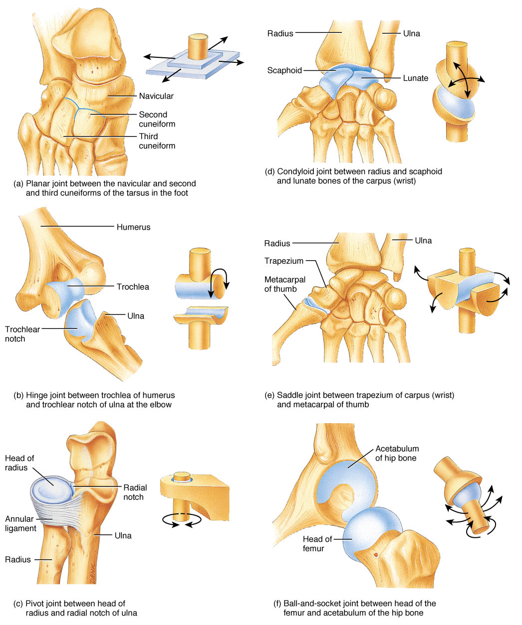
Based on the shape of the articular surfaces, joints are classified as spherical, cup-shaped, flat, ellipsoidal, saddle-shaped, condylar, trochlear, and rotational (cylindrical).
Movements in the joints are possible around the frontal, sagittal and vertical axes. 1) Around the frontal axis, movements are defined as flexion, flexio, and extension, extensio. 2) Around the sagittal axis – abduction, abductio, and adduction, adductio. 3) Movement around the vertical axis is called rotation, rotatio; a distinction is made between outward rotation - supination, supinatio, and inward rotation - pronation, pronatio. Circumduction, circumductio, is a circular movement, a transition from one axis to another. Based on the number of axes of motion, joints are classified into uniaxial, biaxial and multiaxial joints. Ball and socket joints are multiaxial. A typical spherical joint is the shoulder joint, movements in which are possible around 3 axes - frontal (flexion and extension), sagittal (abduction and adduction) and vertical (outward and inward rotation). The hip joint has a cup-shaped shape - it differs from the spherical joint in its deeper articular cavity. In flat joints, the movements are sliding in different directions. Ellipsoidal, condylar and saddle joints have 2 axes of movement: flexion and extension occur around the frontal axis, and adduction and abduction occur around the sagittal axis. Block and rotation joints have one axis of rotation. In the trochlear joint, movements occur around the frontal axis - flexion and extension. In a cylindrical joint, movement occurs around a vertical axis - rotation.
Based on their functional characteristics, combined joints are distinguished, articulations combinatae; - these are 2 or more joints that are anatomically separate (that is, have separate capsules), but participate in movements together. For example, two temporomandibular joints, the proximal radioulnar joint and the distal radioulnar joint.
Classification of joints by form and function
Single-spinous joints
Biaxial joints
|
Condylar, art. condylaris |
Frontal, sagittal |
Atlanto-occipital joints, art. atlantooccipitalis |
|
|
Saddle-shaped, art. sellaris |
Frontal, sagittal |
Flexion, flexio, extension, extension, abduction, abduction, adduction, adductio |
Carpometacarpal joint of the thumb, art. Carpometacarpea pollicis |
|
Elliptical, art. ellipsoidea |
Frontal, sagittal |
Flexion, flexio, extension, extension, abduction, abduction, adduction, adductio |
Wrist joint, art. radio-carpea |
Triaxial (multi-axial) joints
|
Globular, art. spheroidea |
Flexion, flexio, extension, extension, abduction, abduction, adduction, adductio |
Shoulder joint, art. humeri |
|
|
Flat, art. plana |
Frontal, sagittal, vertical |
Flexion, flexio, extension, extension, abduction, abduction, adduction, adductio |
Facet joints, art. zygapophysialis |
|
Cup-shaped, art. cotylica |
Frontal, sagittal, vertical |
Flexion, flexio, extension, extension, abduction, abduction, adduction, adductio |
Hip joint, art. coxae |
The human skeleton consists of more than 200 bones, most of which are movably connected by joints and ligaments. It is thanks to them that a person can move freely and perform various manipulations. In general, all joints are structured the same. They differ only in shape, nature of movement and the number of articulating bones.
Joints simple and complex
Classification of joints by anatomical structure
According to their anatomical structure, joints are divided into:
- Simple. The joint consists of two bones. An example is the shoulder or interphalangeal joints.
- Complex. A joint is formed by 3 or more bones. An example is the elbow joint.
- Combined. Physiologically, the two joints exist separately, but function only in pairs. This is how the temporomandibular joints are designed (it is impossible to lower only the left or right side of the jaw, both joints work simultaneously). Another example is the symmetrically located facet joints of the spinal column. The structure of the human spine is such that movement in one of them entails displacement of the other. To understand more precisely the principle of operation, read the article with beautiful illustrations about.
- Complex. The joint space is divided into two cavities by cartilage or meniscus. An example is the knee joint.
Classification of joints by shape
The shape of the joint can be:
- Cylindrical. One of the articular surfaces looks like a cylinder. The other has a recess of suitable size. The radioulnar joint is a cylindrical joint.
- Block-shaped. The head of the joint is the same cylinder, on the lower side of which a ridge is placed perpendicular to the axis. On the other bone there is a depression - a groove. The comb fits the groove like a key to a lock. This is how the ankle joints are designed.
A special case of trochlear joints is the helical joint. Its distinctive feature is the spiral arrangement of the groove. An example is the shoulder-elbow joint. - Ellipsoidal. One articular surface has an ovoid convexity, the second has an oval notch. These are the metacarpophalangeal joints. When the metacarpal sockets rotate relative to the phalangeal bones, complete bodies of rotation are formed - ellipses.
- Condylekov. Its structure is similar to the ellipsoidal one, but its articular head is located on a bony protrusion - the condyle. An example is the knee joint.
- Saddle-shaped. In its form, the joint is similar to two nested saddles, the axes of which intersect at right angles. The saddle joint includes the carpometacarpal joint of the thumb, which among all mammals is present only in humans.
- Spherical. The joint articulates the ball-shaped head of one bone and the cup-shaped notch of the other. A representative of this type of joint is the hip. When the socket of the pelvic bone rotates relative to the femoral head, a ball is formed.
- Flat. The articular surfaces of the joint are flattened, the range of motion is insignificant. The flat one includes the lateral atlantoaxial joint, connecting the 1st and 2nd cervical vertebrae, or lumbosacral joints.
A change in the shape of the joint leads to dysfunction of the musculoskeletal system and the development of pathologies. For example, against the background of osteochondrosis, the articular surfaces of the vertebrae shift relative to each other. This condition is called spondyloarthrosis. Over time, the deformity becomes fixed and develops into a permanent curvature of the spine. Instrumental examination methods (computed tomography, radiography, MRI of the spine) help to detect the disease.
Division by nature of movement
The movement of bones in a joint can occur around three axes - sagittal, vertical and transverse. They are all mutually perpendicular. The sagittal axis is located in the front-to-back direction, the vertical axis is from top to bottom, and the transverse axis is parallel to the arms extended to the sides.
Based on the number of axes of rotation, joints are divided into:
- uniaxial (these include block-shaped),
- biaxial (ellipsoidal, condylar and saddle-shaped),
- multi-axial (spherical and flat).
Summary table of joint movements
Number of axes Joint shape Examples
One Cylindrical Median Antlantoaxial (located between the 1st and 2nd cervical vertebrae)
One trochlear ulna
Two Ellipsoid Atlanto-occipital (connects the base of the skull with the upper cervical vertebra)
Two Condylar Knee
Two Saddle Carpometacarpal Thumb
Three Ball Shoulder
Three Flat Facet Joints (included in all parts of the spine)
Classification of types of movements in joints:
Movement around the frontal (horizontal) axis - flexion (flexio), i.e. decreasing the angle between the articulating bones, and extension (extensio), i.e. increasing this angle.
Movements around the sagittal (horizontal) axis - adduction (adductio), i.e. approaching the median plane, and abduction (abductio), i.e. moving away from it.
Movements around the vertical axis, i.e. rotation (rotatio): inward (pronatio) and outward (supinatio).
Circular movement (circumductio), in which a transition is made from one axis to another, with one end of the bone describing a circle, and the entire bone - the figure of a cone.
articulationes synoviales are the most advanced types of bone connections. They are distinguished by great mobility and a variety of movements.
Joint structure
Each joint includes articular surfaces of bones covered with cartilage, an articular capsule, and an articular cavity with a small amount of synovial fluid. Some joints also have auxiliary formations in the form of articular discs, menisci and articular labrum.
The articular surfaces, fades articulares, in most cases of articulating bones correspond to each other - they are congruent (from the Latin congruens - corresponding, coinciding).
Articular cartilage, cartilago articularis, is usually hyaline, in individual joints (temporomandibular) it is fibrous, and has a thickness of 0.2-6.0 mm.
The articular capsule, capsula articularis, is attached to the articulating bones near the edges of the articular surfaces or at some distance from them; it firmly fuses with the periosteum, forming a closed articular cavity.
The articular cavity, cavum articulare, is a slit-like space between the articular surfaces covered with cartilage.
Articular discs and menisci, disci et menisci articulares, are cartilaginous plates of various shapes that are located between articular surfaces that do not completely correspond to each other (incongruent). The disc is usually a solid plate, fused along the outer edge with the articular capsule, and, as a rule, divides the articular cavity into two chambers (two floors).
Menisci
These are semi-lunar-shaped, continuous cartilaginous or connective tissue plates that are wedged between the articular surfaces.
The articular lip, labrum articulare, is located along the edge of the concave articular surface, complements and deepens it (for example, in the shoulder joint). It is attached with its base to the edge of the articular surface, and with its inner concave surface facing the joint cavity.
Shapes of articular surfaces
resemble segments of the surfaces of various geometric bodies: a cylinder, an ellipse, a ball. Accordingly, joints are distinguished according to the shape of the articular surfaces: cylindrical, ellipsoidal and spherical. There are also variants of the indicated forms of joints. For example, a type of cylindrical joint would be a trochlear joint, a spherical joint would be a cup-shaped and flat joint.
The shape of the articular surfaces determines the number of axes, around which movement occurs at a given joint. Thus, the cylindrical shape of the articular surfaces allows movement only around one axis, and the ellipsoidal shape allows movement around two axes. In joints with spherical articular surfaces, movements are possible around three or more mutually perpendicular axes.
Thus, there is a certain interdependence between the shape of the articulating surfaces and the number of axes of motion.
Biomechanical classification of joints:
1) joints with one axis of movement (uniaxial);
2) joints with two axes of movement (biaxial);
3) joints with many axes of movement, of which three are main (multiaxial, or triaxial).
Joints unite the bones of the skeleton into a single whole. More than 180 different joints help a person move. Together with bones and ligaments, they are classified as the passive part of the musculoskeletal system.
Joints can be compared to hinges, the task of which is to ensure smooth sliding of bones relative to each other. In their absence, the bones will simply rub against each other, gradually collapsing, which is a very painful and dangerous process. In the human body, joints play a triple role: they help maintain body position, participate in the movement of body parts relative to each other, and are organs of locomotion (movement) of the body in space.
Each joint has various elements that facilitate the mobility of some parts of the skeleton and ensure strong coupling of others. In addition, there are non-bone tissues that protect the joint and soften interosseous friction. The structure of the joint is very interesting.
Main elements of the joint:
Joint cavity;
Epiphyses of bones forming a joint. The epiphysis is a rounded, often widened, end section of a tubular bone that forms a joint with the adjacent bone through the articulation of their articular surfaces. One of the articular surfaces is usually convex (located on the articular head), and the other is concave (formed by the articular fossa)
Cartilage is the tissue that covers the ends of bones and softens their friction.
The synovial layer is a kind of bag that lines the inner surface of the joint and secretes synovial fluid that nourishes and lubricates the cartilage, since joints do not have blood vessels.
The joint capsule is a sleeve-like fibrous layer that envelops the joint. It gives bones stability and prevents them from moving excessively.
The menisci are two hard cartilages shaped like crescents. They increase the area of contact between the surfaces of two bones, such as the knee joint.
Ligaments are fibrous formations that strengthen interosseous joints and limit the range of bone movement. They are located on the outside of the joint capsule, but in some joints they are located inside to provide better strength, such as the round ligaments in the hip joint.
A joint is an amazing natural mechanism for the movable connection of bones, where the ends of the bones are connected in the articular capsule. Bag the outside is made up of fairly strong fibrous tissue - this is a dense protective capsule with ligaments that help control and hold the joint, preventing displacement. The inside of the articular capsule is synovial membrane.
This membrane produces synovial fluid - the lubricant of the joint, a viscoelastic consistency, which even in a healthy person does not have much of, but it occupies the entire cavity of the joint and is capable of performing important functions:
1. It is a natural lubricant that provides the joint with freedom and ease of movement.
2. It reduces the friction of bones in the joint, and thus protects cartilage from abrasion and wear.
3. Acts as a shock absorber and shock absorber.
4. Works as a filter, providing and maintaining nutrition for cartilage, while protecting it and the synovial membrane from inflammatory factors.
Synovial fluid a healthy joint has all these properties, largely due to hyaluronic acid found in the synovial fluid, as well as in cartilage tissue. It is this substance that helps your joints fully perform their functions and allows you to lead an active life.
If the joint is inflamed or painful, then the synovial membrane of the joint capsule produces more synovial fluid, which also contains inflammatory agents that increase swelling, edema, and pain. Biological inflammatory agents destroy the internal structures of the joint.
The ends of the bone joints are covered by an elastic thin layer of smooth substance - hyaline cartilage. Articular cartilage does not contain blood vessels or nerve endings. Cartilage, as mentioned, receives nutrition from the synovial fluid and from the bone structure located under the cartilage itself - the subchondral bone.
Cartilage mainly acts as a shock absorber - it reduces pressure on the mating surfaces of bones and ensures smooth sliding of bones relative to each other.

Functions of cartilage tissue
1. Reduce friction between joint surfaces
2. Absorb shocks transmitted to the bone during movement
Cartilage is made up of special cartilage cells - chondrocytes and intercellular substance - matrix. The matrix consists of loosely arranged connective tissue fibers - the main substance of cartilage, which are formed by special compounds - glycosaminoglycans.
It is the glycosaminoglycans, connected by protein bonds, that form larger structures of cartilage - proteoglycans - that are the best natural shock absorbers, since they have the ability to restore their original shape after mechanical compression.
Due to its special structure, cartilage resembles a sponge - absorbing fluid in a calm state, it releases it into the articular cavity under load and thereby additionally “lubricates” the joint.
Such a common disease as arthrosis upsets the balance between the formation of new and the destruction of old building material that forms cartilage. Cartilage (the structure of the joint) changes from strong and elastic to dry, thin, dull and rough. The underlying bone thickens, becomes more irregular, and begins to grow away from the cartilage. This limits movement and causes joint deformation. The joint capsule thickens and becomes inflamed. Inflammatory fluid fills the joint and begins to stretch the capsule and articular ligaments. This creates a painful feeling of stiffness. Visually, you can observe an increase in the volume of the joint. Pain, and subsequently deformation of the joint surfaces with arthrosis, leads to stiff joint mobility.

Joints are distinguished by the number of articular surfaces:
- simple joint (lat. articulatio simplex) - has two articular surfaces, for example the interphalangeal joint of the thumb;
- complex joint (lat. articulatio composita) - has more than two articular surfaces, for example the elbow joint;
- complex joint (lat. articulatio complexa) - contains intra-articular cartilage (meniscus or disc), dividing the joint into two chambers, for example the knee joint;
- combined joint - a combination of several isolated joints located separately from each other, for example the temporomandibular joint.
According to their shape, the articular surfaces of the bones are compared with geometric figures and, accordingly, joints are distinguished: spherical, ellipsoidal, trochlear, saddle-shaped, cylindrical, etc.
Joints with movement
. Shoulder joint: the articulation that provides the greatest amplitude of movement of the human body is the articulation of the humerus with the scapula using the glenoid cavity of the scapula.
. Elbow joint: the connection of the humerus, ulna and radius bones, allowing rotation of the elbow.
. Knee-joint: a complex articulation that provides flexion and extension of the leg and rotational movements. At the knee joint, the femur and tibia articulate - the two longest and strongest bones, on which, together with the patella, located in one of the tendons of the quadriceps muscle, almost the entire weight of the skeleton presses.
. Hip joint: connection of the femur with the pelvic bones.
. Wrist joint: formed by several joints located between numerous small flat bones connected by strong ligaments.
. Ankle joint: The role of ligaments is very important in it, which not only ensures the movement of the lower leg and foot, but also maintains the concavity of the foot.

The following main types of joint movements are distinguished:
- movement around the frontal axis - flexion and extension;
- movements around the sagittal axis - adduction and abduction movements around the vertical axis, that is, rotation: inward (pronation) and outward (supination).
The human hand contains: 27 bones, 29 joints, 123 ligaments, 48 nerves and 30 named arteries. We move our fingers millions of times throughout our lives. The movement of the hand and fingers is provided by 34 muscles; only when moving the thumb, 9 different muscles are involved.
Shoulder joint
It is the most mobile in humans and is formed by the head of the humerus and the articular cavity of the scapula.
The articular surface of the scapula is surrounded by a ring of fibrocartilage - the so-called articular lip. The tendon of the long head of the biceps brachii muscle passes through the joint cavity. The shoulder joint is strengthened by the powerful coracohumeral ligament and surrounding muscles - deltoid, subscapularis, supra- and infraspinatus, teres major and minor. The pectoralis major and latissimus dorsi muscles also take part in shoulder movements.
The synovial membrane of the thin joint capsule forms 2 extra-articular inversions - the tendons of the biceps brachii and subscapularis. The anterior and posterior arteries that envelop the humerus and the thoracoacromial artery take part in the blood supply to this joint; the venous outflow is carried out into the axillary vein. The outflow of lymph occurs in the lymph nodes of the axillary region. The shoulder joint is innervated by branches of the axillary nerve.

The shoulder joint is capable of movement around 3 axes. Flexion is limited by the acromion and coracoid processes of the scapula, as well as the coracobrachial ligament, extension by the acromion, coracobrachial ligament and joint capsule. Abduction in the joint is possible up to 90°, and with the participation of the upper limb belt (when the sternoclavicular joint is included) - up to 180°. Abduction stops when the greater tuberosity of the humerus rests on the coracoacromial ligament. The spherical shape of the articular surface allows a person to raise his arm, move it back, and rotate the shoulder along with the forearm and hand in and out. This variety of hand movements was a decisive step in the process of human evolution. The shoulder girdle and shoulder joint in most cases function as a single functional formation.
Hip joint
It is the most powerful and heavily loaded joint in the human body and is formed by the acetabulum of the pelvic bone and the head of the femur. The hip joint is strengthened by the intraarticular ligament of the femoral head, as well as the transverse ligament  acetabulum, which surrounds the neck of the femur. From the outside, the powerful iliofemoral, pubofemoral and ischiofemoral ligaments are woven into the capsule.
The blood supply to this joint is through the circumflex femoral arteries, branches of the obturator and (variably) branches of the superior perforating, gluteal and internal pudendal arteries. The outflow of blood occurs through the veins surrounding the femur into the femoral vein and through the obturator veins into the iliac vein. Lymphatic drainage occurs in the lymph nodes located around the external and internal iliac vessels. The hip joint is innervated by the femoral, obturator, sciatic, superior and inferior gluteal and pudendal nerves.
The hip joint is a type of ball-and-socket joint. It allows movements around the frontal axis (flexion and extension), around the sagittal axis (abduction and adduction) and around the vertical axis (external and internal rotation).
This joint experiences a lot of stress, so it is not surprising that its lesions occupy first place in the general pathology of the articular apparatus.
Knee-joint
One of the largest and most complex human joints. It is formed by 3 bones: the femur, tibia and fibula. Stability of the knee joint is provided by intra- and extra-articular ligaments. The extra-articular ligaments of the joint are the fibular and tibial collateral ligaments, the oblique and arcuate popliteal ligaments, the patellar ligament, and the medial and lateral suspensory ligaments of the patella. The intra-articular ligaments include the anterior and posterior cruciate ligaments.
The joint has many auxiliary elements, such as menisci, intra-articular ligaments, synovial folds, and bursae. Each knee joint has 2 menisci - the outer and the inner. The menisci look like crescents and play a shock-absorbing role. The auxiliary elements of this joint include synovial folds, which are formed by the synovial membrane of the capsule. The knee joint also has several synovial bursae, some of which communicate with the joint cavity.
Everyone had to admire the performances of artistic gymnasts and circus performers. People who are able to climb into small boxes and bend unnaturally are said to have gutta-percha joints. Of course, this is not true. The authors of The Oxford Handbook of Body Organs assure readers that “their joints are phenomenally flexible”—medically known as joint hypermobility syndrome.

The shape of the joint is a condylar joint. It allows movements around 2 axes: frontal and vertical (with a bent position in the joint). Flexion and extension occur around the frontal axis, and rotation occurs around the vertical axis.
The knee joint is very important for human movement. With each step, by bending, it allows the foot to step forward without hitting the ground. Otherwise, the leg would be carried forward by raising the hip.
According to the World Health Organization, every 7th person on the planet suffers from joint pain. Between the ages of 40 and 70 years, joint diseases are observed in 50% of people and in 90% of people over 70 years of age.
Based on materials from www.rusmedserver.ru, meddoc.com.ua
