Diagnosis and prognosis of postpartum diseases in cows. Postpartum acute paresis in cows - description of the disease, symptoms and possible causes, as well as prevention and treatment of acute illness in cattle. Diseases of the udder zone
There are a number of non-infectious diseases that you should pay attention to when breeding. These include postpartum paresis in a cow. Treatment is not very difficult and is not expensive. However, the disease brings considerable losses. Such a loss hits those farms where cow breeding is organized as a business the hardest. The death of even one head in a small farm significantly affects the profitability of production.
Postpartum paresis in cows
A severe, acute nervous disease, which is accompanied by partial paralysis of the tongue, pharynx, limbs, intestines, up to loss of consciousness, is called postpartum paresis in a cow. Treatment must be started immediately to prevent death.
At-risk groups
The exact cause of the disease has not yet been established. It is known that it is provoked by an acute lack of calcium in the animal’s body against the background of a sharp drop in blood sugar. A calving cow directs all its forces to the formation of the fetus, including calcium. A diet that is not balanced in minerals can provoke maternity paresis in cows.
Mostly highly productive animals are at risk. In all likelihood, this is due to the fact that high milk yields require more calcium. Well-fed animals, for which concentrated feed is not spared, are also at risk.
The stall period and the age of 5-8 years, when the cow is at the peak of her productivity, colds are concomitant factors of the disease.
Signs of illness
As a rule, the development of the disease occurs in the first three days after a quick and easy calving. Although, as practice shows, in most cases giving birth to a cow requires human help and intervention. Less commonly observed is the development of paresis from several weeks to 1-2 months. The absolute exception is the manifestation of the disease in calving cows and during calving itself.
The first signs of the onset of the disease:
- lethargy of the animal;
- poor appetite;
- lack of chewing gum;
- noticeable shaking;
- there is no peristalsis of the rumen and intestines;
- pain sensitivity is practically absent.
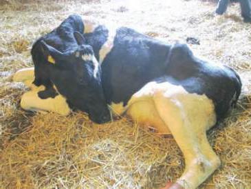
If the condition deteriorates significantly, the cow cannot rise to her feet. The position in which the animal lies is also specific: the legs are tucked under the stomach and the head is turned to the side. The neck is arched in a characteristic S-shaped curvature. The legs and horns are cold to the touch, the eyes are half-closed and watery, the pupils are dilated. In severe cases, the overall body temperature drops to 35 o -36 o.
The animal does not urinate or excrete feces. When the pharynx is paralyzed, the tongue falls out of the mouth and profuse salivation occurs. Some individuals are in an excited state for a short time and can sharply shake their head, throwing it back. They roll over, grind their teeth, hit the walls, and moo loudly. After short-term activity, the cow calms down and goes into a depressed state.
Treatment
Postpartum paresis in a cow should not be left to chance. Treatment started immediately reduces mortality from the disease by up to 4%. For comparison: if no measures are taken in the first hours, 70% of animals die.
Using a special Evers apparatus, air is blown into all four lobes of the udder through the nipples. To do this, they are pre-treated with a 70% ethanol solution. Before the procedure, the cow is milked in a dorsolateral position. Air is introduced using sterile mammary catheters. Injection is carried out until the folds of skin on the udder are straightened. Be sure to massage the udder to distribute air evenly.

To prevent air from escaping, the nipples are tied at the base with a bandage or strips of gauze. The animal is left in this position for 30-45 minutes. If after 8 hours there is no improvement, the procedure is repeated. After the cow gets up, after 1-2 hours she can be milked, without squeezing the air out of the udder.
Instead of air, you can introduce 200-500 ml of fresh milk into each lobe of the udder. It must come from a healthy cow.
To alleviate the animal’s condition, a 20% glucose solution up to 200 ml and a 10% calcium chloride solution up to 150 ml are administered intravenously. A 10% caffeine solution is injected subcutaneously. Active rubbing of the sides and limbs, wrapping the animal, and a hot enema (up to 45°C) are recommended.
Complications
With a rapid and severe course of the disease, the life of the animal may be threatened by tympany; this is one of the complications that causes postpartum paresis in the cow. Treatment boils down to piercing the scar with a thick needle or trocar, after which up to 400 ml of a 5% alcohol solution of ichthyol or up to 40 ml of a 40% formaldehyde solution is injected into the cavity.
Under no circumstances should liquid medicine be administered through the mouth; due to partial paralysis of the pharynx, it may enter the trachea.
The measures taken will completely cure the animal in 2-3 days without any consequences for its health. Perhaps this will never happen again, or perhaps the cow will experience postpartum paresis every time after calving.
Prevention of postpartum paresis
To avoid illness, adhere to the following rules:
- do not allow overfeeding of cows in and during the waning stage of lactation;
- carefully monitor the balance of the diet in micro- and macroelements;
- the percentage of roughage and concentrated feed in the animal diet must be strictly observed;
- control the timely start of cows;
- provide the livestock with regular active exercise.
Farms for cows must be equipped with special birthing boxes where the animal is placed before calving. There should be no drafts in the maternity ward.
For highly productive cows that have previously suffered from postpartum paresis, the following preventive measures are provided: vitamin D 3 is administered intramuscularly twice, 7-10 days before calving, at a dose of 3-4 million units. Sugar is introduced into the diet, 200-300 grams per day for several days before and after calving.
Deposit. Cows, sheep, goats and pigs get sick when they are fed insufficiently and monotonously and kept in cramped rooms with a strongly sloping floor. The disease often occurs in old and emaciated queens, pregnant with several fetuses, as well as after a difficult birth, when the ligaments and bones of the pelvis are damaged, As a result, the animal is unable to rise several days or weeks before and after birth.
Helping an animal comes down to good feeding, maintenance and treatment of bedsores. They provide food rich in protein, carbohydrates, vitamins and mineral salts. The animals are placed on ample bedding, turned from side to side 2-3 times daily and rubbed with strands of straw. You can lift the animal by tying a rope along its body (Fig. 41). If there are bedsores, they are washed with disinfectant solutions, lubricated with ichthyol ointment, boiled vegetable oil or petroleum jelly.
Premature attempts. In pregnant animals, due to a blow to the stomach, a fall, eating poor-quality or frozen food, or after drinking cold water, labor may occur much earlier than the normal delivery date. The animal begins to worry, looks back, moves from foot to foot, and often excretes urine and feces. Sometimes partially amniotic membranes come out into the vagina and there may be a miscarriage (abortion) or fetal death.
If premature attempts occur, the animal is given complete rest, vodka diluted in half with water is given inside (cows and mares - 500-800 g, sheep, goats, pigs - 200-300 g), and the sacrum and lower back are wrapped warmly.
Abortion. Abortions can be of contagious or non-contagious origin and are accompanied by the premature expulsion of a non-viable or dead fetus from the uterus, when during abortion the dead fetus lingers in the uterus, liquefies (maceration), dries out and thickens, or putrefactive decomposition occurs.
Abortion at the beginning of pregnancy may be accompanied by absorption of the fetus or its release along with the amniotic membranes. With later abortions, the animals become restless, attempts appear, the cervix is opened and a bloody-dark liquid is released from it, and then the fetus.
Non-contagious abortions are observed when feeding poor quality, frozen or poisonous food, drinking cold water, bruises, diseases of the stomach and intestines, uterus, ovaries, lungs, as well as as a result of erroneously carried out natural insemination of pregnant animals; sometimes as a result of the administration of potent drugs.
Edema in pregnant animals. In pregnant animals, due to increased porosity of the blood vessels, weak heart or kidney disease, fluid accumulates in the subcutaneous tissue. In sick animals, 1-2 months before birth, large edema forms in the area of the dewlap, lower abdominal column, and limbs.
Such animals are given less water, succulent food and table salt are reduced or excluded from the diet, massage is given, especially in the area of edema, and the animal is systematically taken for a walk.
Vaginal prolapse. The disease is observed during the last period of pregnancy and after birth in cows, goats, sheep and rarely in other animals. It can occur as a result of weakening of the ligaments that support the uterus and vagina, difficult childbirth and extraction of the fetus using excessive force, keeping animals on too sloping floors, lack of exercise, damage to the spinal cord and other reasons.
Vaginal prolapse is detected in the form of a round pink swelling protruding from the vulva when the animal is lying down. When the entire vagina prolapses, a spherical mass protrudes from the vulva with the cervix in the middle. Prolonged vaginal prolapse is accompanied by the formation of ulcers, cracks and tissue necrosis.
The prolapsed vagina must be put back in place and washed daily with disinfectant and astringent solutions (potassium permanganate diluted 1:5000, 2% Lysol solution, oak bark decoction, tannin). The animal is placed on a wooden platform with a forward slope so that the back of the body is higher than the front. To avoid repeated vaginal prolapse, the vulva is sutured or strengthened with a special loop (Fig. 42). The sutures and loop are removed before birth.
To protect animals from vaginal prolapse, they are kept on a floor with a low posterior slope and take daily walks; 10-15 days before birth, they are given less succulent food.

Vaginal rupture. The main causes of vaginal rupture are difficult childbirth, a large fetus, incorrect positioning of the fetal limbs, violent pushing, and rupture with instruments or by hand due to careless assistance during childbirth. When ruptured, wounds of varying sizes and depths form on the walls of the vagina.
For wounds of the vagina, cotton swabs soaked in a disinfectant solution are inserted into it, or carefully lubricated with ichthyol ointment. Do not wash the vagina with solutions if the vaginal wall is completely ruptured. Veterinary specialists provide assistance.
Uterine prolapse. The disease occurs immediately after expulsion of the fetus or in the first hours after birth, when the cervix has not yet contracted. The prolapsed uterus turns the mucous membrane outward and hangs in the form of a large pear-shaped red formation. In ruminants, caruncles are visible on its surface. Uterine prolapse occurs more often in cows, goats, sheep and less often in other animals when the fetus is quickly removed using great force or when strong efforts continue after birth, and also if the animal stands for a long time on a strongly sloping floor and is not allowed to go for a walk.
First aid. The prolapsed uterus must be straightened as quickly as possible by carefully separating the pieces of the placenta and washing with a warm 2-3% solution of alum or a weak solution of potassium permanganate at a dilution of 1:10,000. When straightening, the washed uterus is supported on a clean towel or sheet, and the person providing assistance with clean hands grasps the part of the uterus near the vulva and pushes it inside. When only the top of the uterus remains outside, carefully pressing with a fist, push the uterus into the pelvic cavity and hold it in place for some time. You can adjust the uterus from the top of the horn, pressing with a fist wrapped in a towel. To weaken the animals, they are then given vodka, diluted half and half with water. To prevent recurrent uterine prolapse, sutures or a rope loop are placed on the vulva, and a shield or straw is placed under the back of the animal's body.
Retention of placenta. After birth, the placenta is separated immediately after the fetus or is delayed in cows, sheep, goats for 2-6 hours, and in mares and pigs for up to 1 hour. Sometimes it takes longer, especially in cows, goats and sheep due to the special structure of their placenta. Retention of the placenta mainly occurs in the absence of regular walks and lack of vitamins and mineral salts in the diet, weak pushing, difficult childbirth, early contraction of the cervix and fusion of the placenta with the uterine mucosa. The uncalved placenta hangs from the vagina. After 12-16 hours, its decomposition begins, which can cause inflammation of the uterus and poisoning of the entire body. In mares and pigs, retained placenta often leads to general blood poisoning (sepsis) and death of the animal.
Livestock breeders must monitor the timing of separation of the placenta and promptly provide assistance in maintaining it, as well as systematically provide walks for pregnant animals and provide them with nutritious feed.
Eating the afterbirth. After giving birth, some females swallow the afterbirth, as a result, their digestion is upset, milk production decreases, and pigs develop a tendency to eat piglets. For preventive purposes, the separated placenta is immediately burned or buried. If animals eat it, their diet is reduced and laxative salts are given.
Eating piglets by a sow. The reason for eating newborns is nervous excitement due to diseases of the digestive system, uterus, vagina, udder, as well as injury to the nipples by the sharp teeth of piglets during sucking, eating the placenta and feeding the queens with raw meat. Therefore, in order to prevent this abnormal phenomenon, they stop giving meat to sows 1-2 months before giving birth; they remove the placenta, monitor the condition of the nipples and udder, massage the udder 5-10 days before birth, and do not leave the piglets under the uterus after she feeds them.
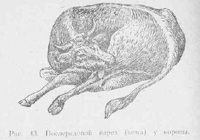
Postpartum paresis. Dairy cows often get sick and goats, sheep and pigs get sick less often, mainly when kept in stalls and underfed during pregnancy, when the diet contains a lot of concentrates and little roughage and succulent feed. This helps reduce calcium and glucose levels in the body of pregnant animals. The disease occurs in the first days after childbirth, and sometimes several weeks later. Animals quickly become depressed and weak in the backside; they lie down and cannot get up. With a mild course of the disease, the body temperature drops to 37.5-37°, the skin of the torso and limbs becomes cold, the head is held suspended, the neck is curved. In more severe cases, the head is thrown back onto the chest; if you lift it, it returns to the same position. The animal does not react to touching the skin and eyelids. Body temperature drops to 35-36°. Sometimes there is lacrimation, swelling of the eyelids, wheezing and moaning, the tongue is paralyzed and hangs out of the mouth (Fig. 43). The illness lasts 2-3 days. If the animal is not helped, it may die.
Treatment. To help the animal, it is recommended to pump air into all teats of the udder using a special Evers apparatus; which should be on every farm. It consists of two rubber balls, a metal can, a rubber tube and a milk catheter, which is inserted into the udder nipple. Before insufflation, you need to milk the milk from the udder, wipe the nipples and milk catheter with alcohol or vodka. The air is pumped slowly until the udder feels elastic. To hold it in the udder, lightly bandage the ends of the nipples with a bandage, and after 1 hour the bandage is removed. You should not pull the nipples too tightly, as they may become dead. You don't have to bandage your nipples. At the same time, the entire body of the animal is massaged with strands of straw and covered with a blanket. Medicinal substances and other liquids cannot be given orally, since the animal cannot swallow due to pharyngeal paresis. The animal's recovery occurs after 3-4 hours; it is not recommended to lift it. Sometimes it is necessary to re-inhale air for complete recovery.
For the treatment of maternity paresis, a new method has been proposed, based on stopping the movement of blood through the milk veins. To do this, take a rubber tube with a diameter of 1-2 cm and a length of 2-3 m (a rope can be used) and pass it around the body so that it passes in front of the udder. The ends of the rubber tube are tied on the animal's back. After 20-40 minutes, the pressure of the tube is gradually (over 3-5 minutes) weakened and removed. If the disease recurs, a tourniquet is applied again. At the same time, rub the surface of the animal’s body with bunches of straw. After recovery, animals are fed only hay and warm water for 2-3 days, and then other foods are gradually introduced into the diet.
For maternity paresis in pigs, they are wrapped warmly, the udder is massaged, and enemas are given.
To prevent maternity paresis, pregnant animals are allowed to go for walks every day; in the last month of pregnancy, the amount of concentrates is reduced. Cows, sheep, and goats that have had postpartum paresis in the past are given a sugar solution or a 10% calcium chloride solution within 4-5 days before birth.
Postpartum intoxication and infection. A serious disease of the whole body that occurs in the first hours after childbirth as a result of penetration of microbes and their toxins into the blood through the mucous membranes of the birth canal. The development of the disease is facilitated by injuries to the mucous membranes during difficult childbirth, rough assistance during childbirth and unsanitary conditions for keeping animals.
Signs of the disease: general weakness, increased body temperature, refusal to feed, indigestion and increased breathing, disheveled fur. The animal quickly loses weight, lies down and greatly reduces milk yield, sometimes diarrhea occurs; bloody fluid with an unpleasant odor is released from the vagina; The vaginal mucosa may have ulcers and dark red spots covered with gray-yellow scabs. The disease lasts 8-10 days and can result in the death of the animal.
Treatment of postpartum intoxication is carried out by a veterinarian, and livestock breeders must ensure that this disease does not occur. To do this, you need to keep the maternity rooms clean, wash the rear part of the animal’s body with disinfectant solutions before and after childbirth, carefully provide assistance during difficult births, lubricate wounds and cracks in the birth canal with ichthyol or creolin ointment, tincture of iodine, monitor the timely separation of the placenta and isolate sick from healthy
is a joyful event for any farmer, but at the same time it can bring a lot of trouble.
For example, during this period the cow is weakened and her immune system is not able to properly fight infections and various diseases that can occur in these animals.
Postpartum illnesses in cows must be treated immediately so that the animal is able to care for both itself and the newborn calf as soon as possible.
After calving, the cow may feel weak and unable to stand on her feet or stay on them for long. Sometimes an animal begins to crawl because it needs movement, but does not find the strength to do more. Complications after childbirth can occur due to the following reasons:
- If you ate insufficiently nutritious and healthy food during pregnancy.
- In case of injuries in the pelvic area during childbirth.
- If the sciatic and obturator nerves have been damaged.
- In case of incorrect behavior of veterinarians during the birth of the fetus, or if the calf is large.
- If the ligaments in the pelvic area have been stretched.
- Due to inflammatory processes and injuries, muscle defects have formed.
How can you determine
Postpartum complications can be identified by the following signs:
- A cow cannot stand on her feet without help.
- Even having risen, the animal cannot stay standing for long and soon falls again.
- The temperature and pain threshold remain unchanged, but the functions of the motor system of the legs malfunction.
- If the animal moves, it does so very slowly.
- If you give an injection into a painful area, the cow will reflexively move sharply.
- Possible swelling of diseased areas.
Postpartum complications can be diagnosed only when there is no doubt about the possibility of mechanical damage to the limbs.
The essence of the treatment of this disease lies in two principles of action:
- Relieve painful sensations.
- Prevent the possibility of complications.
To avoid swelling and bedsores, the cow needs to be provided with a soft floor covering. You also need to make sure that it turns over on different sides from time to time.
Areas of the body where the animal leans must be treated with ointments or camphor alcohols. The lower back is wrapped with a warming compress.
If bedsores appear, you need to rub the skin every three hours with special products, such as a solution of brilliant green.
You should help the animal get up on its feet at least occasionally. For this purpose, auxiliary mechanisms can be built. In this case, it is necessary to take into account that it first raises the pelvic area, and then the forelimbs.
How to prevent the development of the disease
To ensure that the cow does not suffer from postpartum complications, it is necessary to take care of her health even before pregnancy and during gestation. The animal must regularly be in the fresh air and eat well. In addition, it is necessary to ensure the cow’s safety from mechanical damage.
Development of paresis after childbirth
The development of paresis after the birth of a calf occurs due to an acute lack of calcium microelements.
The balance of hormones in the animal changes greatly, and the natural processes of processing and absorption of vitamins begin to slow down.
According to some observant farmers, the development of paresis after calving may be explained by a genetic tendency.
This may be true, but science has not yet confirmed this hypothesis.
By what criteria is it determined
The development of this disease can begin during pregnancy. And sometimes the first signs are visible only after calving. Animals that give birth more than once are more prone to illness.
The first signs of the disease:
- The cow loses her appetite.
- She becomes slow and looks tired (in some cases the reaction is the opposite - the animal is too active).
- Body temperature decreases.
- The animal walks unsteadily.
- Sometimes short-term cramps may occur.
The course of paresis after childbirth can be:
- In a light form. Only the first symptoms are noticed, but there will be no complications leading to the animal being unable to stand on its feet. Another sign of a mild form of the disease is tension in the cervical area and a head posture that is predominantly inclined towards the back of the body.
- In severe form. The angle of inclination of the head is always the same. The animal has blurred vision. Due to numbness or paralysis of the muscles in the mouth area, the tongue often falls out. The cornea of the eyeballs may dry out. In the case of paralysis of the muscles of internal organs, the gastrointestinal tract and bladder malfunction, which is obvious from decreased appetite and problems with urination.
How to approach treatment correctly
Treatment of paresis after childbirth involves increasing the amount of calcium, magnesium and phosphorus trace elements in the blood. To do this, calcium chloride gluconates, as well as preparations containing magnesium, are injected into the vein of the cow. It is also necessary to nourish the animal’s body with substances that help absorb and process these phytamines.
- The development of laminitis often occurs during pregnancy;
- Problems with lameness arise when the cow is kept in a stall for a long time and her lifestyle is immobile.
- An animal's joints weaken most often when it is exposed to injury.
The placenta is retained
If the pregnancy proceeded successfully, the placenta and membranes of the fetus will be released two to six hours after calving. The norm allows a delay of up to twelve hours. But if after this time the placenta has not been released, it is urgent to call a veterinarian.
There are several reasons for the development of this disease:
- Lack of vitamins and beneficial microelements in the animal’s body.
- A cow's too immobile lifestyle leads to many of her muscles weakening and even atrophying. And in cases where this is necessary, they do not work as expected.
- If the calf was large, or there were two of them, then the uterus could stretch, causing the placenta to be retained.
- An inflammatory process in the uterine area can also lead to this disease.
Usually, after childbirth, the fetal membrane remains suspended in the vaginal area along with the vessels of the circulatory system. Safe removal of the placenta is possible only on the first day after birth.
If this does not happen, over the next few days the outer part of the placenta decomposes, and the inner part gradually rots. A persistent unpleasant odor is heard, tissues become weak and lose their elasticity.
Due to decay, trace elements are released, which are absorbed into the body's circulatory system and distributed throughout the body, which can lead to sepsis.
This disease is a complex infection of a cow that is almost untreatable. If these processes occur, the animal’s body temperature rises, the desire to eat food disappears, problems with the gastrointestinal tract arise, it strains, draws in the abdominal area and hunches over.
To prevent such developments, it is necessary to help the cow in the first few hours after calving.
You can give her a special remedy, with the help of which the condition of the uterine muscles is activated and the body will be able to remove the placenta in time.
And in order to prevent the occurrence of diseases associated with weakening of the animal’s muscles, it is necessary to provide the cow with the opportunity to exercise daily (and several times). This should be done for about forty minutes.
Diseases of the udder zone
After the cow has calved, she begins to produce milk to feed her baby. Many systems of the animal’s body are associated with the work of the udder glands, so you should monitor whether this organ is healthy and whether it has any defects. Every farmer should know the following diseases in cows and their treatment:
- Mastitis
- Udder swelling
- Injury to the udder area
- Furunculosis in cows
- Papillomas on the surface of the udder
- Involuntary release of milk
- A cow's udder is tight
It is impossible to fight all these illnesses. To do this, you need to contact a veterinarian to correctly determine the diagnosis and prescribe treatment. You should not try to solve the problem yourself, so as not to complicate the situation and create a risk of relapse.

In the case of pathological childbirth, as well as with widespread retention of the placenta and postpartum complications, it is necessary:
During childbirth, if possible, collect fetal fluid in a bucket and after the cow licks the calf (within 1 hour), drink 2-3 liters, adding the same amount of warm water.
Take into account the fact that the removal of the fetus from the birth canal of a woman in labor after the rupture of amniotic fluid lasts 1.0-1.5 hours.
3. If the fetus has not been delivered within the specified period, proceed to qualified surgical obstetric care in compliance with the rules of asepsis and antisepsis (operations in order: lay clean straw under the woman in labor, wash her rump and external genitalia with warm water and soap, and then treat with 3% - solution of hydrogen peroxide or potassium permanganate (1:3000-5000), wash your hands thoroughly with soap up to your shoulders and treat with a 3% solution of hydrogen peroxide or potassium permanganate (1:3000-5000), ropes and other tools for obstetrics should be pre-clean and disinfected (stored in a freshly prepared potassium permanganate solution).
4. When providing obstetric care, the following rules must be observed:
4.1. The obstetrician's hands and instruments must be sterile.
4.2. The external genitalia of the animal must be clean.
4.3. All presenting parts of the fetus, which may, when pushed away, take on incorrect positions in the uterus, should be secured with sterile obstetric tape or rope.
4.4. If the birth canal is dry, it is necessary to pour several liters of a sterile decoction of flax seed into the uterine cavity, and lubricate the presenting parts of the fetus with sterile petroleum jelly.
4.5. Childbirth is easier if the animal lies on its left side, since in this position the progress of the fetus is not interfered with by the proventriculus, especially the scar.
4.6. The use of force when removing the fetus is permissible only during contractions and pushing, otherwise prolapse or rupture of the uterus is possible.
4.7. After obstetric care and delivery of the fetus, introduce antimicrobial uterine agents into the uterine cavity and intramuscularly inject oil forms of vitamin preparations.
5. After giving birth, give the woman in labor 2-3 liters of colostrum, after diluting it by half with warm water. Actively wipe the woman's croup with a straw tourniquet.
6. After the birth of the calf, 30-40 units of oxytocin should be administered intramuscularly 2-3 hours later.
After birth, cut the cow's umbilical cord stump with sharp sterile scissors, retreating 10 cm from the rupture. This ensures the outflow of blood from the fetal membranes and the separation of the villi of the fetal placenta from the maternal crypts.
Inject prostaglandins F 2b intramuscularly in a dose of 2 ml.
Introduce a 10% solution of white hellebore tincture with fish oil in a dose of 100 ml into the uterine cavity. In this case, you can use a device for inseminating sows (POS-5), the flexible catheter of which is advanced between the uterine wall and the placenta.
If no preventive measures were carried out in the first 5-6 hours and the placenta did not separate during this time, proceed as follows:
1. Inject intramuscularly 3-4 ml of agofolline or 3-5 ml of sinestrol,
3. Intramuscularly administer PGF 2b (estrophan, bioestrophan, magestrophan, estrone, etc.) at a dose of 2 ml in combination with 30 units of oxytocin.
4. Colostrum subcutaneously in a dose of 25-30 ml, with the addition of 1 million antibiotics.
If, after taking measures, the placenta does not separate spontaneously within 24-30 hours after birth, proceed to prompt separation of the placenta. Wherein:
1. Before separating the placenta, inject 400-500 ml of a 3% solution of hydrogen peroxide into the uterine cavity using an Esmarch mug.
2. After separation of the placenta, introduce antimicrobial uterine agents into the uterine cavity (septimethrin 4-5 capsules, exuter M 1-2 tablets, furazolidone sticks 4-5 pieces, metromax or endoxer 3 sticks, uterosan-FT 4-5 suppositories, endodioxide 4- 5 sticks, iodopen, furapen, gynobiotic, geomycin F, etc.). Introduce the drugs into the uterine cavity again at intervals of 12-24 hours.
3. On the day of separation of the placenta, and then after 10 and 20 days, administer intramuscularly 10 ml of trivitamin, tetravit or 5 ml of trivit in combination with 1.5-2.0 ml of ASD f2.
Postpartum endometritis in cows
Endometritis is an inflammation of the uterine mucosa, predominantly of a purulent-catarrhal nature, occurring more often at 8 - 10th, (sometimes at 3 - 6th) day after birth.
Postpartum endometritis occupies a significant place among obstetric and gynecological pathologies in cows and leads to temporary or permanent infertility.
The most common is purulent-catarrhal endometritis (86.1 -94.7%), catarrhal endometritis (1.9-4.8%), fibrinous (2.7-5.8%). Postpartum necrotic metritis occupies (0.7-2.8%), gangrenous - 0.2%.
Endometritis and metritis are diseases of a polyetiological nature, which are based on infection of the genital organs when the integrity of the mucous membrane is impaired, the contractile function of the uterus and involutional processes in the postpartum period are reduced, against the background of low nonspecific immunity of the animal body.
Clinical signs of acute purulent-catarrhal endometritis usually appear on the 5-6th day after birth in the form of discharge from the uterus of altered lochia. Their color can be brown, yellowish or grayish-white. Sometimes crumbs and small flakes of disintegrating caruncles and fragments of decomposing placenta are released. Subsequently, the exudate becomes mucopurulent or purulent in nature. It is released from the uterus when the animal strains, when lying down, and also when the uterus is massaged with the hand through the rectum. Often exudate can be found on the ventral surface of the tail in the form of dried crusts.
A rectal examination reveals that the uterus hangs into the abdominal cavity, the walls are flabby, doughy in consistency, and its contractility is reduced. With a large accumulation of exudate, fluctuation is noted. There are usually no changes in the general condition of the animal.
Acute fibrinous endometritis is characterized by the release of yellow-brown exudate with fibrin flakes. A sick animal is characterized by a depressed general condition, fever, and decreased productivity. Rectal examination reveals thickening of the uterine wall, atony, pain on palpation, and sometimes crepitus.
Necrotic metritis is characterized by the discharge from the uterus of a reddish exudate mixed with crumbly masses (necrotic tissue), and a chorionic odor.
On rectal examination, the uterus does not contract, is thickened, sometimes has a pasty consistency, is painful, and crepitus is often felt.
The disease occurs as a septic process: the cow stands hunched over, there is no appetite or chewing cud, hypotension of the forestomach and intestines is observed, sometimes profuse diarrhea, fever, rapid breathing, rapid pulse of weak filling.
Treatment of cows with postpartum endometritis begins immediately after detection of the pathology.
When treating cows, it is necessary to solve 5 main problems:
1. Provide the sick animal with proper feeding and housing conditions.
2. Free the uterus from accumulated exudate.
3. Increase the tone and contractile function of the uterus.
4. Suppress the vital activity of microflora in the area of inflammation.
5. Activate the body's protective functions.
Treatment must be timely, course-based, comprehensive and continue until complete cure, which is determined by successful insemination.
1. Sick animals must be housed separately from healthy ones and provided with good-quality, high-calorie food. The microclimate must meet the zoohygienic requirements for livestock buildings.
2. The release of the uterine cavity from exudate is achieved by massaging the uterus in the direction from the apex of the uterine horns to the vagina for 2-3 minutes with an interval of 48 hours (for gangrenous and necrotic metritis, massage is contraindicated).
3. Use drugs that increase the contractile function of the uterus (oxytocin in a dose of 8-10 units per 100 kg of animal body weight 2 times a day before milking, prozerin 0.5% solution in a dose of 2 ml). It should be taken into account that oxytocin must be administered only after 2 injections of a 1-2% oil solution of sinestrol at a dose of 0.8-1 ml per 100 kg of body weight with an interval of 12 hours.
4. To suppress the vital activity of microflora in the uterine cavity, a large number of drugs with bactericidal and bacteriostatic effects are proposed. The following drugs have high therapeutic efficacy: tylosinocar, metritil, richomethrin, floxamethrin, rifacycline. The listed drugs are administered intrauterinely at a dose of 20 ml per 100 kg of live weight with an interval of 48 hours using a polystyrene pipette for the recto-cervical method of insemination of cows and heifers and a Janet syringe. It is inappropriate to administer intrauterine medications without achieving the release of the uterus from accumulated exudate.
5. The protective functions of the body can be activated by using vitamin preparations (trivit, tetravit, nitamine, etc.) and using nonspecific therapy (ASD f2, PDE, Ihglukovit, etc.).
6. In case of deviations in the general condition of sick cows, symptomatic therapy is used: 20% solution of caffeine sodium benzoate subcutaneously in a dose of 20 ml, 10% calcium chloride solution and 40% glucose solution intravenously in a dose of 100-200 ml and etc.
Cattle diseases can be of different nature, manifestations and causes. To determine a specific species, it is necessary to know not only the biology of animals, but also the possible causes of the main ailments. Postpartum paresis in cows is considered one of the complex diseases that occurs suddenly, is acute and most often leads to death. Complete or partial paralysis of the limbs, as well as frequent loss of consciousness occurs in female cattle after calving and is practically untreatable. In some cases, livestock survive, but this entails a loss of productivity and a decrease in the quality of milk. Preventive measures related to the preparation of a proper diet and the use of fortified feeds, especially during the launch period, help prevent symptoms and stop the development of complications in the postpartum period.
Paresis in a cow is characterized as a severe nervous illness that occurs acutely and rapidly. It is associated with the transfer of large amounts of calcium to offspring, as well as milk production.
Advice! A deficiency of the element manifests itself due to poor nutrition of the animal, excess or poor quality of feed.
The study of postpartum paresis shows that the disease most often occurs:
- in dairy cows, which require a lot of calcium to produce lactose fluid after calving;
- in cattle with excess body weight fed on succulent feed;
- in young individuals (4-6 years old), characterized by high milk productivity;
- with a rapid or difficult labor process;
- complications of the nervous system;
- when the animal is exposed to the cold for a long time or kept in a stall for a long time.
Manifestation
Postpartum acute paresis is manifested mainly by loss of consciousness of the animal, immobilization for a long time, as well as paralysis of the limbs, pharynx, tongue and gastrointestinal tract. The disease begins with general weakness, which is caused by a disorder of the nervous system. Then anemia occurs, characterized by a strong decrease in blood sugar and calcium. The pathology extends to the thyroid gland.

The first symptoms and signs of the disease are observed mainly immediately after calving. But sometimes the disease becomes noticeable before labor begins. This type of development is characterized by a sudden cessation of the process of producing offspring, a decrease in body temperature and a lethargic state of the individual cattle. With quick veterinary intervention, the animal’s condition can be normalized and labor can resume. In rare cases, paresis develops a week before calving. The disease is characterized by cow falls and other main signs of the disease. There is no effective therapy for this type of prenatal paralysis, so in most cases the cow dies or is slaughtered to reduce farm losses.
Symptoms and signs
The first symptoms of cattle paresis are considered to be a slowdown in all vital processes. Immediately after the birth of a calf, the cow moves restlessly or, conversely, freezes in place. Partial paralysis of parts of the body and general malaise of the animal’s body may occur. The pathology then extends to loss of appetite and gait. Body temperature drops, which becomes clearly noticeable if you touch the limbs or horns. You can observe clouding of the individual's vision and lacrimation. In addition, signs of postpartum complications may include difficulty breathing with characteristic wheezing. Blue veins appear on the female's udder, and milk production is minimized or disappears. Such symptoms belong to the first phase of the disease, which lasts about twelve hours.
The consequences of a worsening condition are that the cow takes a recumbent position, lying on its stomach, stretching its legs forward and throwing its head back. If you raise the head of an individual during postpartum paresis, the cattle will still not be able to hold it. When the tongue is paralyzed, it falls out of the mouth and becomes covered with mucus. Often the disease is associated with impaired urination and difficulty in bowel movements. Also, the very first symptoms can be considered grinding of teeth and sudden shaking of the head.
Development
Postpartum acute paresis develops for the following reasons:
- leaching of phosphorus and calcium from bone tissue during pregnancy;
- low blood sugar levels in female cattle (glucose levels drop during childbirth due to the release of large amounts of insulin);
- weak muscle elasticity;
- disturbance of carbohydrate or protein metabolism.
These factors slow down the functioning of the central nervous system, resulting in paralysis.
First aid
With the early diagnosis of postpartum paresis, the animal should be given immediate assistance, the timeliness and quality of which will determine the recovery of the individual and further productivity.

Urgent action consists of several intramuscular or intravenous injections (depending on the medicine) with special vaccines:
- 300 ml calcium chloride (solution);
- glucose solution (40%);
- vitamin D;
- 40 ml magnesium sulfate;
- 15 ml caffeine sodium benzoate (under the skin).
You can also use complex substances - Glucal and Kamagsol. With rapid medical intervention, an improvement in the cow's condition is observed almost immediately.
Treatment
In addition to increasing pressure with the help of special drugs, treatment is accompanied by blowing air into the female’s udder. This method was developed at the end of the nineteenth century and is characterized by the use of a special Everas apparatus or a conventional bicycle pump. The procedure begins with complete expression of milk from the ducts, as well as disinfection of instruments and teats of the cow. Smooth air injection with careful straightening of folds is accompanied by massage movements. You should be careful that the udder does not inflate too much, otherwise these actions may lead to injury. The end of air pumping of each nipple during postpartum paresis is bandaging. With proper assistance, improvements can occur within half an hour. The procedure must be repeated if paralysis persists for about 8 hours.
Since treatment methods are based on triggering the nervous system by influencing the receptors of the mammary glands, instead of oxygen, you can pump in fresh milk from another female cattle. The intervention technology is characterized by the same actions as using a pump.

Giving the cow medications and influencing the nerve endings during postpartum paresis should be combined with proper care. Due to the low body temperature, it is recommended to further warm the animal by covering it with warm clothes and using a heating pad. Problems with the intestines are eliminated with the help of warm enemas, and problems with urination are eliminated by pumping out non-exhausting fluid with a catheter. In addition, you should massage your nipples with camphor oil.
Advice! You cannot give the vaccine to a cow through the oral cavity, as due to numbness of the tongue and swallowing system, liquid can enter the lungs.
Feeding
A large amount of protein concentrates often leads to rapid weight gain in cattle, metabolic and gastric disorders, which can cause postpartum paresis. A properly formulated diet, aimed at using a variety of foods, including coarse grass, contributes to the normal functioning of body systems. A balanced diet for livestock leads to a stable supply of all vitamins and microelements, especially during the pregnancy period.
Prevention
Prevention of postpartum paresis involves walking the cow in the fresh air, including minerals (bone meal) and sugar water in the food before calving. A warm room and clean bedding will help warm the cattle and prevent colds. Additional fortification of the female during pregnancy is considered useful measures.
It is better to prevent any pathology in livestock in advance than to treat it later. Acute paresis of cows most often ends in the death of the animal and damage to the farm. Therefore, good cattle care and a properly formulated feeding diet will help avoid complications in the postpartum period.
