The structure and functions of the cell. structures of a living cell. Basic functions of the cell
At the dawn of the development of life on Earth, all cellular forms were represented by bacteria. They sucked organic matter dissolved in the primordial ocean through the surface of the body.
Over time, some bacteria adapted to produce organic substances from inorganic ones. To do this, they used the energy of sunlight. The first ecological system emerged in which these organisms were producers. As a result, oxygen released by these organisms appeared in the Earth's atmosphere. With it, you can get much more energy from the same food, and use the additional energy to complicate the structure of the body: dividing the body into parts.
One of the important achievements of life is the separation of the nucleus and cytoplasm. The nucleus contains hereditary information. A special membrane around the core made it possible to protect against accidental damage. As necessary, the cytoplasm receives commands from the nucleus that direct the vital activity and development of the cell.
Organisms in which the nucleus is separated from the cytoplasm formed the super-kingdom of the nuclear (these include plants, fungi, animals).
Thus, the cell - the basis of the organization of plants and animals - arose and developed in the course of biological evolution.
Even with the naked eye, and even better under a magnifying glass, you can see that the pulp of a ripe watermelon consists of very small grains, or grains. These are cells - the smallest "bricks" that make up the bodies of all living organisms, including plants.
The life of a plant is carried out by the combined activity of its cells, creating a single whole. With the multicellularity of plant parts, there is a physiological differentiation of their functions, specialization of various cells depending on their location in the plant body.
A plant cell differs from an animal cell in that it has a dense shell that covers the inner contents from all sides. The cell is not flat (as it is usually portrayed), it most likely looks like a very small vial filled with slimy contents.
The structure and functions of a plant cell
Consider a cell as a structural and functional unit of an organism. Outside, the cell is covered with a dense cell wall, in which there are thinner sections - pores. Under it is a very thin film - a membrane that covers the contents of the cell - the cytoplasm. In the cytoplasm there are cavities - vacuoles filled with cell sap. In the center of the cell or near the cell wall is a dense body - the nucleus with the nucleolus. The nucleus is separated from the cytoplasm by the nuclear envelope. Small bodies, plastids, are distributed throughout the cytoplasm.
The structure of a plant cell
The structure and functions of plant cell organelles
| Organoid | Drawing | Description | Function | Peculiarities |
Cell wall or plasma membrane | Colourless, transparent and very durable | Passes into the cell and releases substances from the cell. | The cell membrane is semi-permeable |
|
Cytoplasm | Thick viscous substance | It contains all other parts of the cell. | Is in constant motion |
|
Nucleus (important part of the cell) | round or oval | Ensures the transfer of hereditary properties to daughter cells during division | Central part of the cell |
|
Spherical or irregular shape | Takes part in protein synthesis | |||
 | A reservoir separated from the cytoplasm by a membrane. Contains cell sap | Spare nutrients and waste products that are unnecessary to the cell accumulate. | As the cell grows, small vacuoles merge into one large (central) vacuole |
|
plastids | Chloroplasts | Use the light energy of the sun and create organic from inorganic | The shape of discs separated from the cytoplasm by a double membrane |
|
Chromoplasts | Formed as a result of the accumulation of carotenoids | Yellow, orange or brown |
||
 | Leucoplasts | Colorless plastids | ||
nuclear envelope | Consists of two membranes (outer and inner) with pores | Separates the nucleus from the cytoplasm | Enables exchange between nucleus and cytoplasm |
The living part of the cell is a membrane-limited, ordered, structured system of biopolymers and internal membrane structures involved in the totality of metabolic and energy processes that maintain and reproduce the entire system as a whole.
An important feature is that there are no open membranes with free ends in the cell. Cell membranes always limit cavities or areas, closing them from all sides.

Modern generalized diagram of a plant cell
plasmalemma(outer cell membrane) - an ultramicroscopic film 7.5 nm thick., Consisting of proteins, phospholipids and water. This is a very elastic film that is well wetted by water and quickly restores integrity after damage. It has a universal structure, i.e. typical for all biological membranes. Plant cells outside the cell membrane have a strong cell wall that creates an external support and maintains the shape of the cell. It is made up of fiber (cellulose), a water-insoluble polysaccharide.
Plasmodesmata of a plant cell, are submicroscopic tubules penetrating the membranes and lined with a plasma membrane, which thus passes from one cell to another without interruption. With their help, intercellular circulation of solutions containing organic nutrients occurs. They also transmit biopotentials and other information.
Poromy called holes in the secondary membrane, where the cells are separated only by the primary membrane and the middle plate. The areas of the primary membrane and the middle plate that separate the adjacent pores of adjacent cells are called the pore membrane or the closing film of the pore. The closing film of the pore is pierced by plasmodesmenal tubules, but a through hole is usually not formed in the pores. Pores facilitate the transport of water and solutes from cell to cell. In the walls of neighboring cells, as a rule, one against the other, pores are formed.
Cell wall has a well-defined, relatively thick shell of a polysaccharide nature. The plant cell wall is a product of the cytoplasm. The Golgi apparatus and the endoplasmic reticulum take an active part in its formation.

The structure of the cell membrane
The basis of the cytoplasm is its matrix, or hyaloplasm, a complex colorless, optically transparent colloidal system capable of reversible transitions from sol to gel. The most important role of hyaloplasm is to unite all cellular structures into a single system and ensure interaction between them in the processes of cellular metabolism.
Hyaloplasm(or the matrix of the cytoplasm) makes up the internal environment of the cell. It consists of water and various biopolymers (proteins, nucleic acids, polysaccharides, lipids), of which the main part is proteins of various chemical and functional specificities. The hyaloplasm also contains amino acids, monosugars, nucleotides and other low molecular weight substances.
Biopolymers form a colloidal medium with water, which, depending on the conditions, can be dense (in the form of a gel) or more liquid (in the form of a sol), both in the entire cytoplasm and in its individual sections. In the hyaloplasm, various organelles and inclusions are localized and interact with each other and with the environment of the hyaloplasm. Moreover, their location is most often specific to certain cell types. Through the bilipid membrane, the hyaloplasm interacts with the extracellular environment. Consequently, hyaloplasm is a dynamic environment and plays an important role in the functioning of individual organelles and the vital activity of cells as a whole.
Cytoplasmic formations - organelles
Organelles (organelles) are the structural components of the cytoplasm. They have a certain shape and size, are mandatory cytoplasmic structures of the cell. In their absence or damage, the cell usually loses the ability to continue to exist. Many of the organelles are capable of division and self-reproduction. They are so small that they can only be seen with an electron microscope.
Core
The nucleus is the most visible and usually the largest organelle of the cell. It was first studied in detail by Robert Brown in 1831. The nucleus provides the most important metabolic and genetic functions of the cell. It is quite variable in shape: it can be spherical, oval, lobed, lenticular.
The nucleus plays a significant role in the life of the cell. A cell from which the nucleus has been removed no longer secretes a shell, stops growing and synthesizing substances. The products of decay and destruction intensify in it, as a result of which it quickly dies. The formation of a new nucleus from the cytoplasm does not occur. New nuclei are formed only by fission or crushing of the old one.
The internal content of the nucleus is karyolymph (nuclear juice), which fills the space between the structures of the nucleus. It contains one or more nucleoli, as well as a significant number of DNA molecules connected to specific proteins - histones.

The structure of the nucleus
nucleolus
The nucleolus, like the cytoplasm, contains mainly RNA and specific proteins. Its most important function is that the formation of ribosomes takes place in it, which carry out the synthesis of proteins in the cell.
golgi apparatus
The Golgi apparatus is an organoid that has a universal distribution in all types of eukaryotic cells. It is a multi-tiered system of flat membrane sacs, which thicken along the periphery and form vesicular processes. It is most often located near the nucleus.

golgi apparatus
The Golgi apparatus necessarily includes a system of small vesicles (vesicles), which are laced from thickened cisterns (discs) and are located along the periphery of this structure. These vesicles play the role of an intracellular transport system of specific sectoral granules and can serve as a source of cellular lysosomes.
The functions of the Golgi apparatus also consist in the accumulation, separation and release of intracellular synthesis products, decay products, and toxic substances outside the cell with the help of bubbles. The products of the synthetic activity of the cell, as well as various substances that enter the cell from the environment through the channels of the endoplasmic reticulum, are transported to the Golgi apparatus, accumulate in this organoid, and then enter the cytoplasm in the form of droplets or grains and are either used by the cell itself or excreted. . In plant cells, the Golgi apparatus contains enzymes for the synthesis of polysaccharides and the polysaccharide material itself, which is used to build the cell wall. It is believed that it is involved in the formation of vacuoles. The Golgi apparatus was named after the Italian scientist Camillo Golgi, who first discovered it in 1897.
Lysosomes
Lysosomes are small vesicles, limited by a membrane, the main function of which is the implementation of intracellular digestion. The use of the lysosomal apparatus occurs during the germination of the plant seed (hydrolysis of reserve nutrients).

The structure of the lysosome
microtubules
Microtubules are membrane, supramolecular structures consisting of protein globules arranged in spiral or straight rows. Microtubules perform a predominantly mechanical (motor) function, providing mobility and contractility of cell organelles. Located in the cytoplasm, they give the cell a certain shape and ensure the stability of the spatial arrangement of organelles. Microtubules facilitate the movement of organelles to locations that are determined by the physiological needs of the cell. A significant number of these structures are located in the plasmalemma, near the cell membrane, where they are involved in the formation and orientation of cellulose microfibrils of plant cell membranes.

Microtubule structure
Vacuole
The vacuole is the most important component of plant cells. It is a kind of cavity (reservoir) in the mass of the cytoplasm, filled with an aqueous solution of mineral salts, amino acids, organic acids, pigments, carbohydrates and separated from the cytoplasm by a vacuolar membrane - the tonoplast.
The cytoplasm fills the entire internal cavity only in the youngest plant cells. With the growth of the cell, the spatial arrangement of the initially continuous mass of the cytoplasm changes significantly: small vacuoles filled with cell sap appear in it, and the entire mass becomes spongy. With further cell growth, individual vacuoles merge, pushing the cytoplasmic layers to the periphery, as a result of which there is usually one large vacuole in the formed cell, and the cytoplasm with all organelles are located near the membrane.
Water-soluble organic and mineral compounds of vacuoles determine the corresponding osmotic properties of living cells. This solution of a certain concentration is a kind of osmotic pump for controlled penetration into the cell and the release of water, ions and metabolite molecules from it.
In combination with the cytoplasm layer and its membranes, which are characterized by semipermeability properties, the vacuole forms an effective osmotic system. Osmotically determined are such indicators of living plant cells as osmotic potential, suction force and turgor pressure.

The structure of the vacuole
plastids
Plastids are the largest (after the nucleus) cytoplasmic organelles, inherent only in plant cells. They are not found only in fungi. Plastids play an important role in metabolism. They are separated from the cytoplasm by a double membrane membrane, and some of their types have a well-developed and ordered system of internal membranes. All plastids are of the same origin.
Chloroplasts- the most common and most functionally important plastids of photoautotrophic organisms that carry out photosynthetic processes that ultimately lead to the formation of organic substances and the release of free oxygen. Chloroplasts of higher plants have a complex internal structure.
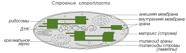
The structure of the chloroplast
The sizes of chloroplasts in different plants are not the same, but on average their diameter is 4-6 microns. Chloroplasts are able to move under the influence of the movement of the cytoplasm. In addition, under the influence of illumination, an active movement of amoeboid-type chloroplasts to the light source is observed.
Chlorophyll is the main substance of chloroplasts. Thanks to chlorophyll, green plants are able to use light energy.
Leucoplasts(colorless plastids) are clearly marked bodies of the cytoplasm. Their sizes are somewhat smaller than the sizes of chloroplasts. More uniform and their shape, approaching the spherical.
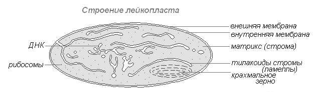
The structure of the leucoplast
They are found in the cells of the epidermis, tubers, rhizomes. When illuminated, they very quickly turn into chloroplasts with a corresponding change in the internal structure. Leucoplasts contain enzymes, with the help of which starch is synthesized from excess glucose formed during photosynthesis, the bulk of which is deposited in storage tissues or organs (tubers, rhizomes, seeds) in the form of starch grains. In some plants, fats are deposited in leukoplasts. The reserve function of leukoplasts occasionally manifests itself in the formation of storage proteins in the form of crystals or amorphous inclusions.
Chromoplasts in most cases they are derivatives of chloroplasts, occasionally - leukoplasts.

The structure of the chromoplast
Ripening of rose hips, peppers, tomatoes is accompanied by the transformation of chloro- or leukoplasts of pulp cells into carotenoids. The latter contain predominantly yellow plastid pigments - carotenoids, which, upon maturation, are intensively synthesized in them, forming colored lipid drops, solid globules or crystals. Chlorophyll is destroyed.
Mitochondria
Mitochondria are organelles found in most plant cells. They have a variable shape of sticks, grains, threads. They were discovered in 1894 by R. Altman using a light microscope, and the internal structure was later studied using an electronic one.
The structure of the mitochondria
Mitochondria have a two-membrane structure. The outer membrane is smooth, the inner one forms outgrowths of various shapes - tubules in plant cells. The space inside the mitochondria is filled with semi-liquid content (matrix), which includes enzymes, proteins, lipids, calcium and magnesium salts, vitamins, as well as RNA, DNA and ribosomes. The mitochondrial enzyme complex accelerates the work of a complex and interconnected mechanism of biochemical reactions, as a result of which ATP is formed. In these organelles, cells are provided with energy - the energy of chemical bonds of nutrients is converted into high-energy bonds of ATP in the process of cellular respiration. It is in the mitochondria that the enzymatic breakdown of carbohydrates, fatty acids, amino acids occurs with the release of energy and its subsequent conversion into ATP energy. The accumulated energy is spent on growth processes, on new syntheses, etc. Mitochondria reproduce by division and live for about 10 days, after which they are destroyed.
Endoplasmic reticulum
Endoplasmic reticulum - a network of channels, tubules, vesicles, cisterns located inside the cytoplasm. Opened in 1945 by the English scientist K. Porter, it is a system of membranes with an ultramicroscopic structure.

The structure of the endoplasmic reticulum
The entire network is integrated into a single whole with the outer cell membrane of the nuclear envelope. Distinguish ER smooth and rough, carrying ribosomes. On the membranes of the smooth EPS there are enzyme systems involved in fat and carbohydrate metabolism. This type of membrane prevails in seed cells rich in reserve substances (proteins, carbohydrates, oils), ribosomes are attached to the membrane of the granular ER, and during the synthesis of a protein molecule, the polypeptide chain with ribosomes is immersed in the ER channel. The functions of the endoplasmic reticulum are very diverse: the transport of substances both inside the cell and between neighboring cells; division of a cell into separate sections in which various physiological processes and chemical reactions take place simultaneously.
Ribosomes
Ribosomes are non-membrane cellular organelles. Each ribosome consists of two unequal-sized particles and can be divided into two fragments that continue to retain the ability to synthesize protein after combining into a whole ribosome.

The structure of the ribosome
Ribosomes are synthesized in the nucleus, then leave it, passing into the cytoplasm, where they are attached to the outer surface of the membranes of the endoplasmic reticulum or are located freely. Depending on the type of protein synthesized, ribosomes can function alone or combine into complexes - polyribosomes.
Cell structure
The human body, like any other living organism, is made up of cells. They play one of the main roles in our body. With the help of cells, growth, development and reproduction occur.
Now let's recall the definition of what is usually called a cell in biology.
A cell is such an elementary unit that is involved in the structure and functioning of all living organisms, with the exception of viruses. It has its own metabolism and is able not only to exist independently, but also to develop and reproduce itself. In short, we can conclude that the cell is the most important and necessary building material for any organism.
Of course, with the naked eye, you are unlikely to be able to see the cage. But with the help of modern technologies, a person has a great opportunity not only to examine the cell itself under a light or electron microscope, but also to study its structure, isolate and cultivate its individual tissues, and even decode the genetic cellular information.
And now, with the help of this figure, let's visually consider the structure of the cell:
Cell structure
But interestingly, it turns out that not all cells have the same structure. There is some difference between the cells of a living organism and the cells of plants. Indeed, in plant cells there are plastids, a membrane and vacuoles with cell sap. In the image you can see the cellular structure of animals and plants and see the difference between them:

For more information about the structure of plant and animal cells, you will learn by watching the video
As you can see, cells, although they have microscopic dimensions, but their structure is quite complex. Therefore, we will now move on to a more detailed study of the structure of the cell.
Plasma membrane of a cell
To give shape and to separate the cell from its kind, a membrane is located around the human cell.
Since the membrane has the ability to partially pass substances through itself, due to this, the necessary substances enter the cell, and waste products are removed from it.
Conventionally, we can say that the cell membrane is an ultramicroscopic film, which consists of two monomolecular layers of protein and a bimolecular layer of lipids, which is located between these layers.
From this we can conclude that the cell membrane plays an important role in its structure, as it performs a number of specific functions. It plays a protective, barrier and connecting function between other cells and for communication with the environment.
And now let's look at a more detailed structure of the membrane in the figure:

Cytoplasm
The next component of the internal environment of the cell is the cytoplasm. It is a semi-liquid substance in which other substances move and dissolve. The cytoplasm consists of proteins and water.
Inside the cell, there is a constant movement of the cytoplasm, which is called cyclosis. Cyclosis is circular or reticulate.
In addition, the cytoplasm connects different parts of the cell. In this environment, the organelles of the cell are located.
Organelles are permanent cellular structures with specific functions.
Such organelles include structures such as the cytoplasmic matrix, endoplasmic reticulum, ribosomes, mitochondria, etc.
Now we will try to take a closer look at these organelles and find out what functions they perform.

Cytoplasm
cytoplasmic matrix
One of the main parts of the cell is the cytoplasmic matrix. Thanks to it, biosynthesis processes take place in the cell, and its components contain enzymes that produce energy.

cytoplasmic matrix
Endoplasmic reticulum
Inside, the cytoplasmic zone consists of small channels and various cavities. These channels, connecting with each other, form the endoplasmic reticulum. Such a network is heterogeneous in its structure and can be granular or smooth. 
Endoplasmic reticulum
cell nucleus
The most important part, which is present in almost all cells, is the cell nucleus. Cells that have a nucleus are called eukaryotes. Each cell nucleus contains DNA. It is the substance of heredity and all the properties of the cell are encrypted in it.

cell nucleus
Chromosomes
If we look at the structure of a chromosome under a microscope, we can see that it consists of two chromatids. As a rule, after nuclear division, the chromosome becomes single chromatid. But by the beginning of the next division, another chromatid appears on the chromosome.

Chromosomes
Cell Center
When considering the cell center, one can see that it consists of a maternal and daughter centrioles. Each such centriole is a cylindrical object, the walls are formed by nine triplets of tubules, and in the middle there is a homogeneous substance.
With the help of such a cell center, the division of animal and lower plant cells occurs.

Cell Center
Ribosomes
Ribosomes are universal organelles in both animal and plant cells. Their main function is protein synthesis in the functional center.

Ribosomes
Mitochondria
Mitochondria are also microscopic organelles, but unlike ribosomes, they have a two-membrane structure, in which the outer membrane is smooth, and the inner one has variously shaped outgrowths called cristae. Mitochondria play the role of a respiratory and energy center

Mitochondria
golgi apparatus
But with the help of the Golgi apparatus, the accumulation and transportation of substances occurs. Also, thanks to this apparatus, the formation of lysosomes and the synthesis of lipids and carbohydrates occur.
In structure, the Golgi apparatus resembles individual bodies, which are crescent-shaped or rod-shaped.

golgi apparatus
plastids
But plastids for a plant cell play the role of an energy station. They tend to change from one species to another. Plastids are divided into such varieties as chloroplasts, chromoplasts, leukoplasts.

plastids
Lysosomes
The digestive vacuole, which is capable of dissolving enzymes, is called a lysosome. They are microscopic single-membrane organelles with a rounded shape. Their number directly depends on how viable the cell is and what its physical condition is.
In the event that the destruction of the lysosome membrane occurs, then in this case the cell is able to digest itself.

Lysosomes
Ways to feed the cell
Now let's look at how cells are fed:

How the cell is fed
It should be noted here that proteins and polysaccharides tend to penetrate the cell by phagocytosis, but liquid drops - by pinocytosis.
The method of nutrition of animal cells, in which nutrients enter it, is called phagocytosis. And such a universal way of feeding any cells, in which nutrients enter the cell already in a dissolved form, is called pinocytosis.
The science that studies the structure and function of cells is called cytology.
Cell- an elementary structural and functional unit of the living.
Cells, despite their small size, are very complex. The internal semi-liquid content of the cell is called cytoplasm.
The cytoplasm is the internal environment of the cell, where various processes take place and the components of the cell - organelles (organelles) are located.
cell nucleus
The cell nucleus is the most important part of the cell.
The nucleus is separated from the cytoplasm by a membrane consisting of two membranes. Numerous pores are present in the shell of the nucleus so that various substances can enter from the cytoplasm into the nucleus, and vice versa.
The internal contents of the kernel are called karyoplasms or nuclear juice. located in the nuclear sap chromatin And nucleolus.
Chromatin is a strand of DNA. If the cell begins to divide, then the chromatin threads are tightly coiled around special proteins, like threads on a spool. Such dense formations are clearly visible under a microscope and are called chromosomes.
Core contains genetic information and controls the vital activity of the cell.
nucleolus is a dense rounded body inside the nucleus. Usually, there are from one to seven nucleoli in the cell nucleus. They are clearly visible between cell divisions, and during division they are destroyed.
The function of the nucleoli is the synthesis of RNA and proteins, from which special organelles are formed - ribosomes.
Ribosomes involved in protein synthesis. In the cytoplasm, ribosomes are most often located on rough endoplasmic reticulum. Less commonly, they are freely suspended in the cytoplasm of the cell.
Endoplasmic reticulum (ER) participates in the synthesis of cell proteins and the transport of substances within the cell.
A significant part of the substances synthesized by the cell (proteins, fats, carbohydrates) is not consumed immediately, but through the ER channels it enters for storage in special cavities, stacked in kind of stacks, “tanks”, and delimited from the cytoplasm by a membrane. These cavities are called apparatus (complex) Golgi. Most often, the tanks of the Golgi apparatus are located near the nucleus of the cell.
golgi apparatus takes part in the transformation of cell proteins and synthesizes lysosomes- digestive organelles of the cell.
Lysosomes are digestive enzymes, are “packed” into membrane vesicles, bud off and spread through the cytoplasm.
The Golgi complex also accumulates substances that the cell synthesizes for the needs of the whole organism and which are removed from the cell to the outside.
Mitochondria- energy organelles of cells. They convert nutrients into energy (ATP), participate in cell respiration.
Mitochondria are covered with two membranes: the outer membrane is smooth, and the inner one has numerous folds and protrusions - cristae.
plasma membrane
For a cell to be a single system, it is necessary that all its parts (cytoplasm, nucleus, organelles) be held together. For this, in the process of evolution, plasma membrane, which, surrounding each cell, separates it from the external environment. The outer membrane protects the inner contents of the cell - the cytoplasm and nucleus - from damage, maintains a constant shape of the cell, provides communication between cells, selectively passes the necessary substances into the cell and removes metabolic products from the cell.
The structure of the membrane is the same in all cells. The basis of the membrane is a double layer of lipid molecules, in which numerous protein molecules are located. Some proteins are located on the surface of the lipid layer, others penetrate both layers of lipids through and through.
Special proteins form the thinnest channels through which potassium, sodium, calcium ions and some other ions with a small diameter can pass into or out of the cell. However, larger particles (nutrient molecules - proteins, carbohydrates, lipids) cannot pass through the membrane channels and enter the cell with the help of phagocytosis or pinocytosis:
- In the place where the food particle touches the outer membrane of the cell, an invagination is formed, and the particle enters the cell, surrounded by a membrane. This process is called phagocytosis (plant cells over the outer cell membrane are covered with a dense layer of fiber (cell membrane) and cannot capture substances by phagocytosis).
- pinocytosis differs from phagocytosis only in that in this case, the invagination of the outer membrane captures not solid particles, but liquid droplets with substances dissolved in it. This is one of the main mechanisms for the penetration of substances into the cell.
We invite you to familiarize yourself with the materials and.
: cellulose membrane, membrane, cytoplasm with organelles, nucleus, vacuoles with cell sap.The presence of plastids is the main feature of the plant cell.
Cell wall functions- determines the shape of the cell, protects against environmental factors.
plasma membrane- a thin film, consists of interacting lipid and protein molecules, delimits the internal contents from the external environment, provides transport of water, mineral and organic substances into the cell by osmosis and active transfer, and also removes waste products.
Cytoplasm- the internal semi-liquid environment of the cell, in which the nucleus and organelles are located, provides connections between them, participates in the main processes of life.
Endoplasmic reticulum- a network of branching channels in the cytoplasm. It is involved in the synthesis of proteins, lipids and carbohydrates, in the transport of substances. Ribosomes - bodies located on the EPS or in the cytoplasm, consist of RNA and protein, are involved in protein synthesis. EPS and ribosomes are a single apparatus for the synthesis and transport of proteins.
Mitochondria-organelles separated from the cytoplasm by two membranes. Organic substances are oxidized in them and ATP molecules are synthesized with the participation of enzymes. An increase in the surface of the inner membrane on which enzymes are located due to cristae. ATP is an energy-rich organic substance.
plastids(chloroplasts, leukoplasts, chromoplasts), their content in the cell is the main feature of the plant organism. Chloroplasts are plastids containing the green pigment chlorophyll, which absorbs light energy and uses it to synthesize organic substances from carbon dioxide and water. Delimitation of chloroplasts from the cytoplasm by two membranes, numerous outgrowths - grana on the inner membrane, in which chlorophyll molecules and enzymes are located.
Golgi complex- a system of cavities delimited from the cytoplasm by a membrane. The accumulation of proteins, fats and carbohydrates in them. Implementation of the synthesis of fats and carbohydrates on membranes.
Lysosomes- bodies separated from the cytoplasm by a single membrane. The enzymes contained in them accelerate the reaction of splitting complex molecules into simple ones: proteins to amino acids, complex carbohydrates to simple ones, lipids to glycerol and fatty acids, and also destroy dead parts of the cell, whole cells.
Vacuoles- cavities in the cytoplasm filled with cell sap, a place of accumulation of reserve nutrients, harmful substances; they regulate the water content in the cell.
Core- the main part of the cell, covered on the outside with a two-membrane, pierced by pores nuclear envelope. Substances enter the core and are removed from it through the pores. Chromosomes are carriers of hereditary information about the characteristics of an organism, the main structures of the nucleus, each of which consists of one DNA molecule in combination with proteins. The nucleus is the site of the synthesis of DNA, i-RNA, r-RNA.

The presence of an outer membrane, cytoplasm with organelles, a nucleus with chromosomes.
Outer or plasma membrane- delimits the contents of the cell from the environment (other cells, intercellular substance), consists of lipid and protein molecules, provides communication between cells, transport of substances into the cell (pinocytosis, phagocytosis) and out of the cell.
Cytoplasm- the internal semi-liquid environment of the cell, which provides communication between the nucleus and organelles located in it. The main processes of vital activity take place in the cytoplasm.
Cell organelles:
1) endoplasmic reticulum (ER)- a system of branching tubules, involved in the synthesis of proteins, lipids and carbohydrates, in the transport of substances in the cell;
2) ribosomes- bodies containing rRNA are located on the ER and in the cytoplasm, and are involved in protein synthesis. EPS and ribosomes are a single apparatus for protein synthesis and transport;
3) mitochondria- "power stations" of the cell, delimited from the cytoplasm by two membranes. The inner one forms cristae (folds) that increase its surface. Enzymes on cristae accelerate the reactions of oxidation of organic substances and the synthesis of energy-rich ATP molecules;
4) golgi complex- a group of cavities delimited by a membrane from the cytoplasm, filled with proteins, fats and carbohydrates, which are either used in life processes or removed from the cell. The membranes of the complex carry out the synthesis of fats and carbohydrates;
5) lysosomes- bodies filled with enzymes accelerate the reactions of splitting proteins to amino acids, lipids to glycerol and fatty acids, polysaccharides to monosaccharides. In lysosomes, dead parts of the cell, whole cells and cells are destroyed.
Cell inclusions- Accumulations of spare nutrients: proteins, fats and carbohydrates.
Core- the most important part of the cell. It is covered with a double-membrane membrane with pores through which some substances penetrate into the nucleus, while others enter the cytoplasm. Chromosomes are the main structures of the nucleus, carriers of hereditary information about the characteristics of an organism. It is transmitted in the process of division of the mother cell to daughter cells, and with germ cells - to daughter organisms. The nucleus is the site of DNA, mRNA, rRNA synthesis.
Exercise:
Explain why organelles are called specialized structures of the cell?
Answer: organelles are called specialized cell structures, since they perform strictly defined functions, hereditary information is stored in the nucleus, ATP is synthesized in mitochondria, photosynthesis proceeds in chloroplasts, etc.
If you have questions about cytology, you can ask for help from
STRUCTURE AND FUNCTIONS OF THE CELL
A cell is an elementary unit of the structure and vital activity of all organisms (except for viruses, which are often referred to as non-cellular life forms), which has its own metabolism, is capable of independent existence, self-reproduction and development. All living organisms either consist of many cells (multicellular animals, plants and fungi), or are single-celled organisms (many protozoa and bacteria). The branch of biology that studies the structure and activity of cells is called cytology. Recently, it has also become customary to talk about the biology of the cell, or cell biology.
Usually, the sizes of plant and animal cells range from 5 to 20 microns in diameter. A typical bacterial cell is much smaller - approx. 2 µm, and the smallest known is 0.2 µm.
Some free-living cells, such as protozoa such as foraminifera, can be several centimeters long; they always have many nuclei. The cells of thin plant fibers reach a length of one meter, and the processes of nerve cells reach several meters in large animals. With such a length, the volume of these cells is small, and the surface is very large.
The largest cells are unfertilized bird eggs filled with yolk. The largest egg (and, therefore, the largest cell) belonged to an extinct huge bird - epiornis (Aepyornis). Presumably its yolk weighed approx. 3.5 kg. The largest egg in living species belongs to the ostrich, its yolk weighs approx. 0.5 kg
At one time, the cell was considered as a more or less homogeneous droplet of organic matter, which was called protoplasm or living substance. This term became obsolete after it became clear that the cell consists of many clearly separated structures, called cellular organelles ("small organs").
The first person to see cells was the English scientist Robert Hooke (known to us thanks to Hooke's law). In 1665, trying to understand why the cork tree floats so well, Hooke began to examine thin sections of cork with an improved immicroscope. He found that the cork was divided into many tiny cells, which reminded him of the honeycombs in honey bee hives, and he called these cells cells (in English, cell means "cell, cell").

In 1675, the Italian doctor M. Malpighi, and in 1682 - an English botanist N. Gru confirmed the cellular structure of plants. They began to talk about the cell as a "bubble filled with nutritious juice." In 1674 a Dutch master Anthony van Leeuwenhoek(Anton van Leeuwenhoek, 1632-1723) using a microscope for the first time saw in a drop of water "animals" - moving living organisms (ciliates, amoeba, bacteria). Leeuwenhoek was also the first to observe animal cells - erythrocytes and spermatozoa. Thus, already by the beginning of the 18th century, scientists knew that under high magnification plants have a cellular structure, and they saw some organisms, which later were called unicellular. In 1802-1808, the French explorer Charles-Francois Mirbel established that all plants consist of tissues formed by cells. Zh. B. Lamarck in 1809
extended Mirbel's idea of the cellular structure to animal organisms. In 1825, the Czech scientist J. Purkyne discovered the nucleus of the egg cell of birds, and in 1839 introduced the term "protoplasm". In 1831, the English botanist R. Brown first described the nucleus of a plant cell, and in 1833 established that the nucleus is an essential organelle of a plant cell. Since then, the main thing in the organization of cells is not the membrane, but the content.
Cell research methods
For the first time, cells could be seen only after the creation of light microscopes; from that time to the present, microscopy has remained one of the most important methods for studying cells. Light (optical) microscopy, despite its relatively low resolution, made it possible to observe living cells. In the twentieth century, electron microscopy was invented, which made it possible to study the ultrastructure of cells.
In the study of cell shape and structure, the first instrument was the light microscope. Its resolution is limited to dimensions comparable to the wavelength of light (0.4–0.7 µm for visible light). However, many elements of the cellular structure are much smaller in size.
Another difficulty is that most cellular components are transparent and their refractive index is almost the same as that of water. To improve visibility, dyes are often used that have different affinities for different cellular components. Staining is also used to study the chemistry of the cell. For example, some dyes bind predominantly to nucleic acids and thereby reveal their localization in the cell. A small amount of dyes
- they are called in vivo - can be used to stain living cells, but usually the cells must be pre-fixed (using substances that coagulate the protein) and only after that they can be stained.
Prior to examination, cells or pieces of tissue are usually embedded in paraffin or plastic and then cut into very thin sections using a microtome. This method is widely used in clinical laboratories to detect tumor cells. In addition to conventional light microscopy, other optical methods for studying cells have also been developed: fluorescence microscopy, phase-contrast microscopy, spectroscopy, and X-ray diffraction analysis.

optical microscopy
In an optical microscope, magnification of an object is achieved through a series of lenses through which light passes. The maximum magnification that can be achieved with an optical microscope is about 1000. Another important characteristic is
resolutions are only about 200 nm; such permission was obtained at the end
XIX century. Thus, the smallest structures that can be observed under an optical microscope are mitochondria and bacteria, the linear size of which is approximately 500 nm. However, objects smaller than 200 nm are visible in a light microscope only if they themselves emit light. This feature is used in fluorescence microscopy when cellular structures or individual proteins bind to special fluorescent proteins or antibodies with fluorescent labels. The quality of the image obtained with an optical microscope is also affected by contrast - it can be increased using various cell staining methods. Phase-contrast, differential interference-contrast, and dark-field microscopy is used to study living cells. Confocal microscopes can improve the quality of fluorescent images.
electron microscopy
In the 1930s, an electron microscope was constructed in which, instead of light, a beam of electrons is passed through an object. The theoretical resolution limit for modern electron microscopes is about 0.002 nm, however, for practical reasons, only about 2 nm resolution is achieved for biological objects. An electron microscope can be used to study the ultrastructure of cells. There are two main types of electron microscopy:
scanning and transmission.
Scanning (raster) electron microscopy (SEM) is used to study the surface of an object. Samples are often coated with a thin film of gold. REM
allows you to get 3D images. Transmission (transmission) electron microscopy (TEM) - used to study the internal
cell structures. An electron beam is passed through an object pre-treated with heavy metals that accumulate in certain structures, increasing their electron density. The electrons scatter to areas of the cell with a higher electron density, causing these areas to appear darker in the images.
Fractionation of cells. To establish the functions of individual components of the cell, it is important to isolate them in their pure form, most often this is done using the differential method. centrifugation. Techniques have been developed to obtain pure fractions of any cell organelles. The production of fractions begins with the destruction of the plasma membrane and the formation of a cell homogenate. The homogenate is sequentially centrifuged at different speeds, at the first stage, four fractions can be obtained: (1) nuclei and large fragments of cells, (2) mitochondria, plastids, lysosomes and peroxisomes, (3) microsomes - Golgi vesicles and endoplasmic reticulum, (4) ribosomes, proteins and smaller molecules will remain in the supernatant. Further differential centrifugation of each of the mixed fractions makes it possible to obtain pure preparations of organelles, to which a variety of biochemical and microscopic methods can be applied.
cell structure
All cellular life forms on Earth can be divided into two kingdoms based on the structure of their constituent cells:
prokaryotes (pre-nuclear) - simpler in structure;
eukaryotes (nuclear) are more complex. The cells that make up the human body are eukaryotic.
Despite the variety of forms, the organization of the cells of all living organisms is subject to uniform structural principles.
prokaryotic cell
Prokaryotes (lat. pro - before, before Greek κάρῠον - core, nut) - organisms that, unlike eukaryotes, do not have a formed cell nucleus and other internal membrane organelles (with the exception of flat cisterns in photosynthetic species, for example, ucyanobacteria). The only large circular (in some species - linear) double-stranded DNA molecule, which contains the main part of the cell's genetic material (the so-called nucleoid) does not form a complex with histone proteins (the so-called chromatin). Prokaryotes include bacteria, including cyanobacteria (blue-green algae), and archaea. The main content of the cell, which fills its entire volume, is viscous granular
cytoplasm.
eukaryotic cell
Eukaryotes (Eukaryotes) (Greek ευ - good, completely and κάρῠον - core, nut)
Organisms that, unlike prokaryotes, have a well-shaped cell nucleus, delimited from the cytoplasm by the nuclear membrane. The genetic material is enclosed in several linear double-stranded DNA molecules (depending on the type of organisms, their number per nucleus can vary from two to several hundred), attached from the inside to the membrane of the cell nucleus and forming in the vast majority of them a complex with histone proteins, called chromatin.

The structure of a eukaryotic cell. Schematic representation of an animal cell.
Some cells, mainly plant and bacterial, have an outer cell wall. In higher plants, it consists of cellulose. The cell wall plays an extremely important role: it is an outer frame, a protective shell, provides turgor of plant cells: water, salts, and molecules of many organic substances pass through the cell wall. Animal cells usually do not have cell walls.
Located under the cell wall of plants plasma membrane or plasmalemma. The thickness of the plasma membrane is about 10 nm, the study of its structure and functions is possible only with the help of an electron microscope.
Inside the cell is filled with cytoplasm, in which various organelles and cell inclusions are located, as well as genetic material in the form of a DNA molecule. Each of the organoids of the cell performs its own special function, and together they all determine the vital activity of the cell as a whole.
The plasma membrane provides primarily a delimiting function in relation to the external for
cells in the environment. It is a double layer of molecules (bimolecular layer, or bilayer). Basically, these are molecules of phospholipids and other substances close to them. Lipid molecules have a dual nature, manifested in the way they behave in relation to water. The heads of the molecules are hydrophilic, i.e. have an affinity for water, and their hydrocarbon tails are hydrophobic. Therefore, when mixed with water, lipids form a film on its surface, similar to an oil film; at the same time, all their molecules are oriented in the same way: the heads of the molecules are in the water, and the hydrocarbon tails are above its surface.
IN the cell membrane has two such layers, and in each of them the heads of the molecules are turned outward, and the tails are turned inside the membrane, one to the other, thus not coming into contact with water.
In addition to the main lipid components, it contains large protein molecules that are able to “float” in the lipid bilayer and are located so that one of their sides is turned inside the cell, and the other is in contact with the external environment. Some proteins are located only on the outer or only on the inner surface of the membrane, or are only partially immersed in the lipid bilayer.
The main function of the cell membrane is to regulate the transport of substances into and out of the cell.
There are several mechanisms for the transport of substances across the membrane:
Diffusion - the penetration of substances through the membrane along the concentration gradient (from the area where their concentration is higher to the area where their concentration is lower). Diffuse transport of substances is carried out with the participation of membrane proteins, in which there are molecular pores (water, ions), or with the participation of the lipid phase (for fat-soluble substances).
Facilitated diffusion- special membrane carrier proteins selectively bind to one or another ion or molecule and transfer them through the membrane.
active transport. This mechanism is associated with energy costs and serves to transport substances against their concentration gradient. It is carried out by special
carrier proteins that form the so-called ion pumps. The most studied is the Na+ /K+ pump in animal cells, which actively pumps out Na+ ions while absorbing K+ ions.
IN In combination with active transport of ions into the cell, various sugars, nucleotides, and amino acids penetrate through the cytoplasmic membrane.
Such selective permeability is physiologically very important, and its absence
– first evidence of cell death. This can be easily illustrated with the example of beets. If a live beet root is immersed in cold water, it retains its pigment; if the beets are boiled, then the cells die, become easily permeable and lose the pigment, which turns the water red.
Large molecules such as protein cells can "swallow". Under the influence of certain proteins, if they are present in the fluid surrounding the cell, an invagination occurs in the cell membrane, which then closes, forming a bubble - a small vacuole containing water and protein molecules; after that, the membrane around the vacuole breaks, and the contents enter the cell. This process is called pinocytosis (literally "cell drinking"), or endocytosis.

Larger particles, such as food particles, can be absorbed in a similar way during the so-called. phagocytosis. As a rule, the vacuole formed during phagocytosis is larger, and the food is digested by the enzymes of the lysosomes inside the vacuole until the membrane surrounding it ruptures. This type of nutrition is typical for protozoa, for example, for amoebas that eat bacteria.
Exocytosis (exo - out), thanks to it, the cell removes intracellular products or undigested residues enclosed in vacuoles, or vesicles. The vesicle approaches the cytoplasmic membrane, merges with it, and its contents are released into the environment. This is how digestive enzymes, hormones, hemicellulose, etc. are secreted.
The structure of the cytoplasm.
The liquid component of the cytoplasm is also called the cytosol. Under a light microscope, it seemed that the cell was filled with something like a liquid plasma or sol, in which the nucleus and other organelles “floated”. Actually it is not. The internal space of a eukaryotic cell is strictly ordered. The movement of organelles is coordinated with the help of specialized transport systems, the so-called microtubules, which serve as intracellular "roads", and special proteins, dyneins and kinesins, which play the role of "engines". Separate protein molecules also do not freely diffuse throughout the entire intracellular space, but are directed to the necessary compartments using special signals on their surface, recognized by the cell's transport systems.
Endoplasmic reticulum
In a eukaryotic cell, there is a system of membrane compartments (tubes and tanks) that pass into each other,
which is called endoplasmic reticulum(or endoplasmic reticulum, EPR or EPS). That part of the EPR, to the membranes of which ribosomes are attached, is referred to as granular (or rough) endoplasmic

reticulum, on its membranes protein synthesis occurs. Those compartments, on the walls of which there are no ribosomes, are classified as smooth ER, which takes part in the synthesis of lipids. The internal spaces of the smooth and granular ER are not isolated, but pass into each other and communicate with the luminal membrane. The tubules also open on the cell surface, and the endoplasmic reticulum thus plays the role of an apparatus through which the external environment can directly interact with all the contents of the cell.
Tiny bodies called ribosomes cover the surface of the rough endoplasmic reticulum, especially near the nucleus. Ribosome diameter is about 15 nm. Each ribosome consists of two particles of different sizes, small and large. Their main function is the synthesis of proteins; matrix (information) RNA and amino acids associated with transfer RNA are attached to their surface. The synthesized proteins are first accumulated in the channels and cavities of the endoplasmic reticulum and then transported to the organelles and cell sites where they are consumed.
golgi apparatus
Golgi apparatus (Golgi complex)
is a stack of flat membrane sacs, somewhat expanded closer to the edges. In the tanks of the Golgi apparatus, some proteins synthesized on the membranes of the granular ER and intended for secretion or the formation of lysosomes mature. The Golgi apparatus is asymmetric - the tanks located closer to the cell nucleus (cis-Golgi) contain the least mature proteins, membrane vesicles, vesicles, budding from the endoplasmic reticulum, continuously join these tanks. Apparently, with the help of the same vesicles, the further movement of maturing proteins from one tank to another takes place. Finally from the opposite end of the organelle
(trans-Golgi) vesicles containing fully mature proteins bud off.
Lysosomes
Lysosomes (Greek "Liseo" - dissolve, "Soma" - body) are small round bodies. These membranous cell organelles are oval in shape and 0.5 µm in diameter. They bud from the Golgi apparatus and possibly from the endoplasmic reticulum. Lysosomes contain a variety of enzymes that break down large molecules: proteins, fats, carbohydrates, nucleic acids. Due to their destructive action, these enzymes are, as it were, "locked" in lysosomes and are released only as needed. But if the lysosome

damaged from any external influences, then the whole cell or part of it is destroyed.
During intracellular digestion, enzymes are released from lysosomes into digestive vacuoles.
During starvation, lysosome cells digest some organelles without killing the cell. Such partial digestion provides the cell with the necessary minimum of nutrients for a while.
Possessing the ability to actively digest nutrients, lysosomes are involved in the removal of parts of cells, whole cells and organs that die in the process of vital activity. For example, the disappearance of the tail of a frog tadpole occurs under the action of lysosome enzymes. In this case, this is normal and beneficial for the body, but sometimes such cell destruction is pathological. For example, when asbestos dust is inhaled, it can enter the cells of the lungs, and then lysosomes rupture, cells are destroyed, and lung disease develops.
The information center of the cell, the place of storage and reproduction of hereditary information that determines all the signs of a given cell and the organism as a whole, is the nucleus. Removal of the nucleus from the cell, as a rule, leads to its rapid death. The shape and size of the cell nucleus is very variable depending on the type of organism, as well as on the type, age and functional state of the cell. Overall plan
The structure of the nucleus is the same in all eukaryotic cells. The cell nucleus consists of the nuclear membrane, the nuclear matrix (nucleoplasm), chromatin and the nucleolus (one or more). The contents of the nucleus are separated from the cytoplasm by a double membrane or the so-called nuclear envelope. The outer membrane in some places passes into the channels of the endoplasmic reticulum; ribosomes are attached to it. The cell nucleus contains DNA molecules on which the genetic information of the organism is recorded. . This determines the leading role of the cell nucleus in heredity. In the nucleus, replication occurs - the duplication of DNA molecules, as well as transcription - the synthesis of RNA molecules on the DNA template. The assembly of caribosomes also takes place in the nucleus, in special formations called nucleoli. The nuclear envelope is permeated with many pores, the diameter of which is about 90 nm. Due to the presence of pores that provide selective permeability, the nuclear membrane controls the exchange of substances between the nucleus and the cytoplasm.
fibrillar structures located in the cytoplasm of the cell: microtubules, actin and intermediate filaments. Microtubules are involved in the transport of organelles, are part of the flagella, and the mitotic spindle is built from microtubules. Actin filaments are essential for maintaining

cell shape, pseudopodial reactions. The role of intermediate filaments also seems to be to maintain the structure of the cell. Proteins of the cytoskeleton make up several tens of percent of the mass of the cellular protein.
Centrioles
Centrioles are cylindrical protein structures located near the nucleus of animal cells (plants do not have centrioles, with the exception of lower algae). The centriole is a cylinder, the lateral surface of which is formed by nine sets of microtubules. The number of microtubules in a set
fluctuate for different organisms from 1 to 3.
Around the centrioles is the so-called center of organization of the cytoskeleton, the area in which the minus ends of the microtubules of the cell are grouped.
Before dividing, the cell contains two centrioles located at right angles to each other. During mitosis, they diverge to different ends of the cell, forming the spindle poles of division. After cytokinesis, each daughter cell receives one centriole, which doubles for the next division. Doubling of centrioles occurs not by division, but by the synthesis of a new structure perpendicular to the existing one.
Mitochondria
Mitochondria - special organelles of the cell, the main function of which is the synthesis ATP - universal carrier of energy. In mitochondria, the oxidation of organic substances occurs, coupled with the synthesis
adenosine triphosphate (ATP). The breakdown of ATP with the formation of adenosine diphosphate (ADP) is accompanied by the release of energy, which is spent on various life processes, such as the synthesis of proteins and nucleic acids, the transport of substances into and out of the cell, the transmission of nerve impulses, or muscle contraction.
Mitochondria, therefore, are energy stations that process "fuel" - fats and carbohydrates - into a form of energy that can be used by the cell, and therefore the body as a whole.
