Presentation on the topic of allergic reactions. Allergy. Immunopathological reactions Department of Pathophysiology Siberian State Medical University Assistant of the Department, Dr. honey. Sciences, immunologist-allergist E.G. Churina. Emergency care for swelling, urticaria and general urticaria
go to places where it has already passed or has not yet arrived. If this is not possible, stick to a “city” lifestyle, do not go out into nature, do not walk where there are flowering trees or grasses. 2. Returning from the street, take off your “street” clothes and rinse your nasopharynx with water. 3. At this time, do not open the windows, hang cotton curtains on them and periodically moisten them with a spray bottle. 4. Use household chemicals and new cosmetics with extreme caution. First, do a skin test by applying a small amount of cosmetic to the inside of your elbow. 5. Drive in a car without opening the windows. 6. Be sure to wear sunglasses outside. 7. During flowering, you should not carry out planned operations or perform work that requires great physical and intellectual effort. 8. Eliminate alcohol, smoked foods, delicacies, chocolate from your diet - foods that promote the release of histamine. 9. Do not forget that antibiotics and other medications can cause a violent reaction. Warn the doctor you contacted for any help. 10. Always carry prescribed medications with you - they may be needed for emergency assistance if your condition worsens.

Lecture plan Relevance of the problem and prevalence of allergic diseases Definition of allergy, its connection with immunity Etiology of allergy Classification of allergic reactions Stages of development of allergic reactions General characteristics of 1-4 types of immunopathological reactions Pseudoallergic reactions


The reasons for the increase in the incidence of allergic diseases are man-made phenomena. Significant increase in foreign substances (xenobiotics) in the environment Use of vaccines and serums, including mass uncontrolled vaccination of the adult population “Hygienic theory” - artificially limiting the immune system from contact with pathogens

Definition of allergy Allergy (hypersensitivity) is a state of increased sensitivity of the body to substances of an antigenic nature. Allergy is a qualitatively altered (pathological) form of the body's immunological reactivity, which is accompanied by damage to its own cells and tissues. Allergy is an immunologically dependent damage to body tissues. Allergy is a typical pathological process.

ALLERGY is a manifestation of immunological reactivity, just like immunity. This is an example of increased reactivity and reduced resistance. Allergy and immunity are protective in nature and are aimed at maintaining homeostasis of the body (protect against genetically foreign information) Allergy and immunity have common development mechanisms


Classification of allergens by origin Allergens Endoallergens Exoallergens Acquired (secondary) Congenital (primary) Infectious origin Non-infectious origin thyroid colloid testicular tissue organ of vision nervous tissue tumor cells necrosis cells denatured proteins Household Food Medicinal Plant Industrial


Pollen allergens. Hay fever is a classic allergic disease. It develops when a sensitized organism comes into repeated contact with plant pollen. Characterized by acute allergic inflammation of the mucous membranes of the respiratory tract, eyes, and skin.

Household allergens. House dust mite Allergy symptoms appear all year round when indoors High concentration of antigen Often associated with pet allergens In 70% - 80% of cases of asthma is associated with household mites Mites are µm in size 1 gram of house dust contains 240 thousand mites


Classification of allergic reactions: immediate hypersensitivity; delayed hypersensitivity 1. According to the classification proposed by Cooke (1930), according to the speed and mechanisms of development, allergic reactions are divided into 2 types:

2. According to the classification of Gell and Coombs (1969), depending on the nature of immune damage to tissues and organs, allergic reactions are divided into 4 types: cell-mediated (HRT) reagin (anaphylactic) cytotoxic immune complex HNT

3. Andrei Dmitrievich Ado (1963), according to the mechanisms of development, divided allergic reactions into 2 types: true false (pseudo-allergic) Pseudo-allergic reactions have only 2 stages - pathochemical and pathophysiological. The main – immunological stage – is absent. There are no antibodies, no immune complexes, the pathogenic factor independently stimulates the formation of damage mediators


1. Immunological stage (stage of immune reactions) 1. Immunological stage (stage of immune reactions) from the moment the allergen first enters the body until it is encountered again. The result is the formation of an immune complex. 1. The period of sensitization, antibodies or sensitized T-lymphocytes are formed 2. The period of re-entry of the allergen into the sensitized body, the immune complex antigen + antibody antigen + sensitized T-lymphocyte is formed

2. The pathochemical stage (stage of biochemical reactions) is characterized by the formation and release of biologically active substances (allergy mediators) entering the blood and tissues. Immune complexes serve as the trigger for these processes. 3. The pathophysiological stage (stage of clinical manifestations) is characterized by the damaging effect of mediators on cells, tissues and organs. This stage includes the clinical manifestations of disorders occurring in the body in the form of allergic reactions and diseases


1. 2. Th2 lymphocytes produce IL4 and IL13, which activate B lymphocytes, which leads to their differentiation into plasma cells and memory B cells. 3. Plasmocytes (antibody-producing cells) secrete allergen-specific IgE and IgG4, which bind to mast cells. 4. Atopic antibodies attach to the mast cell Allergen Macrophage Th2 lymphocyte Histocompatibility complex II IgE antibodies Fixation of antibodies to the mast cell B lymphocyte IL-4 Immunological stage

... causes itching and hyperemia within a few minutes. Further, eosinophils, neutrophils, monocytes are involved in the process, which, in turn, produce late-phase mediators that contribute to further damage to surrounding tissues (4-6 hours after the allergen enters the nose, lungs, eyes ...). IL-1 IL-4 TNF-α ICAM-1 PAF Tryptase Heparin Histamine 5. Mast cell degranulation and release of inflammatory mediators occurs... Pathochemical stage

ConjunctivaAllergic conjunctivitis Distribution of mast cells in tissues Lungs - bronchial epithelium Nasal mucosa - epithelium TK tryptase TK tryptase, chemase Skin, atopic dermatitis Lungs - alveoli Small intestinal mucosa

Characteristics of factors contained in mast cell granules (primary mediators of allergy) Factor Chemical nature Effects Histamine Amine, histidine metabolite Dilation and increased vascular permeability, spasm of smooth muscles, increased mucus secretion, irritation of nerve endings (itching) Heparin Peptidoglycan Dilation and increased vascular permeability, spasm of smooth muscles Serotonin min , metabolite of tryptophan Dilation and increase in vascular permeability, spasm of smooth muscles Himase, tryptase Proteins Proteolysis, increased mucus secretion, epithelial remodeling Chemotactic factor of eosinophils Protein Chemotaxis of eosinophils Chemotactic factor of neutrophils Protein Chemotaxis of neutrophils

Humoral factors released by allergy effector cells Group of factors Mast cells Eosinophils Preformed factors Histamine, heparin, chondroitin sulfate, serotonin, proteases Major basic protein (MBP), eosinophil cationic protein (ECP), eosinophil peroxidase (EPO), eosinophil neurotoxin (EDT) Rapidly synthesized factors Leukotrienes (LTC4, LTD4), prostaglandins (D2), thromboxanes, platelet activating factor (PAF) Leukotrienes (LTC4, LTD4) - SASR, prostaglandins (E, F), platelet activating factor (PAF) Slowly synthesized factors IL-5, GM-CSF, TNFα, IL-8IL-3, IL-5, GM-CSF, TGFβ


The scratch skin test is a method for diagnosing atopic diseases in vivo. The reaction is taken into account in minutes (GNT) after introducing an allergen into the scratch: - with a positive reaction, a blister (2-10 mm) with hyperemia appears; - in case of a negative reaction, there is no blister and severe hyperemia (the results are compared with the control - the reaction to the allergen solvent and to histamine)


Clinical manifestations of allergic urticaria (a), giant urticaria (b). Urticaria is a common group of diseases characterized by inflammatory changes in the skin and/or mucous membranes, the appearance of a diffuse or limited rash in the form of pronounced itchy papules or blisters of various sizes with areas of erythema around them.


1. Immunological stage Appearance of autoantigens formation of autoantibodies of the IgG and IgM class to them Autoantibody combines with autoantigen on the surface of the cell an immune complex is formed, fixed on the membrane of the altered cell Autoantigen Immune complex Antibody

2. Pathochemical stage. There are 3 mechanisms of implementation: 1. Complement-dependent cytolysis (complement is activated, membrane attack complex C5-C9 is formed, damage to cell membranes, cell death) 2. Phagocytosis (IgM and IgG fixed on cells activate phagocytes, phagocytes absorb and destroy these cells using lysosomal enzymes )

Complement-dependent cytolysis and phagocytosis (A. A. Vorobyov et al., 2006) Antibodies attach to cell surface antigens. Complement (C) is attached to the Fc fragment, which is activated along the classical pathway with the formation of anaphylatoxins (C5a, C3a) and MAC, consisting of components C5-9


Antibody-dependent cellular cytotoxicity is the lysis of target cells opsonized by antibodies by NK cells. NK cells attach to Fc fragments of immunoglobulins that have bound to target cell antigens. Destruction of the target cell occurs with the help of perforins and granzymes of NK cells.


Anti-receptor antibodies in malignant myasthenia Gravis - an autoimmune disease In malignant myasthenia, accompanied by severe weakness, antibodies (autoantibodies) are formed against acetylcholine receptors on muscle cells. Antibodies block the binding of acetylcholine receptors, leading to muscle weakness


Allergens (endo- and exoallergens) are free, not associated with the “host” tissues, dissolved in plasma, lymph, tissue fluid. Immune damage is carried out by circulating immune complexes (CIC) - (CIC) - allergen + antibody. Normally, immune complexes are removed from the body with the help of the complement system (components C1-C5), erythrocytes and macrophages of the reticuloendothelial system of the liver.

1. Immunological stage In response to the appearance of an allergen, IgM and IgG are synthesized in the body, they combine with allergens to form immune complexes. When elimination processes are disrupted, immune complexes accumulate and begin to circulate in the body. They are deposited on the vascular wall (vasculitis), on the membranes of the renal glomeruli (glomerulonephritis) or in tissues (local inflammatory reaction like the Arthus phenomenon) Allergen + Antibody Circulating immune complex

2. Pathochemical stage. Under the influence of immune complexes, inflammatory mediators are formed 1. Immune complexes fixed in tissues activate the complement system (C1-C5 components) - anaphylatoxins (C3a and C5a) activate mast cells to produce biologically active substances - macrophages produce TNFα and other pro-inflammatory cytokines - neutrophils 2. Fixed in tissues, immune complexes activate the kallikrein-kinin system (bradykinin)

3. Pathophysiological stage In places where immune complexes are deposited, exudative inflammation develops. Alteration of cells and tissues occurs. Clinically this is manifested by dermatitis, alveolitis, generalized vasculitis, glomerulonephritis, systemic lupus erythematosus

 Reactions of this type do not depend on antibodies and complement - they are cell-mediated. Their course is determined by sensitized T-lymphocytes, i.e. Memory T cells induced by an antigen and targeted for its elimination. HRT is the pathogenetic basis of contact hypersensitivity, some autoimmune diseases, develops during intracellular bacterial, fungal and protozoal infections, and is involved in transplant rejection reactions. general characteristics

1. The immunological stage of the HRT reaction proceeds in a Th1-dependent manner. The allergen is phagocytosed, processed by the macrophage and presented to T helper cells (Th). APCs secrete interleukin-12, which promotes the differentiation of Th0, which recognizes the allergen, into type 1 Th with the CD4+ phenotype - a sensitized lymphocyte. Upon repeated exposure to an allergen, Th1 with the CD4+ phenotype produce cytokines that mediate the inflammatory response

2. Pathochemical stage Caused by either an activating or inhibitory effect of cytokines (they can be pro-inflammatory - IL-1, IL-2, IL-6, TNFα and anti-inflammatory with immunosuppressive activity - IL-10, TGFβ) on: lymphocytes, macrophages and neutrophils target cells

Delayed hypersensitivity reaction (type IV) and stages of its development According to the mechanisms of development, delayed hypersensitivity coincides with the inflammatory type of immune response, only its inductive and effector phases are more clearly separated in time (Yarilin A.A., 2010)

3. The pathophysiological stage of HRT can occur in any organs and tissues, depending on the localization of the allergen. In all cases, inflammation of the productive type develops, which is characterized by powerful cellular infiltration by macrophages and T-lymphocytes. Granuloma forms

Allergic contact dermatitis Allergic inflammation of the skin in response to external factors. Clinically similar to contact dermatitis of a non-immune nature - simple contact dermatitis when exposed to various substances (solvent, cement), plants, physical factors (UV rays), but occurring without pronounced allergic manifestations

Tuberculin test Intradermal injection of tuberculin The size of the papule is measured after an hour Negative Mantoux test - in the complete absence of infiltrate (papules) and hyperemia or in the presence of a prick reaction (0-1 mm); Positive Mantoux test - in the presence of an infiltrate with a diameter of 5 mm or more.

Main types of hypersensitivity reaction (P. Gell, R. Coombs, 1969) Indicator Type I Type II Type III Type IV Name of reaction Anaphylactic (immediate response hypersensitivity) Cytotoxic Immunocomplex Delayed type hypersensitivity Antigen Soluble, usually exogenous Associated with the cell surface Extracellular, soluble Soluble, presented by antigen-presenting cells IgE antibody, IgG4 antibodiesAntibodies of subtypes IgG1, IgG3 Usually IgG antibodiesNo antibodies, TCR Effector mechanism Release of active molecules by mast cells Complement-dependent cytolysis Reaction to deposition of immune complexes Cell-mediated reaction (effectors - macrophages) Duration of reaction development Early phase 5-30 min , last phase - from 2 hours to 2 days 2-5 hours 3-8 hours 24-48 hours Examples Atopic bronchial asthma, allergic rhinitis, hay fever, atopic dermatitis, anaphylactic shock, urticaria, etc. Hemolytic anemia, agranulocytosis, thrombocytopenia, some forms of myocarditis Immune complex grosserulonephritis, systemic lupus erythematosus, periarteritis nodosa Contact dermatitis, some forms of drug allergies, reactions to tuberculin, rheumatoid arthritis, granulomas in schistosomiasis

A type of drug allergy - Lyell's syndrome (fatal in % of cases) - toxic epidermal necrolysis, characterized by deep widespread damage to the skin and mucous membranes with necrolysis and detachment of the epidermis like “socks and gloves”

PSEUDOALLERGY – no immunological stage Reactions associated with the release of mediators from mast cells Anaphylactoid pseudoallergic reactions: disturbance of arachidonic acid metabolism, disturbances of the complement system: - hereditary deficiency of the C1 inhibitor - activation of the alternative pathway

Allergy

Allergy is the body's increased sensitivity to the effects of certain environmental factors (chemicals, microorganisms and their metabolic products, food products, etc.), called allergens. Leads to the development of allergic diseases, among which bronchial asthma, hay fever, urticaria, and contact dermatitis are especially common.


The widespread prevalence of allergies is associated with environmental pollution from exhaust gases, industrial waste emissions, and the rapid development of the chemical industry. Allergens can be various compounds. Some of them enter the body from the outside (exogenous allergens), others are formed in the body itself (endogenous allergens). Ecadogenic allergens are of non-infectious (household dust, animal hair, drugs and other chemicals, plant pollen, animal and plant foods) and infectious (bacteria, viruses, fungi and their metabolic products) origin.

The widespread prevalence of allergies is associated with environmental pollution from exhaust gases, industrial waste emissions, and the rapid development of the chemical industry. Allergens can be various compounds. Some of them enter the body from the outside (exogenous allergens), others are formed in the body itself (endogenous allergens). Ecadogenic allergens are of non-infectious (household dust, animal hair, drugs and other chemicals, plant pollen, animal and plant foods) and infectious (bacteria, viruses, fungi and their metabolic products) origin.

A special group consists of physical factors, such as heat and cold, as a result of which allergy mediators can be formed in the body without the participation of immune mechanisms and pseudo-allergic reactions can develop. Allergens include both substances that have a direct allergenic effect and substances that can strongly potentiate the effect of other allergens. Different people, due to the genetic characteristics of the immune system, have different reactivity towards different groups of allergens.
Upper respiratory tract: bronchospasm, wheezing, and shortness of breath.
Ears: Feeling of fullness, possible pain and hearing loss.
Skin: various rashes. Possible: eczema, urticaria and contact dermatitis. Typical places of localization during the food route of allergen penetration: elbow bends (symmetrically), stomach.
Head: Occasional headache, which occurs with some types of allergies.
Allergy Allergy is a pathological form of immunogenic reactivity of the body, in which there is an increase in the body's sensitivity to repeated exposure to an allergen. Allergies manifest themselves as irritation of the mucous membranes, skin rashes, general malaise, etc. An important role in the development of allergies is played by the state of the nervous, endocrine systems, and gastrointestinal pathology. The cause of allergies can be various substances that, when entering the body, cause an immune response of a humoral or cellular type. The term "allergy", meaning an altered reactivity of the body in response to a number of environmental substances with antigenic properties in response to repeated encounters with the "agent", was proposed in 1906 by von Pirke Allergy is based on the antigen-antibody reaction (AG-AT), during which Ags specifically interact with AT.
Classification of allergic processes All allergies are divided into B-lymphocyte-dependent and T-lymphocyte-dependent. According to the speed of development of clinical manifestations, there are immediate, late (delayed) and delayed types of allergies. An example of an immediate type of allergy is anaphylactic shock, allergic rhinitis or conjunctivitis (several seconds or minutes pass between exposure to the allergen and the appearance of clinical symptoms). Late-type allergic reactions are observed several hours (no more than 5-6 hours) after contact with the allergen (some forms of serum sickness, hemolytic anemia). Delayed allergic reactions are observed several hours or days after exposure to the allergen (transplant rejection reaction, tuberculin test, contact dermatitis). Depending on the characteristics of the immunological mechanisms that mediate certain tissue and cellular damage, 5 types of allergic reactions are distinguished: Allergic reaction type I (immediate type reaction, reaginic, anaphylactic, atopic type); Allergic reaction type II (cytotoxic type); Allergic reaction type III (tissue damage by immune complexes - Arthus type, immune complex type); Allergic reaction type IV, or allergic reaction of a delayed type (cell-mediated, T-lymphocyte-dependent type, delayed type hypersensitivity, cellular hypersensitivity); Allergic reactions of the 5th type (receptor-mediated: “stimulating”, “inhibiting” type).
Allergens are substances that can cause an allergic reaction. some of them enter the body from the outside - exoallergens; non-infectious origin - plant pollen, household dust, animal hair, medicinal substances, food products; infectious origin - viruses, microorganisms, fungi, their metabolic products; through the respiratory tract, digestive tract, skin and mucous membranes, endoallergens are the body’s own, but modified proteins (autoallergens), they are primary (natural) - the lens, thyroglobulin, which normally do not cause an immune response, since, apparently, they do not come into contact with lymphocytes or have innate tolerance to them. Under the influence of infection, enzymes or injury, this physiological isolation is disrupted or the antigenic structure of these organs changes, they begin to be perceived as foreign, antibodies begin to be produced in relation to them, and autoimmune processes develop; There are secondary endoallergens that are formed in the body due to metabolic disorders under the influence of non-infectious and infectious factors (burns, cooling, ionizing radiation, microorganisms, viruses, fungi, etc.). Allergens can be complete antigens and incomplete - haptens. Haptens can cause: an allergic reaction. by combining with macromolecules of the body that induce the production of antibodies, while the specificity of the immune reaction is directed against the hapten, and not against its carrier; the formation of antigenic complexes with body molecules occurs, while antibodies are formed only to the complexes, and not to its components
Complete antigens, haptens Complete antigens, such as pollen antigens, animal epidermal antigens and animal serum antigens, hormone extracts, themselves induce an immune response and the synthesis of IgE. The basis of a complete antigen is a polypeptide chain. Its areas recognized by B lymphocytes are called antigenic determinants. During processing, the polypeptide chain is split into low-molecular fragments, which combine with HLA class II antigens and, in this form, are transferred to the surface of the macrophage. When fragments of processed antigen are recognized in combination with HLA class II antigens and under the influence of cytokines produced by macrophages, T lymphocytes are activated. Antigenic determinants are recognized by B lymphocytes, which begin to differentiate and produce IgE under the influence of activated T lymphocytes. Haptens are low molecular weight substances that become immunogenic only after forming a complex with tissue or serum carrier proteins. Reactions caused by haptens are characteristic of drug allergies. The differences between complete antigens and haptens are important for the diagnosis of allergic diseases. Thus, complete antigens can be determined and used as diagnostic preparations for allergy skin tests. It is almost impossible to determine a hapten and produce a diagnostic drug based on it, with the exception of penicillins. This is due to the fact that low molecular weight substances are metabolized when they enter the body and complexes with the endogenous carrier protein form mainly metabolites.
Allergens Airborne allergens are particles of biological origin; they are fairly large particles of a complex structure (pollen, molds, algae, micromites, insect particles and plant particles, animal epidermis), which can cause allergic reactions when they enter the respiratory tract. Actually, not the entire particle is an allergen, but only some of the substances included in its composition, as a rule, these are proteins and glycoproteins with a molecular weight of 10,000-40,000 Food allergens Household allergens Medicinal substances, etc.
Allergic reaction type I Allergic reaction type I (immediate type reaction, reaginic, anaphylactic, atopic type) It develops with the formation of AT-reagins belonging to the IgE and IgG4 class. These reactions are caused by the interaction of the antigen with specific IgE associated with the corresponding Fc receptors on the surface of mast cells. As a result, degranulation of mast cells occurs, which is accompanied by the release of mediators - histamine, heparin, leukotrienes (B4, C4 and D4), prostaglandins, platelet activating factor, as well as eosinophil chemotactic factor and neutrophil chemotactic factor After contact with a specific allergen, clinical manifestations of an allergic reaction are immediate type appear after 15-20 minutes. A type I allergic reaction develops when the IgE response is directed against normally harmless environmental antigens, such as pollen, house dust mites or animal dander; in this case, usually harmless antigens are called allergens.
Allergic reaction of type I An allergic reaction of immediate type goes through a number of stages: - contact with the antigen; - synthesis of IgE; - fixation of IgE on the surface of mast cells; - repeated contact with the same antigen; - binding of antigen to IgE on the surface of mast cells; - release of mediators from mast cells; - the effect of these mediators on organs and tissues. Thus, the development of immediate allergic reactions is preceded by contact with the antigen, the production of IgE and their fixation on the surface of basophils and mast cells
Allergic reaction type I Clinical manifestations of allergies can be of 2 types: 1. General - anaphylaxis (manifested by a decrease in blood pressure, body temperature, fainting, changes in cellular respiration up to asphyxia, death from vascular-respiratory failure is possible) 2. Local manifestations - atopy. Atopy is hay fever, allergic fever, urticaria, swelling of the skin and mucous membranes - Quincke's edema and bronchial asthma, expressed in the development of attacks of suffocation. Atopy occurs in the shock organs where class E immunoglobulins are localized. Features of atopy: they are often reaginic and hereditarily determined (autosomal recessive polygenic inheritance). Changes in the reactivity of the immune system are inherited, for example, deficiency of T-lymphocytes, excessive synthesis of immunoglobulins of class E. Food allergies are often associated with a deficiency of secretory immunoglobulins of class A. Reagin allergies are usually caused by contact by haptens
Allergic reaction type I In allergic reactions of the immediate type, the skin, gastrointestinal tract, respiratory and cardiovascular systems are usually affected. The clinical picture includes itching, urticaria, nausea, vomiting, intestinal colic, bronchospasm, and laryngeal edema. Anaphylactic shock is possible, which can lead to death. Symptoms may appear either immediately after administration of the drug, or after several hours or days. In the latter case, urticaria and laryngeal edema are typical. Immediate allergic reactions most often occur during treatment with penicillins, the introduction of X-ray contrast agents and are caused by the release of histamine, adenosine, leukotrienes, prostaglandins, platelet activating factor, enzymes and glycoproteins from sensitized mast cells and basophils. The release of all these biologically active substances occurs as a result of cross-linking of the complex consisting of the drug and the carrier protein with IgE fixed on the surface of mast cells and basophils. The clinical picture is due to the action of biologically active substances on target organs - skin, gastrointestinal tract, respiratory and cardiovascular systems. It depends on the route of administration of the drug (symptoms from the gastrointestinal tract usually occur when taken orally, from the cardiovascular system - when administered intravenously)
Allergic reaction type II. Allergic reaction type II (cytotoxic type) - characterized by the fact that AT are formed to tissue cells and are represented by IgG and IgM. This type of reaction is caused only by AT that can activate complement. AT connects with modified cells of the body, which leads to a reaction of complement activation, which also causes damage and destruction of cells, followed by phagocytosis and their removal. It is the cytotoxic type that causes the development of drug allergies.
Allergic reaction type II Features: 1. antigens - endogenous. These are modified structures of cells or tissues - autoantigens 2. cytolysis - damage, splitting, removal of own cells. Can occur through three mechanisms: activation of complement, phagocytosis, antibody-dependent cellular cytotoxicity (ADCC). There are 3 categories of allergens that cause a cytotoxic reaction: 1. components of cell membranes that have antigenic determinants (membranes of blood cells, sperm, spleen, kidney, liver, heart, brain, eye, thyroid gland), these antigens are characteristic of the pathogenesis of such conditions as the autoallergic form of hemolytic anemia, leukopenia, thrombocytopenia, etc. 2. Exogenous non-cellular antigens adsorbed on the cell surface (most often medicinal substances, structural components of microorganisms). During the A-G-A-T reaction on the surface of cells, not only foreign A-G is “neutralized,” but also the normal cells themselves are damaged. Autoallergic forms of myocarditis, encephalitis, thyroiditis, hepatitis develop according to this type. 3. Non-cellular tissue structures (antigens of the basement membrane of the glomeruli of the kidneys, collagen, myelin), the involvement of which in allergic reactions is accompanied by damage and death of nearby cells Etiology: under the influence of environmental factors occur disruption of cell membranes and tissue proteins, which become foreign to the immunocompetent system. This usually happens with blood cells, red blood cells, cells of the kidneys, liver, endocrine organs - those systems that are richly vascularized. this may be due to changes in tissue components such as collagen, elastin, myelin, etc. Structural proteins in the autoimmune reaction involve immunoglobulins of class M and G. NK cells (normal killer cells) are included in the allergic reaction, which exhibit cytotoxicity towards their cells , releasing so-called lymphotoxins
Serum sickness Serum sickness is a manifestation of an allergy to foreign proteins contained in heterogeneous (usually from the blood of horses) sera. Occurs after parenteral (by injection) administration of serum for the purpose of seroprophylaxis or serotherapy. The incubation period for the initial administration of serum is 7-12 days, with repeated administration it is significantly less. Serum sickness is manifested by elevated body temperature and rash (starting from the site of drug administration, spreading throughout the body), which is accompanied by itching, swelling of the lymph nodes, joints, and sometimes diarrhea. Duration of the disease from several hours to 2 weeks, the outcome is usually favorable Treatment: antihistamines (suprastin, etc.); in severe cases - corticosteroids Prevention: adherence to the method of administering heterogeneous serums (after an intradermal test for sensitivity to this protein); limiting the use of anti-tetanus serum (by immunizing the population against tetanus and administering only tetanus toxoid for injuries); replacement of heterogeneous sera with immunoglobulins from human blood; the use of antihistamines before the introduction of heterogeneous serum with an increased risk (repeated administration of the drug, etc.) of occurrence.
Allergic reaction type IV Allergic reaction type IV, or allergic reaction of a delayed type (cell-mediated, T-lymphocyte-dependent type, delayed type hypersensitivity, cellular hypersensitivity) In this type of reaction, the role of AT is performed by sensitized T-lymphocytes, which have receptors on their membranes that can interact specifically with sensitizing antigens. When a lymphocyte combines with an allergen, mediators of cellular immunity - lymphokines - are released. They cause the accumulation of macrophages and other lymphocytes, resulting in inflammation. One of the functions of mediators is their involvement in the process of destruction of antigens (microorganisms or foreign cells), to which lymphocytes are sensitized. Delayed-type reactions develop in a sensitized organism 24-48 hours after contact with an allergen. The cellular type of reaction underlies the development of viral and bacterial infections (tuberculosis, syphilis, leprosy, brucellosis, tularemia), some forms of infectious-allergic bronchial asthma, transplantation and antitumor immunity. The pathogenesis of delayed-type allergic reactions is caused by the interaction of sensitized lymphocytes with a specific allergen. The resulting mediators of cellular immunity affect macrophages, involving them in the process of destruction of antigens, against which lymphocytes are sensitized. Clinically, this is manifested by the development of hyperergic inflammation: a cellular infiltrate is formed, the cellular basis of which is made up of mononuclear cells - lymphocytes and monocytes. Mononuclear infiltration is expressed around small blood vessels. It should be noted that fibrinoid degeneration is most characteristic of this allergic inflammation. Allergic inflammation is regulated by the nervous system, and its intensity depends on the reactivity of the body. An example is allergic skin tests - tuberculin.
Allergic reactions of the 5th type Allergic reactions of the 5th type (receptor-mediated: “stimulating”, “inhibitory” type) The receptor-mediated type of allergy was discovered in the 20s of the last century by Bogomolets. Antigens in reactions of this type are mediators, hormones, and their structural analogues. Interacting with B-lymphocytes, these substances cause the synthesis of class G antibodies, which imitate the actions of mediators or hormones, stimulating or inhibiting various processes. An example of such an inhibitory effect of antibodies is their suppression of the effects of insulin or acetylcholine when such antibodies interact with the corresponding receptor formations, which clinically may manifest as diabetes mellitus and myasthenia gravis.
AUTOALLERGY Autoallergy is an increase in the body's sensitivity to its own proteins and tissue cells that have acquired the properties of allergens. Autoallergy (Greek autos itself + allergy; synonym: endoallergy, autoaggression, autoimmunity) is a pathological process based on the development of immune reactions to antigens of the body's own tissues. There are 2 groups of autoantibodies to any tissue antigens: Natural (primary) - eye lens, thyroglobulin, nervous tissue, testes, mammary gland. Due to their anatomical features, during embryogenesis these tissues do not have contact with immunocompetent cells and immunological tolerance does not arise in relation to them, but they become, as it were, foreign substances for the body. If these antigens subsequently enter the bloodstream and come into contact with cells of the immune system, this leads to the production of antibodies and the development of a pathological reaction: autoantigen-autoantibody. The process is triggered by injury and infectious inflammation. Acquired (secondary) - non-infectious (burn, cold, etc.), infectious (complex: tissue + microbe, tissue + toxin; intermediate: products of tissue damage by microbes) Autoantibodies are involved in the creation of immunological tolerance and do not lead to tissue damage. In the development of autoallergy, a distinction is made between the mechanism associated with the formation of autoallergens in the body and the mechanism caused by damage to the immune system. In the first case, proteins and tissue cells acquire foreign antigenic determinants and become thus autoallergens (autoantigens, endoallergens) or autoallergens are released from cells where they were in an isolated state. In this case, a normally functioning immune system reacts to the appearance of autoallergens by the formation of autoantibodies and (or) sensitized lymphocytes, which cause damage
Stevens-Johnson syndrome The leading clinical symptoms allowing the diagnosis of Stevens-Johnson syndrome were: the appearance of a polymorphic rash (spots, papules, vesicles) mainly on the dorsum of the hands and feet, prone to fusion. The color scheme of the rash elements was dominated by various shades of crimson-violet, especially in the stage of their reduction. Damage to the mucous membranes of the oral cavity, eyes, and less often the genitals was obligatory, with the formation of extensive erosions, rough bleeding crusts, and scabs (Fig. 4). In two patients, the process spread to the larynx with the development of clinical signs of false croup. The described symptoms were combined with high and persistent fever, severe intoxication (up to the development of delirium, stuporous state).
The work can be used for lessons and reports on the subject "Health and Medicine"
Medicine is an important science, so everyone who cares about their health should know its basics. Medical materials are in greatest demand. They are used in lectures in medical schools, schools, life safety classes, and in clinics. You can view and demonstrate any presentation in class by opening it on the website
Definition. Classification Acute allergic reactions - reactions
immediate type hypersensitivity,
requiring emergency assistance.
All acute allergic reactions can be
divided into two main groups.
Generalized - anaphylactic shock and
toxic-allergic reactions (syndromes
Stevens-Johnson and Lyell).
Localized - skin (urticaria, edema
Quincke) and respiratory (“Bronchial asthma”).
Causes
Inhalant allergens from homes (home andlibrary dust, mite)
Epidermal allergens (animal hair,
primarily cats)
Pollen allergens (pollen from various
plants)
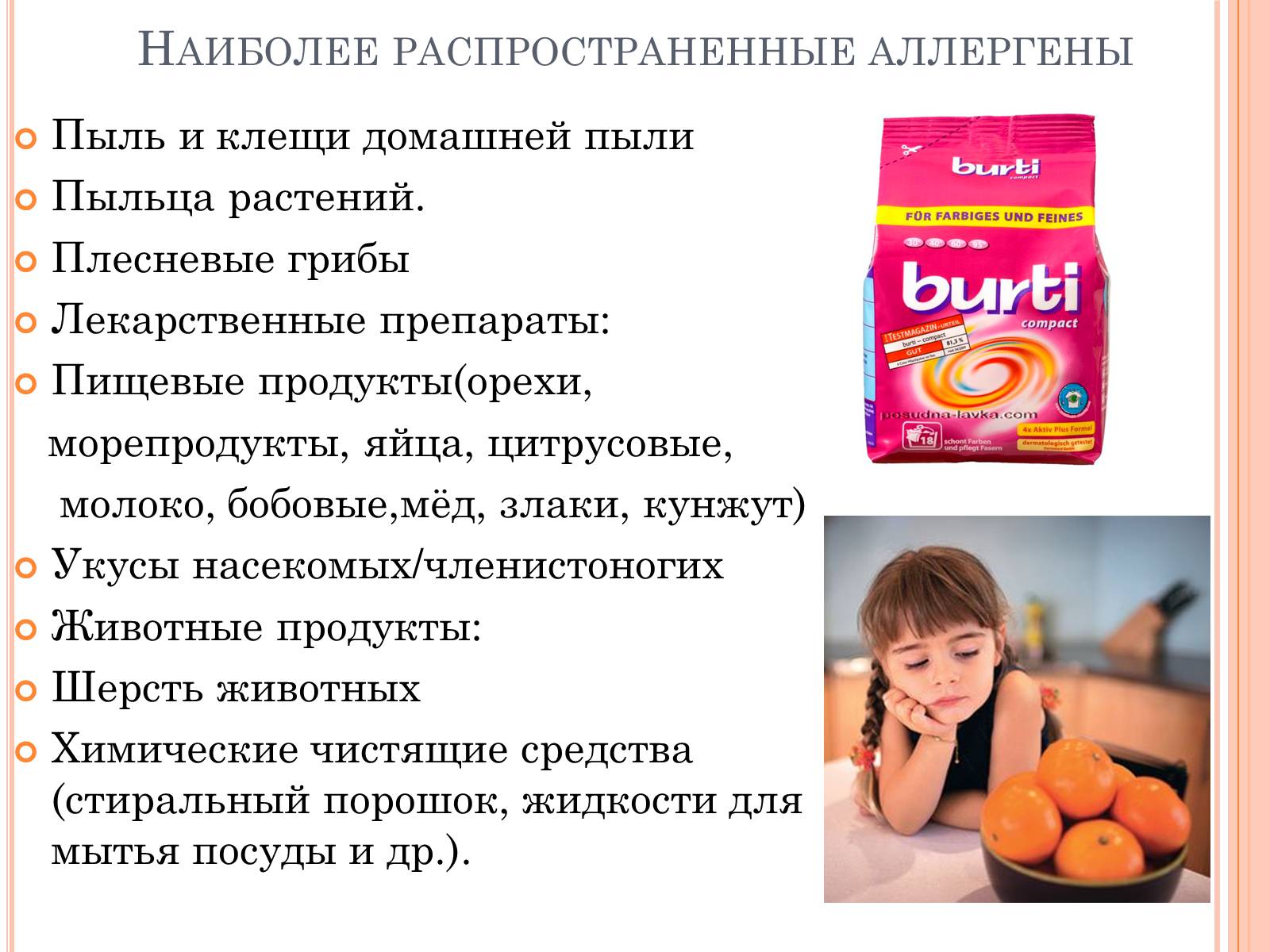
Food allergens (milk, eggs, fish, nuts,
seafood)
Medicines (antibiotics,
antipyretics, anesthetics)
Insect bites (bees, wasps) !!! Allergic reactions may be
immediate (when they develop
very quickly, violently - within
several minutes) and slow
type (can develop during
several days).
Allergic rhinitis
Difficulty in nasal breathing ornasal congestion, swelling of the mucous membrane
nose, copious watery discharge
mucous secretions, sneezing, burning sensation
in the throat
Emergency care for rhinitis
Oral monotherapyantihistamines and II-III
generation: acrivastine 8 mg
or cetirizine 10 mg.
If oral administration is not possible
Medicines are used parenterally
First generation antihistamines:
IM 1-2 ml 2% chloropyramine solution
(suprastina).
Allergic conjunctivitis
Hyperemia, edema, injectionconjunctiva, itching, lacrimation,
photophobia, swelling of the eyelids, narrowing of the eye
cracks
Emergency care for conjunctivitis
1)Instillation into the conjunctival saccorticosteroid solutions (0.1% solution
dexamethasone, 1% hydrocotisone solution),
Lubricating the eyelids with 1% hydrocortisone ointment.
2) Orally - diphenhydramine, suprastin, gluconate
calcium and other desensitizing
drugs. It is recommended to exclude from
food products or not to use products,
causing allergies.
Hives
Sudden onset of damage to a partskin with the formation of sharply defined
round blisters with raised
erythematous scalloped edges and
pale center, accompanied by
severe itching. The rash may persist
within 1-3 days, without leaving pigmentation
Generalized urticaria
Sudden onset of damage to the entire skinwith the formation of sharply defined rounded
blisters with raised erythematous
scalloped edges and pale center,
accompanied by severe itching. Maybe
the appearance of new rashes during
next 2-3 days
Emergency care for swelling, urticaria and general urticaria:
Stop taking medications.For food allergies, take enterosorbents
(white coal, enterosgel, etc.), rinse the stomach,
take a laxative.
For insect bites, remove the source of poison
(for example, a sting).
If contact allergies occur, remove
irritant from the surface of the skin.
!!!The next step of treatment is the appointment
medications.
Algorithm for prescribing antihistamines for urticaria:
Start with a standard dose of second-generation histamine blockers:
Loratadine (Claritin, Lomilan) – 10 mg per day
Fexofenadine (Telfast) – 150 mg per day
Ebastine (xysal) – 10 mg per day
Desloratadine (Erius) – 5 mg per day
Cetirizine (Zyrtec, Zodac) – 10 mg per day
If the first point is not effective, the dose of the prescribed drug should be increased (maximum
up to 4 times, taking into account body weight).
If points 1 and 2 are ineffective, one more should be added to the drug used.
antihistamine of the second generation.
Prescription of first generation drugs
Diphenhydramine (diphenhydramine, allergin) – 25-50 mg, 4 to 6 times a day
Suprastin - 25-50 mg per day in the form of tablets or in the form of injections 20-40 mg Tavegil (clemastine) 2 times a day, 1 mg in tablets or in injections 2 mg 2 times a day.
Acrivastine – 3 times a day, 8 mg
Cyproheptadine 3 times a day, 2-4 mg;
Emergency care for urticaria and urticaria:
If antihistamines are ineffective, you shouldtake hormonal:
Prednisolone 20 mg 2 times a day for 4 days or
prednisolone 50 mg per day for 3 days
Dexamethasone – 4-20 mg per day
Immunosuppressants are prescribed to patients with severe
autoimmune urticaria and in the absence of response to treatment
antihistamines. More often used for urticaria
cyclosporine.
Cyclosporine at the rate of 4 mg per 1 kg of body weight per day; The drug is often
causes side effects.
Quincke's edema
Local swelling of the skin, subcutaneous tissue ormucous membranes. Most often develops in the lip area,
cheeks, eyelids, forehead, scalp, scrotum,
hands, dorsal surface of feet. Simultaneously
swelling may occur with skin manifestations
joints, mucous membranes, including the larynx and
Gastrointestinal tract. Edema of the larynx is manifested by cough, hoarseness
voices, choking, wheezing, possible
death from asphyxia. Swelling of the gastrointestinal mucosa is accompanied by intestinal colic,
nausea, vomiting (cause of diagnostic errors and
unjustified surgical interventions)
Emergency care for Quincke's edema:
1) Adrenaline 0.3-0.5 ml of 0.1% solution subcutaneously;2) Pipolfen 2 ml of 2.5% solution intramuscularly; suprastin - 2 ml 2%
solution or diphenhydramine - 2 ml of 5% solution;
3) Prednisolone - 60-90 mg intramuscularly or intravenously;
4) Salbutamol, Alupent - inhalations;
5) Hot foot baths;
6) Lasix - 2-4 ml of 1% solution intravenously in a stream in isotonic
sodium chloride solution;
7) Aminocaproic acid 100-200 ml of 5% solution intravenously;
8) Contrical (trasylol) - 3 units intravenously in 300 ml isotonic
sodium chloride solution;
9) In case of hereditary Quincke's edema, fresh transfusion is indicated
blood, fresh frozen plasma (contains an inhibitor of the Cl component
complement).
