Topography of the superior mesenteric artery. Surgical interventions on the mesenteric vessels are acute disorders of the mesenteric circulation. Pathology of the portal system
The superior and inferior mesenteric arteries are responsible for the blood supply to certain organs and depart from the main aorta. They have many branches that extend to different parts of the intestines, stomach and kidneys. Disturbances in the mesenteric arteries entail a lack of nutrition, which leads to the development of diseases.
The structure of the superior mesenteric vessel
A large vessel forms in the anterior part of the aorta. Place of origin of the superior mesenteric artery 1-3 cm under the celiac trunk. It goes behind the pancreas, from where it goes down to the right. Next to it - on the right side - is the mesenteric vein. Together they go along the first wall of the duodenum horizontally and across, moving away to the right side from the skinny fracture.
Further, the circulatory element reaches the root of the mesentery and passes between the layers of the small intestine, creating an arc convex to the left. Thus, it passes to the right iliac fossa and is divided into several branches. Arteries depart from it:
- Inferior pancreatoduodenal. It starts at the starting point of the blood vessel and is divided into anterior and posterior. They go down and pass along the anterior wall of the pancreas, bypassing the head in the area of junction with the intestines. Small branches extend to the gland and duodenum, and then diverge from the upper pancreatoduodenal blood elements.
- jejunum. In total, there are from 7 to 8 in the human body, and the blood elements depart one by one from the convex zone. They are sent through the mesentery to the jejunum. Each branch of the mesenteric artery is further divided into 2 trunks and intertwined with the vessels of the intestinal branches.
- ileo-intestinal. Depart to the loops of the ileum. There are 5-6 of them in the body. Like the previous ones, the iliac blood elements are divided into 2 trunks and form arcs of the 2nd order (small size). Even smaller arteries depart from them again and go to the walls of the loops of the small intestine. They also form small branches that are responsible for feeding the lymph nodes of the mesenteric region.
- ileocolic-intestinal. It starts in the zone of the cranial part of the mesenteric vessel and goes to the right side to the ileum along the posterior wall of the abdominal cavity. It is divided into additional branches that go to the caecum and colon, as well as to the ileum region of the intestine.
- Right colon—intestinal. Forms a process on the right side of the main mesenteric artery, starts from the upper third. Goes to the edge of the colon.
- Middle colon—intestinal. It originates in the upper part of the mesenteric artery, passes through the mesentery of the colonic area and is divided into 2 branches. The right one goes to the ascending vessel, and the left one forms a branch through the mesenteric edge of the intestine.
- colica constanta - ascending and descending pair;
- sigmoideae - with several branches forming an arc;
- rectalis superior - descends into the mesentery of the sigmoid colon and goes into the small pelvis, forming several lateral branches to the rectum.
Several large branches are separated from the ileocolic vessel. The first is the ascending artery, which departs from the right to the colon and ascends to the blood branch emanating from this zone. In the same place, it forms an arc, from which the colonic branches are formed. They are responsible for supplying blood to the upper part of the cecum and the ascending part of the colon loop.
From the same blood branch, the caecal arteries depart back and forth, heading to the caecum. They form a vascular network that extends to the ileocecal angle, where they connect with the terminal arteries of the ileo-intestinal arch.
Another feeding element is the appendix, which is responsible for the blood supply to this area. These arteries pass through the mesentery of the appendix.
The superior mesenteric artery is not a separate blood vessel, but a whole system of descending branches with a slope to the right.
The structure of the inferior mesenteric branch
 The lower part of the mesenteric vessel is located on the edge of the third vertebra, just above the aortic division. It goes down to the left and is located behind the abdominal wall on the surface of the psoas muscle. There are several branches in the anatomy of the inferior mesenteric artery:
The formation of vessels from these arteries form anastomoses along the entire length of the rectum.
Main functions
The superior and inferior mesenteric arteries are part of the circulatory system. Since these are rather large vessels, they are considered the main sources of nutrition for the abdominal organs, including all branches. The superior artery supplies blood to more than half of the intestines, as well as the entire pancreas.
Violation of the functions of the superior mesenteric vessel leads to a general deterioration in blood circulation. Because of this, the internal organs located in the peritoneum suffer, most often the large intestine.
Embolism of the circulatory mesentery
 A common disease of the superior artery begins with acute pain in the abdomen, located in the umbilical region. In some patients, symptoms begin in the lower right abdomen. The intensity of pain depends on many factors and can vary greatly.
On palpation, the doctor detects a too soft abdomen, as well as a slight tension in the muscles of the anterior wall. Pain during the examination is practically absent. In some cases, there is increased intestinal peristalsis.
Patients with embolism often suffer from vomiting, nausea, and diarrhea. At the same time, no functional disorders were detected during the examination. In the early stages, occult blood is detected in stool tests, but there are no visible impurities.
The presence of an embolism can be suspected by a combination of symptoms from the gastrointestinal tract, as well as the cardiovascular system. It is not uncommon for an embolism to develop in people who have recently had a heart attack or have rheumatic valve disease.
Features of treatment
Therapy of embolism is possible by conservative methods, but in the acute course of the disease, the best results are observed only after surgical intervention. The laparotomy method is used, in which the upper artery is opened and an embolectomy is performed.
As a result of the operation, blood flow is restored, and the condition of the small intestine is also determined. Sometimes during the procedure, necrosis of part of the tissues of this part of the intestine is detected. Then, during the operation, doctors remove damaged cells. After the operation, an additional autopsy is prescribed 24 hours later to ensure the viability of the intestine.
Abdominal aorta(abdominal aorta), pars abdominalis aortae (aorta abdominalis), is a continuation of the thoracic aorta. It starts at the level of the XII thoracic vertebra and reaches the IV-V lumbar vertebra. Here the abdominal aorta divides into two common iliac arteries, aa. aliacae communes. The place of division is called the bifurcation of the aorta, bifurcatio aortica. A thin branch descends from the bifurcation, lying on the anterior surface of the sacrum - the median sacral artery, a. sacralis mediana.
Two types of branches depart from the abdominal part of the aorta: parietal and splanchnic.
The abdominal part of the aorta is located retroperitoneally. In the upper part, the body of the pancreas and two veins adjoin to its surface, crossing it: the splenic vein lying along the upper edge of the pancreas, v. lienalis, and the left renal vein, v. renalis sinistra, running behind the gland. Below the body of the pancreas, in front of the aorta, is the lower part of the duodenum, and below it is the beginning of the root of the mesentery of the small intestine. To the right of the aorta lies the inferior vena cava, v. cava inferior; behind the initial section of the abdominal aorta is the cistern of the thoracic duct, cisterna chyli, the initial part of the thoracic duct, ductus thoracicus.
Wall branches.
1. Inferior phrenic artery, a. phrenica inferior, is a rather powerful paired artery. It departs from the anterior surface of the initial part of the abdominal aorta at the level of the XII thoracic vertebra and goes to the lower surface of the tendon part of the diaphragm, where it gives off the anterior and posterior branches that supply the latter. In the thickness of the diaphragm, the right and left arteries anastomose with each other and with branches from the thoracic aorta. The right artery passes behind the inferior vena cava, the left one behind the esophagus.
In its course, the artery gives off 5-7 upper adrenal arteries, aa. suprarenales superiores. These are thin branches that extend from the initial section of the inferior phrenic artery and supply blood to the adrenal gland. On the way, several small branches depart from them to the lower parts of the esophagus and to the peritoneum.
2. Lumbar arteries, aa. lumbales, are 4 paired arteries. They depart from the posterior wall of the abdominal part of the aorta at the level of the body of the I-IV lumbar vertebrae. They are directed transversely, to the lateral side, while the two upper arteries pass behind the legs of the diaphragm, the two lower ones - behind the psoas major muscle.
All lumbar arteries anastomose with each other and with the superior and inferior epigastric arteries, which supply blood to the rectus abdominis. In their course, the arteries give a number of small branches to the subcutaneous tissue and to the skin; in the region of the white line, they anastomose here and there with the arteries of the same name on the opposite side. In addition, the lumbar arteries anastomose with the intercostal arteries, aa. intercostales, iliac-lumbar artery, a. iliolumbalis, deep circumflex iliac artery, a. circumflexa ilium profunda, and superior gluteal artery, a. glutea superior.
Having reached the transverse processes of the vertebrae, each lumbar artery gives off a dorsal branch, r. dorsalis. Then the lumbar artery goes behind the square muscle of the lower back, supplying it with blood; then it goes to the anterior wall of the abdomen, passes between the transverse and internal oblique muscles of the abdomen and reaches the rectus abdominis.
The dorsal branch goes to the back surface of the body to the muscles of the back and the skin of the lumbar region. Along the way, she gives a small branch to the spinal cord - the spinal branch, r. spinalis, which enters the spinal canal through the intervertebral foramen, supplying the spinal cord and its membranes with blood.

3. Median sacral artery, a. sacralis mediana, is a direct continuation of the abdominal aorta. It starts from its posterior surface, slightly above the aortic bifurcation, i.e., at the level of the V lumbar vertebra. It is a thin vessel passing from top to bottom in the middle of the pelvic surface of the sacrum and ending at the coccyx in the coccygeal body, glomus coccygeum.
From the median sacral artery along its course branch:
a) inferior lumbar artery, a. lumbalis imae, steam room, departs in the region of the V lumbar vertebra and supplies blood to the iliopsoas muscle. On its way, the artery gives off a dorsal branch, which is involved in the blood supply to the deep muscles of the back and spinal cord;
b) lateral sacral branches, rr. sacrales laterales, depart from the main trunk at the level of each vertebra and, branching on the anterior surface of the sacrum, anastomose with similar branches from the lateral sacral arteries (branches of the internal iliac arteries).
Several branches depart from the lower part of the median sacral artery, which supply blood to the lower parts of the rectum and the loose tissue around it.
Internal branches
I. celiac trunk, truncus celiacus, - a short vessel, 1-2 cm long, departs from the anterior surface of the aorta at the level of the upper edge of the body of the 1st lumbar vertebra or the lower edge of the body of the 12th thoracic vertebra in the place where the abdominal aorta exits the aortic orifice. The artery goes anteriorly and immediately divides into three branches: the left gastric artery, a. gastricasinistra, common hepatic artery, a. hepatica communis, and splenic artery, a. splenica (lienalis).

1. Left gastric artery, a. gastrica sinistra, the smaller of these three arteries. It rises a little up and to the left; approaching the cardial part, gives several branches towards the esophagus - esophageal branches, rr. esophageales, anastomosing with the same-named branches from the thoracic aorta, and descending to the right side along the lesser curvature of the stomach, anastomosing with the right gastric artery, a. gastrica dextra (from the common hepatic artery). On its way along the lesser curvature, the left gastric artery sends small branches to the anterior and posterior walls of the stomach.
2. Common hepatic artery, a. hepatica communis, is a more powerful branch, up to 4 cm long. Moving away from the celiac trunk, it goes along the right crus of the diaphragm, the upper edge of the pancreas from left to right and enters the thickness of the lesser omentum, where it is divided into two branches - its own hepatic and gastroduodenal arteries.
1) Own hepatic artery, a. hepatica propria, moving away from the main trunk, goes to the gates of the liver in the thickness of the hepatoduodenal ligament, to the left of the common bile duct and somewhat anterior to the portal vein, v. portae. Approaching the gates of the liver, the own hepatic artery is divided into the left and right branches, while the gallbladder artery departs from the right branch, a. cystica.
Right gastric artery, a. gastrica dextra, - a thin branch, departs from its own hepatic artery, sometimes from the common hepatic artery. It goes from top to bottom to the lesser curvature of the stomach, along which it goes from right to left, and anastomoses with a. gastric sinistra. The right gastric artery gives rise to a number of branches that supply blood to the anterior and posterior walls of the stomach.
At the gates of the liver, the right branch, r. dexter, own hepatic artery sends to the caudate lobe artery of the caudate lobe, a. lobi caudati, and arteries to the corresponding segments of the right lobe of the liver: to the anterior segment - the artery of the anterior segment, a. segmenti anterioris, and to the posterior segment - the artery of the posterior segment, a. segmenti posterioris.
Left branch, r. sinister, gives the following arteries: artery of the caudate lobe, a. lobi caudati, and arteries of the medial and lateral segments of the left lobe of the liver, a. segmenti medialis et a. segmenti lateralis. In addition, a non-permanent intermediate branch, r, departs from the left branch (less often from the right branch). intermedius, supplying the square lobe of the liver.
2) Gastroduodenal artery, a. gastroduodenalis, is a rather powerful trunk. It is directed from the common hepatic artery downwards, behind the pyloric part of the stomach, crossing it from top to bottom. Sometimes the supraduodenal artery departs from this artery, a. supraduodenalis, which crosses the anterior surface of the head of the pancreas.
The following branches depart from the gastroduodenal artery:
a) posterior superior pancreatoduodenal artery, a. pancreaticoduodenalis superior posterior, passes along the posterior surface of the head of the pancreas and, heading down, gives pancreatic branches along its course, rr. pancreatici, and duodenal branches, rr. duodenales. At the lower edge of the horizontal part of the duodenum, the artery anastomoses with the inferior pancreatoduodenal artery, a. pancreaticoduodenalis inferior (branch of the superior mesenteric artery, a. mesenterica superior);
b) anterior superior pancreatoduodenal artery, a. pancreaticoduodenalis superior anterior, is located arcuately on the anterior surface of the head of the pancreas and the medial edge of the descending part of the duodenum, goes down, giving off duodenal branches in its path, rr. duodenales, and pancreatic branches, rr. pancreatici. At the lower edge of the horizontal part of the duodenum, it anastomoses with the inferior pancreatoduodenal artery, a. pancreatoduodenalis inferior (branch of the superior mesenteric artery).
c) right gastroepiploic artery, a. gastroepiploica dextra, is a continuation of the gastroduodenal artery. It goes to the left along the greater curvature of the stomach between the leaves of the greater omentum, sends branches to the anterior and posterior walls of the stomach - gastric branches, rr. gastrici, as well as omental branches, rr. epiploici to the greater omentum. In the region of greater curvature, it anastomoses with the left gastroepiploic artery, a. gastroepiploica sinistra (branch of the splenic artery, a. splenica);
d) retroduodenal arteries, aa. retroduodenales, are the right terminal branches of the gastroduodenal artery. They surround the right edge of the pancreatic head along the anterior surface.

3. Splenic artery, a. splenica, is the thickest of the branches extending from the celiac trunk. The artery goes to the left and, together with the vein of the same name, lies behind the upper edge of the pancreas. Having reached the tail of the pancreas, it enters the gastrosplenic ligament and breaks up into terminal branches heading to the spleen.
The splenic artery gives off branches that supply the pancreas, stomach, and greater omentum.
1) Pancreatic branches, rr. pancreatici, depart from the splenic artery along its entire length and enter the parenchyma of the gland. They are represented by the following arteries:
a) dorsal pancreatic artery, a. pancreatica dorsalis, follows downward, respectively, the middle section of the posterior surface of the body of the pancreas and, at its lower edge, passes into the inferior pancreatic artery, a. pancreatica inferior, supplying the lower surface of the pancreas;
b) large pancreatic artery, a. pancreatica magna, departs from the main trunk or from the dorsal pancreatic artery, follows to the right and goes along the posterior surface of the body and head of the pancreas. It connects to the anastomosis between the posterior superior and inferior pancreatoduodenal arteries;
c) tail pancreatic artery, a. caude pancreatis, is one of the terminal branches of the splenic artery, supplies blood to the tail of the pancreas.
2) Splenic branches, rr. splenici, only 4 - 6, are the terminal branches of the splenic artery and penetrate through the gate into the parenchyma of the spleen.
3) Short gastric arteries, aa. gastricae breves, in the form of 3-7 small trunks depart from the terminal section of the splenic artery and in the thickness of the gastro-splenic ligament go to the fundus of the stomach, anastomosing with other gastric arteries.
4) Left gastroepiploic artery, a. gastroepiploica sinistra, starts from the splenic artery in the place where the terminal branches to the spleen depart from it, and follows down in front of the pancreas. Having reached the greater curvature of the stomach, it goes along it from left to right, lying between the leaves of the greater omentum. On the border of the left and middle thirds of the greater curvature, it anastomoses with the right gastroepiploic artery (from a. gastroduodenalis). In its course, the artery sends a number of branches to the anterior and posterior walls of the stomach - gastric branches, rr. gastrici, and to the greater omentum - omental branches, rr. epiploici.
5) Posterior gastric artery, a. gastrica posterior, unstable, supplies blood to the back wall of the stomach, closer to the cardial part.
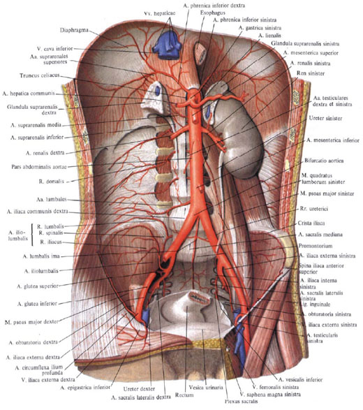
II. superior mesenteric artery, a. mesenterica superior, is a large vessel that starts from the anterior surface of the aorta, slightly lower (1 - 3 cm) of the celiac trunk, behind the pancreas.

Coming out from under the lower edge of the gland, the superior mesenteric artery goes down and to the right. Together with the superior mesenteric vein located to the right of it, it runs along the anterior surface of the horizontal (ascending) part of the duodenum, crosses it across immediately to the right of the duodenal-lean flexure. Having reached the root of the mesentery of the small intestine, the superior mesenteric artery penetrates between the leaves of the latter, forming an arc with a bulge to the left, and reaches the right iliac fossa.
In its course, the superior mesenteric artery gives off the following branches: to the small intestine (with the exception of the upper part of the duodenum), to the caecum with the appendix, ascending and partially to the transverse colon.
The following arteries depart from the superior mesenteric artery.
1. Inferior pancreatoduodenal artery, a. pancreaticoduodenalis inferior (sometimes non-single), originates from the right edge of the initial section of the superior mesenteric artery. Divides into an anterior branch, r. anterior, and posterior branch, r. posterior, which go down and to the right along the anterior surface of the pancreas, go around its head along the border with the duodenum. Gives branches to the pancreas and duodenum; anastomoses with the anterior and posterior superior pancreatoduodenal arteries and with the branches of a. gastroduodenalis.
2. Jejunum arteries, aa. jejunales, only 7 - 8, depart sequentially one after another from the convex part of the arch of the superior mesenteric artery, are sent between the sheets of the mesentery to the loops of the jejunum. On its way, each branch divides into two trunks, which anastomose with the same trunks formed from the division of neighboring intestinal arteries.
3. Ileo-intestinal arteries, aa. ileales, in the amount of 5 - 6, like the previous ones, go to the loops of the ileum and, dividing into two trunks, anastomose with adjacent intestinal arteries. Such anastomoses of the intestinal arteries look like arcs. New branches depart from these arcs, which also divide, forming arcs of the second order (slightly smaller). From the arcs of the second order, the arteries again depart, which, dividing, form the arcs of the third order, and so on. From the last, most distal row of arcs, straight branches extend directly to the walls of the loops of the small intestine. In addition to intestinal loops, these arcs give small branches that supply blood to the mesenteric lymph nodes.
4. Ileocolic-intestinal artery, a. ileocolica, departs from the cranial half of the superior mesenteric artery. Heading to the right and down under the parietal peritoneum of the posterior abdominal wall to the end of the ileum and to the caecum, the artery divides into branches supplying the caecum, the beginning of the colon and the terminal ileum.
A number of branches depart from the iliac-colon-intestinal artery:
a) the ascending artery goes to the right to the ascending colon, rises along its medial edge and anastomoses (forms an arc) with the right colonic artery, a. colic dextra. Colon-intestinal branches depart from the specified arc, rr. colici, supplying the ascending colon and upper caecum;
b) anterior and posterior cecum arteries, aa. cecales anterior et posterior, are sent to the corresponding surfaces of the caecum. Are a continuation of a. ileocolica, approach the ileocecal angle, where, connecting with the terminal branches of the ileo-intestinal arteries, they form an arc, from which branches extend to the caecum and to the terminal ileum - ileo-intestinal branches, rr. ileales;
c) arteries of the appendix, aa. appendiculares, depart from the posterior cecal artery between the sheets of the mesentery of the appendix; blood supply to the appendix.
5. Right colonic artery. a. colica dextra, departs on the right side of the superior mesenteric artery, in its upper third, at the level of the root of the mesentery of the transverse colon, and goes almost transversely to the right, to the medial edge of the ascending colon. Before reaching the ascending colon, it is divided into ascending and descending branches. The descending branch connects to branch a. ileocolica, and the ascending branch anastomoses with the right branch of a. colica media. From the arcs formed by these anastomoses branches extend to the wall of the ascending colon, to the right flexure of the colon, and to the transverse colon.

6. Middle colonic artery, a. colica media, departs from the initial section of the superior mesenteric artery, goes forward and to the right between the sheets of the mesentery of the transverse colon and is divided at the bottom of the branch: right and left.
The right branch connects to the ascending branch a. colica dextra, a the left branch runs along the mesenteric edge of the transverse colon and anastomoses with the ascending branch a. colica sinistra, which departs from the inferior mesenteric artery. Connecting in this way with the branches of neighboring arteries, the middle colon-intestinal artery forms arcs. From the branches of these arcs, arcs of the second and third order are formed, which give direct branches to the walls of the transverse colon, to the right and left bends of the colon.
III. Inferior mesenteric artery, a. mesenterica inferior, departs from the anterior surface of the abdominal aorta at the level of the lower edge of the III lumbar vertebra. The artery goes behind the peritoneum to the left and down and is divided into three branches.

1. Left colonic artery, a. colica sinistra, lies retroperitoneally in the left mesenteric sinus in front of the left ureter and left testicular (ovarian) artery, a. testicularis (ovarica) sinistra; splits into ascending and descending branches. The ascending branch anastomoses with the left branch of the middle colic artery, forming an arc; blood supply to the left side of the transverse colon and the left flexure of the colon. The descending branch joins the sigmoid intestinal artery and supplies the descending colon with blood.
2. Sigmoid-intestinal artery, a. sigmoidea (sometimes there are several), goes down first retroperitoneally, and then between the sheets of the mesentery of the sigmoid colon; anastomoses with the branches of the left colonic artery and the superior rectal artery, forming arcs from which the branches extend, supplying the sigmoid colon.
3. Superior rectal artery, a. rectalis superior, is the terminal branch of the inferior mesenteric artery; heading down, it is divided into two branches. One branch anastomoses with a branch of the sigmoid artery and supplies blood to the lower sections of the sigmoid colon. Another branch goes to the cavity of the small pelvis, crosses in front a. iliaca communis sinistra and, lying in the mesentery of the pelvic section of the sigmoid colon, is divided into the right and left branches, which supply the rectal ampulla with blood. In the intestinal wall, they anastomose with the middle rectal artery, a. rectalis media, a branch of the internal iliac artery, a. iliaca interna.
IV. Middle adrenal artery, a. suprarenalis media, steam room, departs from the side wall of the upper aorta, slightly below the place of origin of the mesenteric artery. It is directed transversely outward, crosses the crus of the diaphragm and approaches the adrenal gland, in the parenchyma of which it anastomoses with branches of the superior and inferior adrenal arteries.
v. renal artery, a. renalis, - paired large artery. It starts from the lateral wall of the aorta at the level of the II lumbar vertebra almost at a right angle to the aorta, 1-2 cm below the origin of the superior mesenteric artery. The right renal artery is somewhat longer than the left, since the aorta lies to the left of the midline; heading towards the kidney, it is located behind the inferior vena cava.
Before reaching the hilum of the kidney, each renal artery gives off a small inferior adrenal artery, a. suprarenalis inferior, which, having penetrated the adrenal parenchyma, anastomoses with the branches of the middle and superior adrenal arteries.
At the hilum of the kidney, the renal artery divides into anterior and posterior branches.
Anterior branch, r. anterior, enters the renal gate, passing in front of the renal pelvis, and branches, sending arteries to the four segments of the kidneys: the artery of the upper segment, a. segmenti superioris, - to the top; artery of the upper anterior segment, a. segmenti anterior superioris, - to the upper anterior; artery of the lower anterior segment, a. segmenti anterior is inferioris, - to the lower anterior and artery of the lower segment, a. segmenti inferioris, - to the bottom. Back branch, r. posterior, the renal artery passes behind the renal pelvis and, heading to the posterior segment, gives off the ureteral branch, r. uretericus, which may originate from the renal artery itself, divides into posterior and anterior branches.
VI. testicular artery, a. testicularis, steam room, thin, departs (sometimes right and left common trunk) from the anterior surface of the abdominal aorta, slightly below the renal artery. It goes down and laterally, goes along the psoas major muscle, crosses the ureter on its way, above the arcuate line - the external iliac artery. Along the way, it gives branches to the fatty capsule of the kidney and to the ureter - ureteral branches, rr. ureterici. Then it goes to the deep inguinal ring and, having joined the vas deferens here, passes through the inguinal canal into the scrotum and breaks up into a number of small branches that go to the parenchyma of the testicle and its epididymis - branches of the epididymis, rr. epididymales.
In its course it anastomoses with a. cremasterica (branch a. epigastrica inferior and with a. ductus deferentis (branch a. iliaca interna).
In women, the corresponding testicular artery is the ovarian artery, a. ovarica, gives off a number of ureteral branches, rr. ureterici, and then passes between the sheets of the broad ligament of the uterus, along its free edge, and gives off branches to the fallopian tube - tubal branches, rr. tubales, and into the hilum of the ovary. The terminal branch of the ovarian artery anastomoses with the ovarian branch of the uterine artery.
Superior mesenteric artery, a. mesenterica superior, is a large vessel that starts from the anterior superficial aorta, slightly lower (1-3 cm) of the celiac trunk, behind the pancreas. Coming out from under the lower edge of the gland, the superior mesenteric artery goes down and to the right. Together with the superior mesenteric vein located to its right, it lies on the anterior surface of the horizontal (or ascending) part of the duodenum, crosses it across, immediately to the right of the flexura duodenojejunalis. Having reached the root of the mesentery of the small intestines, the superior mesenteric artery penetrates between the sheets of the latter, forming an arc with a bulge to the left, and reaches the right iliac fossa. In its course, the superior mesenteric artery gives off the following branches: to the small intestine (with the exception of the upper part of the duodenum) , a cecum with a appendix, ascending and partially to the transverse colon. The following arteries depart from the superior mesenteric artery.
- Inferior pancreatoduodenal artery, a. pancreatico-duodenalis inferior (sometimes non-single), originates from the right edge of the initial section of the superior mesenteric artery, goes down and to the right along the anterior surface of the pancreas, bending around its head along the border with the duodenum. The inferior pancreatoduodenal artery gives off branches to the pancreas and duodenum and anastomoses with the superior pancreatoduodenal artery - branch a. gastroduodenalis.
- Intestinal arteries up to 15 depart sequentially one after the other from the convex part of the arch of the superior mesenteric artery. The intestinal arteries are sent between the sheets of the mesentery to the loops of the jejunum and ileum - these are the jejunal arteries and the ileal arteries, aa .. jejunales et aa. ilei. On its way, each branch divides into two trunks, which anastomose with the same trunks formed from the division of neighboring intestinal arteries. Such anastomoses look like arcs or arcades. New branches depart from these arcs, which also divide, forming arcs of the second order, of a somewhat smaller size. From the arcs of the second order, arteries again depart, which, dividing, form arcs of the third order, and so on. From the last, most distal, series of arcs, straight branches extend directly to the walls of the loops of the small intestines. In addition to intestinal loops, these arcs give small branches that supply blood to the mesenteric lymph nodes.
- Iliocolic artery, a. ileocolica, departs from the cranial half of the superior mesenteric artery, to the right of the root of the mesentery of the small intestine. Heading to the right and down under the parietal peritoneum of the posterior abdominal wall to the end of the ileum and to the cecum, the ileocolic artery divides into two branches supplying the cecum, the beginning of the colon and the end of the ileum. Branches extending from the iliac-colic artery are as follows.
- Anterior and posterior caecal arteries, aa .. cecales anterior et posterior, heading to the corresponding surfaces of the caecum.
- The ileal branch is a continuation of a. ileocolica, goes down to the ileocecal angle, where, connecting with the terminal branches of aa .. ilei, it forms an arc, from which the branches extend to the terminal ileum.
- The branch of the colon goes to the right to the ascending colon. Before reaching the medial edge of this colon, it is divided into two branches, of which one is the ascending branch, g. ascendens, rises along the medial edge of the ascending colon and anastomoses (forms an arc) with a. colica dextra; the other branch descends along the medial edge of the colon and anastomoses (forms an arc) with a. ileocolica. Branches depart from these arcs, supplying the ascending colon and caecum, as well as the appendix through the artery of the appendix, a. appendicularis.
- Right colic artery, a. colica dextra, departs from the right side of the superior mesenteric artery in its upper third, at the level of the root of the mesentery of the transverse colon, and goes almost transversely to the right, to the medial edge of the ascending colon. At some distance from the ascending colon, the right colic artery divides into ascending and descending branches. The descending branch connects to branch a. ileocolica, and the ascending branch anastomoses with the right branch of a. colica media. From the arcs formed by these anastomoses, branches depart to the wall of the ascending colon, to the flexura coli dextra and to the transverse colon.
- Middle colic artery, a. colica media, departs from the initial section of the superior mesenteric artery, heading forward and to the right between the sheets of the mesentery of the transverse colon, and is divided into two branches: right and left . The right branch connects to the ascending branch a. colica dextra, and the left one, going along the mesenteric edge of the transverse colon, anastomoses with the ascending branch a. colica sinistra, which departs from a. mesenterica inferior. Connecting in this way with the branches of neighboring arteries, the middle colic artery forms arcs. From the branches of these arcs, arcs of the second, third order are formed, which give direct branches to the walls of the transverse colon, flexura coli dextra et sinistra.
Page 26 of 34
So far, operations on mesenteric vessels have been performed in a limited number of patients. No more than 200 cases of surgical interventions on mesenteric vessels in this disease are reported in the world literature. The most frequently performed embolectomy was from the superior mesenteric artery, much less frequently - thrombo- and thrombinthymectomy, shunting, vascular plasty, reimplantation, vascular switching, thrombectomy from the portal and superior mesenteric veins.
This section is based on the experience of 46 surgical interventions on mesenteric vessels.
Access to the mesenteric vessels. Exposure of the superior mesenteric artery can be made from two accesses: anterior and posterior.
With an anterior approach (to the right in relation to the root of the mesentery of the small intestine), the transverse colon is brought into the wound and its mesentery is stretched. The mesentery of the small intestine is straightened, the loops of the intestines are moved to the left and downwards. The initial section of the mesentery, in accordance with the beginning of the jejunum, is also stretched. The peritoneum is dissected from the ligament of Treitz along a line connecting the latter with the ileocecal angle. The length of the incision is 8-10 cm. Palpation of the vessel helps to more accurately find the trunk of the superior mesenteric artery with a non-greasy mesentery. In cases where there is a fatty mesentery or its edema is observed, the following technique can be used. By pulling on the mesentery of the transverse colon, the location of the middle colon artery is determined by eye or by palpation and then, exposing it towards the mouth, they reach the trunk of the superior mesenteric artery, after which it is exposed under visual control up and down from the place of origin of the middle colon artery.
Exposure of blood vessels requires the surgeon to be careful and take care of the tissues. Damage to the mesenteric vessels makes further operation on the vessel problematic.
The incision line of the peritoneum of the mesentery is coagulated, after which the peritoneum is carefully dissected with a scalpel. Subsequently, it is advisable to use vascular scissors. The tissues are cut with scissors between anatomical forceps, with which the surgeon and his assistant lift the tissues along the incision line. Tweezers should capture a small amount of tissue in order to see the vascular branches, which coagulate or immediately ligate with thin silk threads. Large branches of the superior mesenteric vein lying above the trunk of the artery (usually 1-3 of them) are mobilized, lifted above the artery, but in no case do they cross. Mobilization of the venous trunks allows them to be further displaced with the help of vascular holders or hooks. If the lymphatic vessels are damaged, they should be ligated or coagulated if possible. The trunk and branches of the superior mesenteric artery are exposed for 6-8 cm.
The superior mesenteric vein is exposed in a similar way. The cut line should be 1-2 cm to the right.
With posterior access to the superior mesenteric artery (to the left in relation to the root of the mesentery of the small intestine), the transverse colon is also brought into the wound and its mesentery is pulled. The loops of the intestines are moved to the right and down. The ligament of Treitz is stretched by pulling on the initial section of the jejunum. Then the ligament is dissected and this section of the jejunum is mobilized up to the duodenum.
Rice. 50. Anterior access to the superior mesenteric artery.
- - the trunk of the superior mesenteric artery;
- - middle colic artery; 3 - intestinal arteries; 4 - iliac colon artery.
Rice. 51. Posterior access to the superior mesenteric artery.
1 - superior mesenteric artery; 2 - left renal vein; 3 - aorta; 4 - inferior mesenteric artery; 5 - lower hollow foam.
Next, the peritoneum is dissected over the aorta so that a curved or L-shaped incision is obtained. It is better to dissect tissues from below: the aorta is exposed, then the left renal vein, which is mobilized and retracted downward with a vascular hook. Above the vein, the mouth of the superior mesenteric artery is exposed. At the same time, it should be remembered that its initial section is covered with fibrous tissue for 1.5-2 cm, which requires not blunt, but sharp dissection. In order to apply a parietal clamp on the aorta, it is necessary to highlight the area of the aorta above and below the mouth of the artery. The trunk of the superior mesenteric artery is exposed for 5-6 cm. When the first segment of the artery is exposed, one should not forget that the inferior pancreatoduodenal artery departs from it and the additional hepatic artery may depart.
The dissection of the peritoneum with underlying tissues can be extended downward along the aorta and expose the inferior mesenteric artery. In case of thrombosis of the orifice of the superior mesenteric artery, it is better to start exposure from below - from the inferior mesenteric artery and go up along the aorta.
After surgery on the vessel, rare silk sutures are applied to the dissected tissues. A polyethylene tube with a diameter of 0.5-1.0 cm is brought to the vessel to control and outflow of blood and lymph (Fig. 50, 51).
Embolectomy
Embolectomy from the superior mesenteric artery with an unfavorable outcome was first performed by Ya. B. Ryvlin in 1940, then by N. I. Blinov (1950), Klass (1951). Embolectomy with recovery of the patient was performed by Steward in 1951. In our country, the first successful embolectomy from the superior mesenteric artery was performed by A. S. Lyubsky in 1961. S. I. Spasokukotsky, this operation was first performed in 1966 by B. D. Komarov, with a favorable outcome - in 1968 by K. G. Kislova together with resection of the intestine, without resection - in 1972 by V. S. Savelyev.
To date, embolectomy from the superior mesenteric artery has been performed in 27 patients, 10 of them in pure form, 17 with bowel resection.
In its pure form, the operation was performed within 4 to 26 hours from the onset of the disease (in 8 patients in the ischemia stage, in 2 in the infarction stage with damage to the intestinal mucosa only).
Table 25
EMBOLECTOMY FROM THE SUPERIOR MESENTERIC ARTERY
artery segment |
||||
Type of embolectomy |
||||
Direct embolectomy |
||||
Indirect embolectomy |
||||
Embolectomy from the artery stump |
||||
Embolectomy together with bowel resection was performed within 9 to 98 hours in 6 patients at the stage of infarction, in 11 patients at the stage of peritonitis.
Direct embolectomy was performed in 14 patients, indirect - in 13 (Table 25).
The technique for performing direct and indirect embolectomy is somewhat different.
Both direct and indirect embolectomy is possible along segment I of the artery; direct embolectomy is preferable during segments II and III. At any localization of the embolus, an anterior access to the artery is indicated.
With anterior access to the artery, the first 2-3 cm of the trunk and its mouth are usually not exposed. When the embolus is localized in the proximal segment of the I segment of the arterial trunk, it can be easily removed with a Fogarty probe. If the embolus is located slightly lower, at the level of the origin of the first intestinal arteries, then direct embolectomy can be performed.
In both types of surgery, the mouth of the middle colic artery, the trunk of the superior mesenteric artery above and below it for about 2 cm in each direction are exposed (direct embolectomy requires exposure of the artery above the upper pole of the embolus) and all intestinal arteries, starting with the first, extending from the selected segment artery trunk (usually no more than 4-5).
Tourniquets made of braid or round rubber are placed on the trunk of the superior mesenteric artery, and silk tourniquets in the form of a loop passed through a rubber tube (silk No. 4 or 5) or vascular clamps are placed on the branches. It is necessary to pay attention to the fact that the introduction of a silk thread under a small-diameter vessel requires great care, since the vessel may tear in case of quick and rough manipulation. The lumen of the vessel should be blocked not so much by pulling on the ends of the silk thread, but by bringing down the rubber tube.
After blocking the trunk of the superior mesenteric artery and branches with the help of tourniquets with vascular scissors or a scalpel, an arteriotomy is performed. You can make a longitudinal arteriotomy (Baue, Austen, 1963; Zuidema et al, 1964) or transverse (Rutledge, 1964). It is better to dissect the artery in the transverse direction, since in this case, after suturing, there is no narrowing of the vessel. The length of the arteriotomy opening should not exceed XU or 1/3 of the perimeter of the artery. The transverse section of the artery must be made taking into account the origin of the branches of the artery, so that, if necessary, it is possible to pass the Fogarty probe from it into the middle colon artery and at least one of the intestinal ones. These requirements usually correspond to a place just above the mouth of the middle colon artery (4-5 mm from it). With a closer location of the arteriotomy opening to the middle colic artery, its mouth may be deformed during suturing. If the embolus is located higher, the arteriotomy hole has to be made 1.5-2 cm from the mouth of the middle colic artery. If in the course of the operation it becomes necessary to revise any branch with the Fogarty probe, the introduction of the probe into which is impossible through the arteriotomy made, a second transverse arteriotomy should be performed.
When performing indirect embolectomy, Fogarty probes are used: 5, 6 or 7 for the arterial trunk, 3 or 4 for branches. By inserting the probe into the proximal segment of the artery, followed by traction with an inflated balloon at the end of the probe, the embolus is removed. Then, a revision of the distal segment of the artery and its branches is performed. At the same time, the tourniquets are first loosened and retrograde blood flow from the distal segment of the artery, intestinal and middle colic arteries is checked. According to the indications, these trunks are inspected with Fogarty probes. It should be remembered that even with passable arteries, retrograde blood flow may be weak or completely absent (Fig. 52).
Direct embolectomy is performed with vascular forceps. With a significant embolus, in order not to injure the arteriotomy hole, the embolus is removed in parts. To do this, it is destroyed through the arteriotomy hole by the branches of the hemostatic clamp. The embolus can be squeezed out with fingers or by pressing the vessel with a tupfer.
Rice. 53. Direct embolectomy from segment I of the trunk of the superior mesenteric artery. 
Rice. 52. Indirect embolectomy from segment I of the trunk of the superior mesenteric artery.
After removing the thrombus, the trunk of the superior mesenteric artery is inspected above and below the arteriotomy orifice and vessel branches. A good restoration of blood flow is judged by a powerful pulsating blood stream from the central segment of the artery (Fig. 53).
When the embolus is localized in the second segment of the artery, the mouth of the middle colon artery, the trunk of the superior mesenteric artery throughout the second segment and slightly lower, the mouth of the iliac-colic artery, and the intestinal arteries in this segment of the artery (both non-pulsating and pulsating) are exposed. Arteriotomy is performed over the embolus. If the embolus is located at the level of the vessel outlet, then the artery is opened 1 cm above the orifice of the iliac-colon artery. During embolectomy, revision of the iliac-colic artery is obligatory (Fig. 54). If the embolus is localized in the III segment of the artery, then the place of its discharge, the trunk above and below the embolus and the intestinal arteries departing in this area should be exposed. If the diameter of the artery is small, the arteriotomy hole can be made higher and an indirect embolectomy can be performed. If embolectomy from the superior mesenteric artery is performed after intestinal resection, then the opening of the vessel stump is used as the arteriotomy opening. Obligatory and with this type of intervention is the exposure of the outgoing branches of the artery. If necessary, an arteriotomy is performed upstream of the artery trunk for revision and removal of thrombotic masses from outgoing branches.
Embolectomy from the stump of the superior mesenteric artery is indicated for extensive intestinal gangrene and a serious condition of the patient. The vascular stage of the operation takes a little time (Fig. 55).
Before suturing the vessel in the distal direction, a polyethylene catheter is inserted and 10,000 units of heparin diluted in 40-50 ml of isotonic sodium chloride solution are infused.
The arteriotomy is sutured with an inverted U-shaped suture: nodal according to Briand and Jabouley (1896) or continuous according to Dorrans (1906). Fine silk is used on an atraumatic needle.
When applying interrupted U-shaped sutures, it is more convenient to first put two sutures without tying on the edges of the arteriotomy opening. By tightening the sutures, the edges are brought together in the form of a lip, which facilitates suturing the middle part of the incision. Usually impose 3-4 U-shaped seam. Only after the imposition of all the seams are they tied. For better sealing, the lip of the artery is stitched with a twisting suture, for which one of the semi-threads of the extreme U-shaped suture is used (Fig. 56). If the artery spasmed before suturing, then novocaine is administered periarterially (infiltration with novocaine is necessary before the artery is exposed), papaverine intravenously (if the patient's condition allows).
Rice. 54. Direct embolectomy from the second segment of the trunk of the superior mesenteric artery. 
Rice. 55. Embolectomy from the stump of the superior mesenteric artery.
Rice. 56. The imposition of U-shaped sutures on the arteriotomy opening of the superior mesenteric artery.
I, II, III - stages of the operation. 
Swabs moistened with hot isotonic sodium chloride solution are placed on the artery. If this is not enough, you can apply the following trick. A clamp is inserted into the arteriotomy opening and, by expanding its branches, the vessel is stretched. This technique can be applied only with unchanged vessel walls.
Restoration of blood flow produced as follows. First, the tourniquet is loosened on the distal part of the trunk, then on the branches of the vessel, and only lastly, in the proximal section.
To prevent angiospasm, a 1% novocaine solution is injected under the adventitia of the artery or periarterial sympathectomy is performed. 60-80 ml of 0.25% novocaine solution is injected into the mesentery.
The effective restoration of blood flow is judged by the pulsation of the trunk and branches of the superior mesenteric artery, direct vessels, the appearance of a pink color of the intestine and peristalsis. Doubtful areas (cyanosis, lack of peristalsis) are warmed by wrapping large swabs moistened with hot isotonic sodium chloride solution (which should not be done in the presence of occlusion).
Sections of the intestine with obvious gangrenous changes are resected. Areas of doubtful viability are either left or resected. If they are left, further relaparotomy is necessary. Let's take an example.
Patient K., aged 46, was admitted on 10/1V 1974 at 22:35 to the surgical department of the 1st City Clinical Hospital of Moscow. He fell ill acutely at 1 h 30 min, when severe pain of a constant nature in the epigastric region, vomiting and a single liquid stool suddenly appeared.
Since 1960, the patient has been suffering from rheumatic heart disease. On admission he was in a state of moderate severity. The skin is pale, cyanosis of the skin of the face and lips. He is restless, tends to lie on his left side, pulls his legs up to his stomach. The boundaries of the heart are expanded in all directions, systolic and diastolic murmurs are heard. Pulse 96 per minute, arrhythmic, without deficiency. BP 190/100 mmHg Art. ECG shows ischemia in the anterior septal region of the left ventricle.
Tongue dry, coated with white. The abdomen is of normal shape, evenly participates in the act of breathing, soft, painless. The liver protrudes from under the costal edge by 5 cm, its edge is even, dense. There are no dullness in sloping places of the abdomen. Peristalsis is somewhat increased.
Body temperature 37 ° C. Leukocytes 11 - 103 in 1 μl of blood.
4 ml of a 2% papaverine solution was injected subcutaneously, after which the abdominal pain significantly decreased. Subsequently, the pain intensified again. An embolism of the superior mesenteric artery was suspected.
At 5 h 11 /IV, 9 h 30 min after the onset of the disease, the operation was started. An incision from the xiphoid process to the middle of the distance between the navel and the pubic bone. There is no effusion. The superior mesenteric artery pulsates for 5 cm, then the pulsation stops. The small intestine is almost all over the pale color with a bluish tinge. The diagnosis of embolism was confirmed.
The superior mesenteric artery was exposed by the anterior approach for 6 cm with four branches branching off from it. Turnstiles installed. Produced transverse arteriotomy over the embolus. A dark red embolus, 2X0.8 cm in size, was removed by the method of "milking". Received pulsating central blood flow, good blood flow from the peripheral segment of the artery and its branches. 10,000 units of heparin diluted in 40 ml of isotonic sodium chloride solution were injected into the artery in the distal direction. The arteriotomy opening was sutured with three interrupted U-shaped sutures (silk threads on an atraumatic needle), then with twist sutures. After the restoration of blood flow, a good pulsation of the arteries was noted, the small intestine turned pink, and peristalsis appeared. 60 ml of 0.25% solution of novocaine was introduced into the root of the mesentery. In the jejunum, approximately 20 cm long, 50 cm from the Treitz ligament, an edema with a bluish tint developed. A polyethylene tube 1 cm in diameter was connected to the root of the mesentery for control.
Due to the significant time elapsed from the moment of occlusion to revascularization (more than 10 hours), and the presence of signs of doubtful viability of the jejunum, a control relaparotomy was performed (30 hours after the first operation). The revision showed a good pulsation of the trunk of the superior mesenteric artery and its branches. The edema of the toshes of the intestine remains for a cm, but there is no cyanotic hue, there is peristalsis and pulsation of the direct arteries. The entire intestine was found to be viable.
Rice. 57. Laparotomy.
Rice. 58. The superior mesenteric artery is exposed. 
Rice. 59. Direct embolectomy from the superior mesenteric artery. The mouths of Emb-ll are visible. 
Rice. 60. Removed parts of the embolus.
Rice. 61. Intestine before revascularization. 
Ryas. 62. Intestine after revascularization.
In the postoperative period, the patient received anticoagulant, antibacterial, antispasmodic and symptomatic therapy. Intestinal paresis persisted for 4 days, then the stool returned to normal. Discharged 17/V in a satisfactory condition (Fig. 57-62).
superior mesenteric artery, a. mesenterica superior, about 9 mm in diameter, departs from the abdominal aorta at an acute angle at the level of the 1st lumbar vertebra, 1–2 cm below the celiac trunk. First, it goes retroperitoneally behind the neck of the pancreas and splenic vein.
Then it comes out from under the lower edge of the gland, crosses the pars horizontalis duodeni from top to bottom and enters the mesentery of the small intestine. Entering the mesentery of the small intestine, the superior mesenteric artery goes in it from top to bottom from left to right, forming an arcuate bend directed by a bulge to the left.
Here, branches for the small intestine depart from the superior mesenteric artery to the left, aa. jejunales etileales. Branches for the ascending and transverse colon depart from the concave side of the bend to the right and upwards - a. colica media and a. colic dextra.
The superior mesenteric artery ends in the right iliac fossa with its terminal branch - a. ileocolica . The vein of the same name accompanies the artery, being to the right of it. A. ileocolica supplies blood to the final section of the ileum and the initial section of the colon.
Branches, a. mesentericae superioris:
a) a.pancreatieoduodeiialis inferior goes to the right along the concave side of the duodeni towards aa. pancreaticoduodenales superiores;
b) aa. intestinales- 10-16 branches that extend from a. mesenterica superior to the left side to the jejunum (aa. jejundles) and ileum (aa. ilei) intestine; along the way, they divide dichotomously and adjacent branches are connected to each other, which is why it turns out along aa. jejunales three rows of arcs, and along aa. ilei - two rows. Arcs are a functional device that provides blood flow to the intestines with any movements and positions of its loops. Many thin branches extend from the arcs, which encircle the intestinal tube in an annular fashion;
V) a. ileocolica departs from a.r mesenterica superior to the right, supplying with branches the lower part of the intestinum ileum and the caecum and sending to the appendix a. appendicularis, passing behind the final segment of the ileum;
G) a. Colica dextra goes behind the peritoneum to the ascending colon and near it is divided into two branches: ascending (going up towards a. colica media) and descending (descending towards a. ileocolica); branches depart from the resulting arcs to the adjacent sections of the large intestine;
e) a. colica media passes between the sheets of the transverse colon and, having reached the transverse colon, is divided into the right and left branches, which diverge in the corresponding directions and anastomose: the right branch - with a. colica dextra, left - with a. colic sinistra.
