Treatment of inflammation and cysts of the salivary glands. Features and methods of treatment of salivary gland cysts
23.6. SALIVARY GLAND CYSTS
Cysts can occur in both major and minor salivary glands. Cysts of minor salivary glands are more common than large ones (respectively: 61.2% and 38.8%). Among cysts of the major salivary glands, the most frequently observed cysts are those of the sublingual (33.6%), much less frequently the parotid (3.4%) and submandibular (1.8%) glands. The age of patients ranged from 12 to 76 years, but they are more common at a young age.
♦ Minor salivary gland cysts
Small cysts salivary gland arise as a result of a violation of the patency of its excretory duct, which is observed as a result of injury or inflammatory phenomena. A traumatic origin is evidenced by the predominant localization of cysts on the lower lip (when biting) and the fact that patients do not have a cystic membrane, and its wall is represented by granulation or fibrous connective (fibrous) tissue. Depending on the histological structure, the following cysts of the minor salivary glands are distinguished (J.D. Harrison, 1975):
true (retention)- they do not contain a cystic membrane, and its role is played by the capsule of the minor salivary gland;
extravasate (post-traumatic) arise as a result of a defect in the capsule of the salivary gland and the release of its contents into the soft tissues, which will later be surrounded by granulation tissue at different stages of its maturity.
As mentioned earlier, most often cysts of the minor salivary glands are localized on the mucous membrane of the lip, less often on the upper lip and cheek (in the area where the teeth meet) and very rarely on the soft palate.
Clinic .. Patients' complaints boil down to the presence of a tumor-like formation on the mucous membrane of the lips or cheek, which interferes with eating or causes discomfort. When examining a patient, a mobile, dense or soft-elastic consistency, translucent, hemispherical protrusion with dimensions from 0.5 to 2 cm in diameter is revealed on the oral mucosa. When the mucous membrane is injured during food intake (biting), the cyst opens and a viscous, usually yellowish liquid is released from it (if the vessel is damaged, the contents of the cyst turn red). When the size of the cyst is small, it is covered with an unchanged mucous membrane, and when its size increases, the mucous membrane becomes thinner and acquires a bluish tint. On histological examination, the cystic membrane is thin and lacks an epithelial lining, i.e. represented by the wall of the capsule of the minor salivary gland. Thus, when we consider cysts of the minor salivary glands, we do not mean a true cyst, but a pseudocyst (false). Therefore, from a clinical point of view, these cysts are more correctly divided only into _retention And post-traumatic, meaning pseudocysts.
Establishing a diagnosis is usually not difficult.
Treatment cysts of minor salivary glands - surgical. Infiltration anesthesia is performed. To create good access to surgical field The medical assistant grabs and squeezes tightly with his large and index fingers with the right and left hands the patient’s lower (or upper) lip and twists it. This not only improves access to the surgical field, but also reduces bleeding of blood vessels in the wound. Two semi-oval converging incisions of the mucous membrane are made above the projection of the cyst along its entire length. In this way, the cyst is isolated from the surrounding soft tissue. If during the isolation of a cyst its shell bursts, then the cyst is removed within the limits of obviously healthy tissue. The edges of the wound are everted, hemostasis is performed, and the lobules of the minor salivary gland that are located in the postoperative wound are necessarily removed. The operation is completed with layer-by-layer suturing. Pressure bandage.
♦ Sublingual gland cysts
Synonym: ranula or frog tumor. So named because the swelling in the sublingual region resembles the pouch-like protrusion of the floor of the mouth in frogs.
There are 2 points of view on the pathogenesis of these cysts. S. Rauch (1959) points to their dysontogenetic origin, i.e. develop from diverticula of the excretory duct (in the anterior section). E.Yu. Simanovskaya (1964) believes that the frequent formation of cysts in the sublingual gland depends on the characteristics anatomical structure and the location of its ducts. Small ducts opening at the top of the sublingual fold create favorable conditions for the penetration of infection, as well as traumatization of the ostial sections of these ducts, which can lead to narrowing and closure of the duct with the formation of a cyst (in the middle and posterior sections).
In my opinion, these two theories complement each other and explain the formation of cysts in different parts of the sublingual gland.
Cysts of the sublingual gland slowly increase in size without causing much concern. When the membrane (gland capsule) breaks through, the ranula is emptied, but recovery does not occur, because the defect heals and the cyst fills with contents. Histological examination of the ranula shell does not reveal an epithelial lining, i.e. We are not talking about true cysts here, but about pseudocysts. Only in some cases can a true cystic ranula be detected, i.e. lined with epithelium (A.I. Struchkov, L.E. Kremenetskaya, 1995).
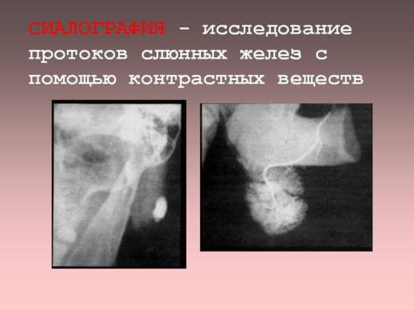
Clinic . On external examination there is no facial asymmetry. Only in cases where the cyst grows into the submental area (pushes apart the fibers of the mylohyoid muscle) can swelling be seen in this area. Mouth opening is free. In the sublingual region there is a hemispherical protrusion of a round or oval shape, dense or soft-elastic consistency, painless (Fig. 23.6.1 - 23.6.3). The mucous membrane over the protrusion is stretched and thinned, translucent with a bluish tint. The cyst cannot be punctured, because after the puncture, it empties (a clear, mucous, viscous yellowish liquid is released). The cyst is located next to the duct of the submandibular gland, but does not compress it. This can be verified by probing the duct (insertion of a polyethylene catheter) or by performing sialography of the submandibular gland
Rice. 23.6.1. View of a patient with a ranula located in the anterior sublingual region.

Rice. 23.6.2. View of a patient with a ranula localized on the left: a) when the tongue is displaced to the side; b) up.

Rice. 23.6.3. A patient with a ranula that has grown into the submental area: a) appearance; b) appearance of ranula in the oral cavity.
Diagnostics ranula usually does not cause difficulties. Only in the case when the cyst of the sublingual gland originates from its deep parts, difficulties may arise in establishing a diagnosis. In this case, it is necessary to puncture the cyst. With ranula we get a translucent viscous liquid yellow color, with an epidermoid cyst - a clear liquid with cholesterol crystals, with a hemangioma - blood.
Treatment cysts of the sublingual gland surgical. The following operations are used: cystotomy, cystectomy and cystsialadenectomy.
Cystotomy consists of excision of the dome (upper wall) of the cyst, followed by suturing the mucous membrane of the sublingual region with the gland capsule or with the cyst wall. The resulting niche quickly flattens.
Cystectomy used only in the presence of a true cyst. With a pseudocyst, this operation cannot be performed due to the fact that it is thin. fibrous tissue, which surrounds the false cyst, is difficult to remove, because it breaks easily and its guidelines are lost.
Cystsialadenectomy- removal of the cyst along with the gland. An incision in the mucous membrane is made bordering the sublingual fold (the mouths of the small sublingual ducts are located on it and it is difficult to dissect them from the mucous membrane). First, the cyst is desquamated, and then the sublingual gland is bluntly removed. You need to be careful because... The duct of the submandibular gland and the lingual nerve pass nearby.
If the cyst grows soft fabrics the bottom of the oral cavity in the form of “sand layers”, then a two-stage one-stage operation is performed. First, the cyst is removed using extraoral access. The isthmus of the cyst is bandaged with a silk ligature, the cyst is cut off, and the wound is sutured in layers. In the second stage of the operation, the cyst is removed along with the sublingual gland using intraoral access.
The cystotomy operation can be used in childhood, in elderly and weakened people (with severe concomitant diseases). The best results were obtained with cystosialadenectomy. There were no relapses of the disease.

Rice. 23.6.4. Sialograms of the parotid gland of patients with epidermoid cysts,
localized in the parenchyma of the gland (indicated by arrows). excretory ducts and
the parenchyma of the gland is shifted away from the cyst (a, b, c).

Rice. 23.6.4.(continuation).
♦ Parotid cysts
They can be congenital (epidermoid), i.e. true and false (retention) - when the interlobular duct is blocked as a result of injury (scar) or chronic inflammation. The growth of the cyst is slow and asymptomatic.
TO  clinic
.
There is facial asymmetry due to swelling of the soft tissues of the parotid-masticatory area. The color of the skin is not changed, it is easily folded. Upon palpation, a mobile formation of a round shape, dense or soft-elastic consistency is determined. Fluctuation with small cyst sizes is difficult to determine, because the cyst is located in the thickness of the parotid gland and is surrounded by dense fascia. Mouth opening is free. The mouth of the excretory duct is not changed. The function of the gland is preserved. The sialogram does not show complete filling of the interlobular ducts with the contrast mass (Fig. 23.6.4). When the cyst is located in the deep part of the gland, its growth into the oropharynx is observed with corresponding complaints.
clinic
.
There is facial asymmetry due to swelling of the soft tissues of the parotid-masticatory area. The color of the skin is not changed, it is easily folded. Upon palpation, a mobile formation of a round shape, dense or soft-elastic consistency is determined. Fluctuation with small cyst sizes is difficult to determine, because the cyst is located in the thickness of the parotid gland and is surrounded by dense fascia. Mouth opening is free. The mouth of the excretory duct is not changed. The function of the gland is preserved. The sialogram does not show complete filling of the interlobular ducts with the contrast mass (Fig. 23.6.4). When the cyst is located in the deep part of the gland, its growth into the oropharynx is observed with corresponding complaints.

Rice. 23.6.5. Appearance of the patient with Rice. 23.6.6. Active drainage with fixation
 active suction drainage. puck offered by I.B. Kindrasem.
Rice. 23.6.7. Polyethylene catheters for draining and washing the cavity of the “salivary” cyst.
Diagnostics. Cysts of the parotid glands must be differentiated from sialosis, lymphadenitis, neck cysts, benign tumors, limited purulent-inflammatory processes in the parotid-masticatory area.
Treatment surgical cyst. Parotidectomy is performed with preservation of the branches facial nerve(for a description of the operation, see the next chapter).
For the treatment of post-traumatic cysts of the parotid gland, especially with relapses of the disease, we use drainage in combination with periodic injection of 10% sterile sodium chloride solution into the cavity. The drug promotes the occurrence of adhesive inflammation, which leads to the disappearance of the cavity.
Active suction drainage is an elastic transparent tube with an internal diameter of 0.2 - 0.3 cm and a length of 30-35 cm. One end of it, inserted into the cyst cavity, has 2-3 additional oval-shaped holes of size 0. 1-0.2 cm, and the other is hermetically connected via a metal adapter to an elastic rubber medical cylinder, which allows creating negative pressure in this system. To insert a drainage tube into the cavity of the cyst, the skin and subcutaneous fatty tissue are pierced with a thick needle. The contents are sucked out. The end of the polyethylene tube, which has additional holes, is inserted into the lumen of the needle and advanced into the cavity, after which the needle is removed (Fig. 23.6.5). As the rubber balloon fills with the contents of the cyst, it is emptied. To ensure stationary fixation of the working part of the drainage tube in the cavity of the cyst and to prevent the drainage holes from being pressed against its walls, as well as to prevent depressurization of the system and loss of drainage I.B. Kindras (1987) designed a locking washer (Fig. 23.6.6). And to prevent overstretching of tissues with the washing liquid, the author proposed inserting a polyethylene catheter of a smaller diameter into the main drainage tube so that there is a gap between them for the flowing liquid (Fig. 23.6.7). Rinsing with a 10% sterile sodium chloride solution is carried out once a day for 3-5 days. Having finished washing the cavity with a hypertonic solution, it is necessary to resume active drainage. After the last rinse, the active drainage is left for a day and then removed. Treatment lasts 5-7 days, there are no relapses after treatment. You just need to remember that the proposed conservative treatment should be carried out for pseudocysts, i.e. without epithelial (true) cystic membrane.

Rice. 23.6.8. Appearance of a patient with a submandibular gland cyst:
a) front view; b) side view.
♦ Submandibular gland cysts
They are located below the diaphragm of the floor of the mouth. Therefore, the swelling spreads from the submandibular region to the lateral surface of the neck, and does not cause noticeable changes in the oral cavity.
The cyst can be false (retention) or true. It grows slowly and painlessly. Upon external examination (Fig. 23.6.8), there is asymmetry of the face due to swelling of the soft tissues of the submandibular region and the upper third of the neck. The skin color is not changed, it gathers into a fold. The swelling is painless, dense or soft-elastic consistency. The mouth of the excretory duct is not changed. The sialogram reveals compression of the ducts and the absence of complete filling of the interlobular ducts with the contrast mass. Treatment is surgical. Cysts of the submandibular gland must be removed along with the gland, because remnants of the gland can cause a relapse of the disease.
♦ Cysts of the main excretory ducts of the major salivary glands
These cysts can be in the form of two options: cyst-like expansion in the area of the mouth of the duct (with sialodochitis) or cysts that arise when the duct wall ruptures when saliva enters the soft tissue, forming a so-called salivary cyst (occurs during injury). In the first case, the wall of the cyst will be the dilated duct of the gland, and in the second, fibrous tissue formed as a result of the body’s response to saliva entering the soft tissue (as during encapsulation of a foreign body).
Clinically a painless elastic protrusion is palpated in the area of the excretory ducts. Fluctuation may be detected. The mouth of the duct may be narrowed, and less saliva is released from the duct. In some cases, obstruction of the duct is observed and the clinical picture of obstructive sialadenitis develops. There may be exacerbations inflammatory process.
Treatment cysts of the excretory duct of the submandibular gland surgical - extirpation of the gland. An exception may be post-traumatic cysts of the initial parts of the excretory duct of the submandibular gland. In this case, by removing the dome of the mucous membrane (above the salivary cyst), it is possible to create an additional orifice of the excretory duct, which can function without causing concern to the patient.
If the wall of the cyst in the area of the mouth of the main excretory duct of the parotid gland is represented by a dilated duct, then after its isolation, the dilated part of the duct is dissected and a deformed part of the excretory duct is plastically formed on a polyethylene catheter by cutting off the cyst-shaped dilated part of it. The mouth of the parotid gland duct is sutured to the buccal mucosa in its original place. When the middle part of the parotid duct expands, it is also plastically formed, but in the postoperative period the catheter is left in the duct for 6-7 days to prevent its fusion or narrowing.
In case of cysts of the excretory duct of the parotid gland (its submucosal section), an additional orifice (internal salivary fistula) can be formed on the mucous membrane of the cheek.
The occurrence of compaction in the oral cavity in most cases is provoked by a salivary gland cyst. Cysts appear in the salivary glands in both children and adults, causing noticeable inconvenience while talking and chewing food. The salivary glands play extremely important functions– they are one of the first to participate in the digestion process, moistening food with saliva.
Most often these are reactive dystrophic diseases are one-sided. As a rule, patients consult a doctor in a timely manner, immediately after a small lump appears. A more precise diagnosis is made after an ultrasound of the salivary gland, and an MRI of the salivary gland is also performed. If a salivary gland cyst is diagnosed, treatment is only surgical.
Inflammation of the glands is diagnosed quite rarely. Most of the reactive inflammatory processes are taken over by the minor salivary glands - approximately half of all pathological changes it flows right there. The sublingual, mandibular and parotid glands are slightly less commonly affected.
 The appearance of salivary gland cysts begins with problems with saliva drainage. Blockage of the duct occurs for the following reasons:
- inflammation, obliteration by purulent contents;
- inflammatory processes in the oral cavity, for example, stomatitis;
- trauma to the mucous membrane from a broken tooth, dental metal plates, removable dentures;
- blockage of the salivary gland by the formed compaction;
- cicatricial deformation processes;
- external compression, for example, a tumor, etc.
Doctors suggest that some people may develop a predisposition to developing a tumor in a given organ even during fetal development.
Classification of formations
Despite the fact that there is a fairly extensive classification of cysts, for many of them it is possible to give general characteristics neoplasms.
The walls of the tumor are formed from a fibrous membrane, and the inner surface of the tumor is lined with flat epithelium or granulation tissue.
 The tumor increases in size due to the accumulation of salivary secretions or the permeation of fluid through the walls of the capillaries.
The most common classification is based on their location. Depending on this, cysts are divided into cysts of small and large glands.
Small SGs are located in the cheeks, palate, tongue, lips and molars. They appear quite rarely, since inflammation in such places passes quickly and does not leave behind complications such as cysts.
Large SGs are affected much more often. These formations include the following varieties:
- A cyst of the submandibular salivary gland usually grows slowly, but soon the submandibular salivary gland becomes so deformed that it is not difficult to notice the neoplasm. When a tumor appears, the duct of the submandibular fluid opens spontaneously, which leads to the outflow of contents and filling with new ones;
- Parotid salivary gland cyst - usually parotid gland is rare and can be congenital. Often the pathology is provoked by sialadenitis of the parotid gland, because the excretory duct of the parotid salivary gland is discharged directly into the oral cavity. A number of cysts grow inward, into the area of the pharyngeal process. The same formations that grow outward lead to noticeable deformation in the ear area. The parotid gland is well visualized using ultrasound; in the same way, the duct of the parotid gland, called the stenon duct, can be examined, so diagnosis is not difficult;
- Sublingual cyst of the salivary gland - plays a decisive role in the diagnosis here ultrasonic method, with the help of which the diagnosis is differentiated. Like other similar tumors, a sublingual cyst can open on its own, but then reappear.
Based on localization, formations are divided into parenchymal tumors and tumors of the gas duct, and based on their structure, doctors distinguish retention and post-traumatic formations. According to this classification, retention cyst of the minor salivary gland is quite rare.
Mixed tumors
 The most severe form of neoplasm is a mixed tumor of the salivary gland, often combined with benign neoplasm. With mixed tumors, the excretory ducts of the gastrointestinal tract also become inflamed. These cysts are largely painless, but cause discomfort due to their rapid growth and involvement in pathological process facial nerve.
Several years ago, interventions for resection of mixed tumors included removal of the facial nerve. This had a negative impact on the patient’s future life. Currently, it is possible to remove such tumors while preserving the facial nerve, which makes it possible to restore digestive functions much faster oral cavity after surgical intervention.
Signs of the disease
Signs of the presence of a salivary gland cyst are quite clear - the salivary glands turn into a painless lump, mobile to the touch, which progresses sluggishly. This is the main symptom that worries patients. But the patient may not notice the manifestations of sialadenitis during its sluggish course. It is with a complaint about a tumor that patients come to the clinic. Depending on the location, the size of the cyst is also noted.
 With a tumor of the small glands, the obstruction barely increases in diameter to one centimeter; it is usually located on the cheeks, palate, and lips. It is rare that such tumors cause inconvenience, but if the integrity of the walls of even a small tumor is damaged, infection and suppuration can occur. But the parotid gland, when suppurated and a tumor forms, can reach impressive sizes.
The sublingual salivary gland cyst is localized in the area of the root of the tongue. Such a neoplasm is not difficult to notice - it is visible through the mucous membrane, and when moving the tongue it interferes with the patient. Because of this, noticeable speech defects and problems with chewing and swallowing food appear. Due to its rather rapid growth, the tumor often breaks out on its own, but is again filled with viscous saliva.
Tumors of significant size, such as a submandibular or parotid cyst, change the face, so deformation of the facial contour is another sign of the appearance of pathology.
Diagnostic methods
Determining the pathology is quite simple, but it is necessary to differentiate a cystic tumor from inflammation lymph nodes, benign or malignant neoplasms and other pathologies.
A sublingual cyst, or ranula, is distinguished from a lipoma or dermoid cyst. Differentiation from a submandibular neoplasm is also important. If a focus of suppuration has formed, differentiation should occur with acute inflammatory diseases of the salivary glands.
 Sialography
 Doctors perform a number of procedures, such as sialography of the salivary glands, ultrasound of the salivary glands, and in controversial cases, a magnetic resonance imaging scanner is used. Usually, tumors can already be seen on ultrasound - they give smooth, but irregular contours when visualized.
If tomography does not definitively resolve the issue of differentiating the diagnosis, a histological examination– biopsy of the salivary gland to determine the malignancy of the neoplasm. This is especially important if a pleomorphic adenoma that is prone to degeneration is suspected.
Surgical care for patients
Salivary gland cyst removal is performed surgically. Conservative treatment of pathology does not make sense, because it does not lead to desired result. Based on the location of the tumor, the doctor determines access to it. Tumors and the SG themselves can be removed different ways. For example, a retention cyst of the salivary gland in the area of the lips, palate or cheek is more often removed using access through the oral cavity, but a tumor in the parotid area is more often operated on using external access.
 The essence of the surgical intervention is to remove the tumor and then apply a suture. Usually, during surgery it is necessary to remove the salivary gland so that the disease does not recur. In this case, the functions of the salivary glands are redistributed among the remaining healthy glands. The operation takes place under local anesthesia. If the patient’s tumor has reached gigantic proportions, for example, if the parotid gland is inflamed, a consultation is possible plastic surgeon and one-time intervention.
After surgery, it is necessary to carefully care for the suture so as not to provoke infection of the wound. Patients are advised to especially monitor the appearance of inflammatory lesions in the oral cavity and regularly consult a dentist so that the lesion does not reappear.
The size of the cyst, its contents, and the structure of the walls are very diverse. All of the above depends on the duration and mechanism of formation, localization, as well as many other factors. There are cysts:
- true - lined with epithelium;
- false - without special lining.
By nature they can be:
- congenital;
- acquired.
All these two sources of their formation imply the occurrence of a cyst in the process of vicious formation of organs and/or tissues. Based on their mechanism of formation, they distinguish:
Now let's take a closer look at each cyst in more detail from the list listed.
Retention
In the vast majority of cases they are acquired. Widely distributed in a variety of glandular-secretory organs. They arise due to difficulty or complete cessation of outflow from the secretory gland, which ultimately leads to blockage of the duct with a kind of microscopic stone, pollen or other debris. The cause of the blockage may be a plug created from thickened secretions, compressed by a scar or tumor.
Accumulating in the glandular lobule and duct, the secretion stretches them and gradually enlarges the cavity with watery, sebaceous, mucous or other contents. The most common cysts are:
glands
- dairy;
- sebaceous;
- salivary;
- prostate;
- pancreas, pancreas
and follicular cyst ovaries and many others. The wall of a retention cyst is lined with the flattened epithelium of the gland itself or its duct. In the case of intrauterine atresia of the glandular duct, retention congenital cysts develop.
Ramola
They got their name from the word “softening”. They are formed in compact tissues during focal necrosis: inflammation, infarction, hemorrhage, followed by softening, liquefaction or resorption of dead tissue. The walls of such a cyst are formed by the tissue of the same organ on which it “grows.” However, in the future the cyst may be replaced by connective tissue. As a rule, they are found in the spinal cord and brain, as well as tumors. The most common are:
- cyst corpus luteum ovaries;
- dental;
- bone (osteoblastoma, osteitis fibrosa).
Traumatic
They are provoked by displacement during injuries epithelial tissue. Among them there are epithelial traumatic cysts:
- palms;
- fingers.
Due to the penetration of the epithelial cover into the underlying tissue with the subsequent accumulation of secretion in the resulting sac. Cysts of the pancreas and iris have the same origin.
They are the larval bladder stage of tapeworms such as:
- cysticercus;
- echinococcus.
Dysontogenetic
As a rule, they are congenital. They are a cyst-like transformation, which sometimes preserves clefts and embryonic canals or occurs during the formation of an embryo in displaced tissues. These include cysts that are preserved from the gill slits, or those remaining from the remains of the vitelline-intestinal tract, on the prostate gland, resulting from a violation of the formation of the paranephrotic ducts, as well as as developmental defects sweat glands: syringoepitheliomas and syringocystadenomas, paraovarian, dermoid, endometriotic ovaries, multiple cysts of the kidneys, lungs, liver, pancreas, central nervous system.
Tumor
They arise due to growing tumor tissues due to metabolic disorders and the development of the process of carcinogenesis, which in turn creates single- and multi-chamber cavities. They are formed, as a rule, in glandular organs:
- salivary gland adenoma;
- cystic amelobastoma or lymphangioma.
Treatment methods for inflammation of the salivary glands
In the following conversation we will try to describe as much as possible possible cases cyst formation and methods of getting rid of them. So.
Salivary gland retention cyst - treatment
It is observed, as a rule, on the mucous membrane of the lips and is a small, elastic, bluish elevation to the touch. spherical, the edges of which are perfectly contoured. Located under the mucous membrane. Consists of a capsule that contains a light liquid. In the dominant case it occurs on inside lips or cheeks. The formation is absolutely painless, sometimes decreasing, sometimes increasing in size. Occurs as a result of teeth biting the lips. The accumulating secretion gradually leads to neoplasm. The cyst stops growing after complete removal of its own tissue. In most cases it is used surgical intervention .
Before surgery, as an alternative, the cyst is punctured. With a syringe, from the skin side, its contents are sucked out, and the cavity is washed with a chlorine solution according to N.I. Krause, which is a physiological solution that is saturated with chlorine gas, as well as its derivatives. Its use does not cause necrosis and completely eliminates the development of the inflammatory process.
In case of absence positive result the doctor resorts to conservative surgical treatment. If the cyst has spread to the suprahyoid area and becomes bright expressed form hourglass, then use combined method. Outside, in inner part, a physiological denaturing solution is injected, and the protruding one is opened and treated surgically.
Minor salivary gland cyst - treatment
The minor salivary glands include:
- mucous-protein;
- alveolar-tubular;
- Merocrine.
They are located in the mucous membrane of the oral cavity, classified according to their location:
- buccal;
- labial;
- palatal;
- lingual;
- molar.
Among the most numerous are the palatal and labial ones. They are the favorite location for tumors. It is extremely rare that a cyst forms on a hard and soft palate. First, a small round formation appears, which increases over time, reaching a diameter of 1.5 cm. In the event of a breakthrough, a viscous fluid is released from the cyst and the tumor disappears. This happens during a meal as a result of biting.
If the cyst reaches a diameter of two centimeters, the configuration of the lip is completely deformed. In the case of extremely large cysts, due to the thinning of the membrane, the cyst acquires a bluish tint. During palpation, it is felt as soft or densely elastic with a well-defined shape and mobility from the surrounding tissues. Treatment of such a cyst is usually always performed by surgery followed by its removal.
Retention cyst of the minor salivary gland - treatment
The peculiarity of this cyst is its formation on inner surface lips or cheeks closer to the corner of the lips or their lower part. As in the case described above, treatment is carried out through surgery - complete removal of the tumor. However, I would especially like to dwell on possible risks such an operation. Quite often the cyst is associated with the branches of the facial nerve. Its removal entails a violation of integrity, which can lead to facial distortion or paralysis of facial muscles. A cyst formed on the lip or cheeks is removed without much risk. To avoid relapse, complete removal of the cyst shell is a prerequisite.
Parotid cyst
The parotid gland is one of the largest salivary glands. Its cysts are quite rare, but they cause a lot of concern, especially if they cause deformation of the natural contour of the face. A parotid salivary gland cyst is identified by a painless swelling. It is surprising that at the site of its formation the color of the skin does not change, although underneath it an oval or round-shaped formation is clearly palpable, has clear boundaries, is not connected and has an elastic consistency. When pressing with fingers, the cyst is mobile. The transfer of pressure from one side to the other is felt, which indicates its filling with liquid contents.
Pain may occur in the event of the development of an abscess, which can occur as a result of inflammation of the cyst or the eruption of a wisdom tooth. In the case of a deep focus of inflammation, there will be no redness, but there will be a characteristic limitation in opening the mouth.
Parotid cyst - treatment
Cyst treatment is carried out exclusively by surgery.. If the cyst is localized in the parotid region, its shell is removed along with a section of adjacent tissue. Any surgical intervention is complicated by the peculiarities of its location due to the risk of damage to the facial nerve.
Sublingual salivary gland cyst
This cyst is called a ranula or frog tumor. The disease got its name due to the fact that the mucous membrane protrudes into the sublingual region, which resembles a sac-like formation in the oral cavity of a frog. Is extremely rare disease. Occurs in young or middle age and in isolated cases in infants. As a rule, the ranula is located closer to the frenulum of the tongue in the sublingual area.
Interferes with eating and talking. It has a slow growth rate. It is possible to disappear after an arbitrary period of time with the next appearance. The cyst has a soft elastic consistency. Due to excessive thin shell bursts under the pressure of a scalpel. Based on the fact that the bundles connective tissue Such cysts penetrate deep into the connecting layers of the lobules of the sublingual gland; their elimination is quite problematic.
Retention cyst of the sublingual salivary gland
The salivary sublingual gland consists of several lobules. Some open into small individual ducts located in the area of the sublingual fold. It is the blockage of the excretory ducts that leads to the formation of a cyst. In my own way appearance such a cyst resembles a frog's laryngeal bladder. As it grows, it moves its tongue up and back. Removal occurs through surgery.
Submandibular salivary gland cyst
It grows slowly, developing in one of the gland lobes. Often reaches impressive sizes. From the clinical side, it is a bulging, fluctuating, painless formation, in the submandibular region, of soft elastic consistency with smooth surface. In rare cases, a cyst develops from the submandibular region, going around back wall The mylohyoid muscle penetrates the oral cavity at the level of the maxillary lingual groove.
Based on the above, such a cyst should be differentiated from a dermoid or lateral cyst, linfangioma, lipoma and cavernous hemangioma. Treatment is carried out through surgery, namely cutting out the cyst along with the submandibular salivary gland.
Salivary gland cyst treatment
As mentioned above, we will not repeat ourselves; treatment of a cyst is impossible with any using medicinal methods. In order not to repeat ourselves and not waste your precious time, we will say again that the treatment of a cyst of any salivary gland is carried out by cutting it out along with the tissues that form it to avoid relapses.
Removal of a salivary gland cyst
Basically, to remove a cyst, two semi-oval incisions are made in the mucosa above and below the tumor. In a semi-blunt way, its shell is separated from the surrounding tissues, and its connecting bridges are cut off with scissors. In this case, the cyst is “selected” completely. Small glands that interfere with suturing are removed and catgut sutures are applied to the wound. The operation is completed.
Treatment of cysts using traditional methods
Traditional medicine claims that cysts can be gotten rid of not only through surgery. Therefore, we present to your attention the most effective recipes.
- 2 tbsp. Stir tablespoons of eucalyptus oil in 1 glass of warm boiled water. Use as a mouth rinse;
- 1 tbsp. Pour a spoonful of eryngium herb into 1 cup of boiling water. Leave for 2 hours. Use as a mouth rinse;
Traditional medicine claims that the following are great help in the fight against salivary gland cysts:
- raspberries;
- immortelle flowers;
- horsetail;
- elderberry flowers, veronica;
- leaves of sage, yarrow, viburnum;
- eucalyptus;
- chamomile.
Salivary gland cysts are single-chamber or multi-chamber neoplasms filled with colorless or yellowish fluid, the appearance of which occurs as a result of obstruction or partial patency salivary ducts without visible symptoms, which makes it much more difficult early diagnosis diseases. The cystic formation looks like a small round sac or nodule, characterized by gradual increase size and discomfort during conversation and eating.
Everyone is at risk of developing a salivary gland cyst - from an infant to an elderly person, but more often the disease is diagnosed in patients whose age is within 30 years. Salivary gland cysts form equally in both men and women.
Causes of formation of salivary gland cysts
Salivary gland cysts appear as a result of blockage of the salivary ducts. Pathology of patency can be caused by various factors:
- various types of injuries;
- Poor, untimely or completely absent oral hygiene;
- unhealthy diet;
- bad habits;
- various types infectious diseases oral cavity and teeth;
- difficulty or disturbance, followed by cessation of secretion outflow;
- the appearance of a plug as a result of thickening of the secretion, disrupting the patency of the excretory canal;
- the presence of various tumors that put pressure on the duct;
- the presence of scars narrowing the canal.
Classification of salivary gland cysts
Depending on the location, salivary gland cysts are divided into two types:
- Minor salivary gland cysts appear on the cheeks, lips, palate, tongue, or molars.
- Cysts of the major salivary glands: sublingual salivary gland, parotid salivary gland and submandibular salivary gland.
In addition, salivary gland cysts can be located either in the duct or in the functional part of the gland. According to the structure of the cyst, there are true (retention) and false (post-traumatic). A mucocele salivary gland cyst with mucoid mucous content is also isolated.
Symptoms of a salivary gland cyst
A cyst of the minor salivary gland is formed in the area of the corners of the mouth inside the lip; in addition, there is a possibility of its occurrence on the mucous membrane of the cheeks. Cyst formation can occur as a result mechanical damage minor salivary gland and areas of the oral cavity during eating or talking. Initially, the neoplasm is small and round, but gradually increases in size. A patient with such a cyst does not feel discomfort, but in some cases he may complain about pain syndrome while talking and eating. Painful palpation of the cyst is possible. During diagnosis, it is important to distinguish a cyst of the minor salivary gland from hemangioma, fibroma and other benign tumors.
The sublingual salivary gland cyst is located at the bottom of the mouth. In its shape, the cystic formation can have the shape of an hourglass, round or oval shape with a characteristic bluish tint. As the disease develops, the frenulum of the tongue is disrupted and displaced, and the patient also feels discomfort while eating and talking. The cyst of the sublingual salivary gland is characterized by independent periodic emptying with subsequent filling clear liquid. When diagnosing a sublingual gland cyst, it is necessary to distinguish it from a submandibular gland tumor, dermoid and lipoma. Possibility of sialolithiasis or viral diseases excluded.
The submandibular salivary gland cyst is fixed in the area of the submandibular glands. A growth forms that is soft and elastic to the touch. As it grows, swelling appears in the tongue area and at the bottom of the mouth. The risk of inflammation is associated with deformation of the oval of the face. In the diagnostic process, it is necessary to distinguish a growth of the submandibular salivary gland from a gill cyst, dermoid and soft tissue sarcoma, as well as diseases associated with purulent processes in the lymph nodes and submandibular salivary canals.
A submandibular gland cyst can be localized in the salivary ducts, it is called congenital cyst The thyroid duct is located in the middle of the neck or at the bottom of the hyoid bones.
A parotid cyst is characterized by slow growth and location near the ear; in rare cases, the cyst can be bilateral. It is smooth and elastic in appearance and has a dense consistency. This very rare form of cyst can be congenital or acquired. It is formed without causing discomfort to the patient. A parotid cyst affects the ducts of the salivary glands and can be located quite deep, which makes it difficult to determine fluctuations. If the cyst has formed in lower section, then it is characterized by internal growth. This is due natural structure oral cavity, which complicates diagnosis and treatment. The patient begins to feel discomfort only if it progresses purulent process. When diagnosing, it is necessary to distinguish a parotid salivary gland cyst from lymphadenitis, lipomas and bronchial cysts.
Diagnosis of salivary gland cysts
Diagnosis of salivary gland cysts involves determining the nature of the neoplasms; they can be benign or malignant. Determining the nature of cysts directly depends on their clinical picture. To do this, it is necessary to interview the patient, identify and evaluate complaints, examine and palpate the cyst. During these manipulations, the doctor determines the size, type, location and mobility of the cyst.
Due to the fact that all cysts have an almost identical clinical picture of the disease, for an accurate diagnosis it is necessary to carry out additional diagnostics with cytological, radiological and biochemical studies.
Cytological diagnosis of the salivary glands involves taking a puncture from the tumor mass. Thanks to this study, it becomes possible to determine the process of tumor development.
X-ray examination allows you to find out how salivary ducts filled with contrasting mass. The diagnostic method consists of conventional radiography and contrast radiography of the salivary ducts.
Also, for an accurate diagnosis, the method of differential diagnosis (method of exclusion) is used. This is necessary to distinguish one cyst from another.
Treatment of salivary gland cyst
Depending on the type of salivary gland cyst, they are used various methods treatment of the disease. For example, for a cyst of the minor salivary gland, surgery is often used, followed by complete removal of the cyst membrane. The operation, as a rule, takes place without complications due to the small size of the tumors. If the cyst is associated with areas of the facial nerve, there is a risk of facial distortion or facial paralysis.
A sublingual salivary gland cyst is characterized by a soft shell, which can cause the cyst to rupture during surgery under scalpel pressure. Removing such a cyst is problematic, since the connecting bundles of the formation are located deep in the lobar layers of the tongue and are connected to the sublingual glands. A retention cyst of the sublingual gland is also removed through surgery.
Cyst of the submandibular salivary gland is subject to complete removal surgical intervention, which results in complete excision of the cyst along with the submandibular gland.
Parotid cyst is removed surgically. If the location of the cyst is parotid, then the membrane of the neoplasm is removed along with the area of tissue to which it is adjacent. Due to the anatomical structure and location of the cyst, surgical intervention is always difficult and has a high risk of damage to the facial nerve.
As it became clear from the above, treatment of a salivary gland cyst of any type involves exclusively surgical intervention. Medication method treatment is not provided due to its ineffectiveness. Excision of the cyst shell along with particles of tissue to which it is adjacent is necessary to avoid relapse of the disease.
Removal of the salivary gland cyst occurs through two semi-oval incisions. Using a blunt object, the cyst is separated from the tissue to which it is attached, after which it is cut off with scissors. Then the wound is sutured. If there are minor salivary glands that interfere with this process, they are removed as a group.
– cavity formations that arise as a result of obliteration of the ducts of the salivary glands. A salivary gland cyst is manifested by the presence of a soft, painless formation, a slow increase in size, fluctuation, difficulty swallowing and speech. Diagnosis of a salivary gland cyst takes into account examination data, ultrasound of the salivary glands, sialography, puncture and fine-needle aspiration biopsy cystic formation, cytological and biochemical research punctate. Treatment of salivary gland cysts is surgical (cystostomy, cystectomy, extirpation of the gland) using intraoral or extraoral access.

General information
A salivary gland cyst is a tumor-like formation of the maxillofacial area in the form of a cavity filled with liquid contents. Salivary gland cysts are relatively uncommon. In most cases, they come from the minor salivary glands (56%), less often - from the sublingual glands (35%), parotid (5%) and mandibular glands - (4%). Salivary gland cysts develop predominantly in individuals young(about 30 years old). Treatment of salivary gland cysts has its own specifics and is within the competence of maxillofacial surgery (dental surgery) and otolaryngology.
Causes of formation of salivary gland cysts
The formation of a cyst may be associated with difficulty or complete cessation of the outflow of salivary gland secretions. The causes of obstruction of the duct may be its blockage with a mucus plug; obliteration as a result of inflammation (sialadenitis, stomatitis), trauma to the gland with a prosthesis or a destroyed tooth; salivary gland stone obstruction; cicatricial narrowing, external compression by a tumor, etc. It is assumed that some salivary gland cysts may be of congenital origin and develop from an accessory rudimentary duct detached during embryogenesis.
In most cases, salivary gland cysts are single, single-chamber formations filled with colorless or yellowish mucous fluid. The cyst capsule is presented fibrous membrane; the inner surface is lined with stratified squamous and columnar epithelium or granulation tissue. An increase in the size of a salivary gland cyst can occur both due to the accumulation of salivary secretions in the obliterated cavity, and due to the transudation of fluid through the walls of the capillaries.
Classification of salivary gland cysts
Based on the place of education they distinguish:
1. Cysts of the minor salivary glands (buccal, labial, palatal, lingual, molar).
2. Cysts of the major salivary glands:
- sublingual salivary gland (ranula)
- parotid salivary gland
- submandibular salivary gland
In addition, cysts of the parenchyma and ducts of the salivary glands are distinguished by location. Depending on the structure, a salivary gland cyst can be retentional (true) or post-traumatic (false). Cysts of the salivary glands with mucoid mucous contents are called mucoceles.
Symptoms of salivary gland cysts
Minor salivary gland cyst
Most often, such cysts are localized on the inner surface of the lower lip, less often in the cheeks or other parts of the oral cavity. A cyst of the minor salivary gland usually does not exceed 0.5-1 cm in diameter and slowly increases in size. A salivary gland cyst is defined as a mobile formation of round shape and elastic consistency, protruding above the surface of the mucous membrane.
A cyst of the minor salivary gland usually does not cause concern to the patient and painful sensations. Sometimes, when accidentally injured by food or biting, the salivary gland cyst opens with the release of a viscous translucent liquid with a yellowish tint; then the contents accumulate in it again. A minor salivary gland cyst must be distinguished from hemangioma, fibroma and other benign tumors of the oral cavity.
Sublingual salivary gland cyst
The sublingual salivary gland cyst (ranula, “frog tumor”) is localized in the floor of the mouth, under the base of the tongue. Usually it shines through the mucosa in the form of a round or oval protrusion of a bluish color. Less commonly (when located above and below the mylohyoid muscle), the cyst has the appearance of an hourglass.
An enlarging cyst of the sublingual salivary gland can cause displacement of the frenulum of the tongue and interfere with eating and speaking. Periodic spontaneous emptying and filling of the sublingual salivary gland cyst with a clear secretion is possible.
Differential diagnosis of a sublingual salivary gland cyst is carried out with a submandibular gland cyst, dermoid cyst, lipoma. If the contents of the cyst become infected, exacerbation of chronic sialadenitis and salivary stone disease should be excluded.
Submandibular salivary gland cyst
Manifested by the presence of a round, soft-elastic, fluctuating formation in the submandibular region; when spreading to the sublingual area - bulging in the bottom of the mouth. When a cyst of the submandibular salivary gland reaches a large size, it can cause deformation of the facial contour.
A cyst of the submandibular salivary gland requires differentiation from a lateral neck cyst, dermoid cyst, soft tissue tumors (hemangioma, lipoma, lymphangioma, etc.), lymphadenitis, submandibulitis.
Parotid cyst
A parotid salivary gland cyst is clinically manifested by a rounded swelling of soft tissue in the preauricular area, usually on one side, causing facial asymmetry. Upon palpation, the soft or dense elastic consistency of the cyst is determined. The skin over it is not changed, there is no pain or fluctuation.
When infected, a parotid cyst can become complicated by an abscess. In this case, skin hyperemia, pain in the parotid region, limited mouth opening, fluctuation, and low-grade fever appear. Differential diagnosis of parotid salivary gland cysts is carried out with chronic lymphadenitis, tumors of the salivary gland.
Diagnosis of salivary gland cysts
Salivary gland cysts are recognized based on the clinical picture, instrumental and laboratory research. Besides, additional methods allow for differential diagnosis of cystic formations with tumors of the salivary glands. To clarify the size, position of the cyst and its connection with the salivary gland, ultrasound of the salivary glands, cystography and sialography, CT and MRI in contrast mode are performed. Crucial importance in confirming the diagnosis belongs to puncture and fine-needle aspiration biopsy of the salivary gland cyst, followed by biochemical and cytological examination content.
Treatment of salivary gland cysts
Treatment of salivary gland cysts at any location conservative methods not provided. Depending on the location of the cyst, surgical intervention is performed through intraoral (for a cyst of the minor salivary gland) or extraoral (external, open) access.
Surgical treatment of retention cysts of the minor salivary glands involves their removal from the oral cavity under local infiltration anesthesia with the application of catgut sutures. The scope of surgery for a sublingual salivary gland cyst may include cystostomy, cystectomy, or cystosialadenectomy.
A cyst of the submandibular salivary gland usually needs to be removed along with the gland. For a parotid cyst optimal method serves to remove the cystic formation along with the adjacent parenchyma of the gland through an external approach (partial, subtotal or total parotidectomy) while preserving the branches of the facial nerve.
Prognosis and prevention of salivary gland cysts
The main risk when removing a parotid salivary gland cyst is the possibility of damage to the branches of the facial nerve, which can lead to paralysis of the facial muscles. In addition, if the shell of the salivary gland cyst is not completely removed, a relapse of the disease may occur. Without treatment, there is always a risk of developing purulent complications(abscess, phlegmon).
Prevention of the formation of acquired salivary gland cysts consists mainly in preventing inflammatory diseases and oral injuries, carrying out
