Postpartum diseases in cows and their prevention. Postpartum complications
Numerous breeding problems in highly profitable herds confirm the fact that there is also a significant loss of livestock. The main reasons are: diseases of cows after calving, perinatal diseases (residual and postpartum paralysis, placental retention and, as a consequence, reproductive disorders and infertility).
Diseases of cows after calving: causes
If animals begin to behave unnaturally, symptoms and nutrition should be analyzed to determine the diagnosis. All mistakes made lead to reproductive disorders and metabolic disorders. The cow must be provided with sufficient nutrients, that is, provide the necessary amount of roughage and change the components of the feed ration as little as possible after the animal has become accustomed to receiving feed. Here are some metabolic disorders caused by nutritional errors.
Ketosis
 Ketosis is one of the most important metabolic diseases. The main cause of this disease in cows after calving is malnutrition of animals, that is, a lack of carbohydrates in feed. To cover the deficit of carbohydrates in the body, the cow begins to use accumulated fat. As a result, there is a so-called incomplete combustion fatty acids and an increase in ketone compounds.
This disease occurs especially:
- in highly productive cows that are in good condition, usually 10-60 days after calving;
- in older cows after the fourth and subsequent lactations.
Depending on the concentration of these compounds in the blood, milk, saliva, urine, ketosis can occur in subclinical (latent) or clinical (with pronounced symptoms of the disease). Apart from loss of milk quantity and quality, ketosis also causes many other complications. This is the reason for the decrease in cow productivity. Then childbirth with complications and retention of the placenta is observed, calves are more often stillborn, inflammation of the uterus occurs (dryness), and a longer period between walks occurs.
Preventing ketosis is based on a healthy, nutritious diet, including:
- providing the cow with enough feed, taking into account energy demand;
- adjusting the feed dose to the performance and genetic potential of each cow;
- avoiding sudden changes in food during feeding;
- do not feed low quality silage, especially those containing butyric acid;
- proper feeding of the cow during the drying period (use low energy intake during the first drying period, gradually introducing concentrate over 2-3 weeks before parturition);
- during the perinatal period, do not use ketogenic feeds containing simple sugars (sugar beets, sugar beets, molasses, beet silage);
- usage prophylactic drugs that regulate carbohydrate metabolism (for example, niacin, propylene glycol).
Rumen acidosis

This is a painful process that involves a significant decrease in the pH of the rumen, causing destabilization of its functioning. A decrease in pH below 6.0 stops fermentation in the rumen and reduces the activity of cellulolytic bacteria. This means that the feed components are not completely used. Harvesting easily digestible carbohydrates produces large amounts of volatile fatty acids, which lowers rumen pH and inhibits the growth of cellulose-degrading bacteria.
A pH below 5.2 effectively removes all cellulolytic bacteria from the rumen.
Below pH 4.7, lactic acid bacteria develop, producing increasing amounts of lactic acid, the excess of which leads to immobilization of rumen motility, disappearance of microflora and cessation of fermentation processes.
The consequences of lowering rumen pH are:
- decreased milk production;
- reduction of fat content in milk;
- deterioration in reproduction rates;
- deterioration of hoof health;
- increase in the content of somatic cells in milk.
This manifests itself in reduced or diminished appetite, deterioration in the general condition of the animals, diarrhea, limb ulcers or laminitis. At the first symptoms of rumen acidosis, feed containing easily fermentable carbohydrates should be removed from the diet, while feeding good quality meadow hay. IN severe cases Rumen acidosis requires veterinary intervention. However, prevention is most important to avoid pestilence.
The most important measures to protect against this disease are:
- avoid frequent and rapid feed changes,
- usage transition periods when changing winter nutrition for the summer and vice versa,
- the feed ration must contain a sufficient amount of structural carbohydrates, that is, unprocessed fiber (it must be at least 17% in the dry matter of the feed ration and at least 23% in the dry matter of the feed roughage),
- systematic introduction of cows to feed concentrate, especially during the last drying period and during the distribution period,
- dividing large doses of concentrated feed into several pastures (up to 2.5 - 3 kg per pasture),
- avoiding feeding excessively fragmented feeds,
- using a proper feeding method, i.e. not feeding concentrate and young green feed in the morning,
- avoid feeding too much acidic feed (beet silage, distillery) and containing easily fermentable sugars (molasses, sugar beet and fodder beet),
- use of rumen pH buffers (eg sodium bicarbonate).
Scar destruction
 Mesothelioma rumen (alkaline indigestion) is a disease process consisting of a change in the acidity of the rumen contents in the alkaline direction due to excessive production of ammonia. The occurrence of the disease occurs mainly when the feed intended for cattle contains a large amount of protein (concentrate feed, fresh alfalfa, clover) with insufficient amounts of digestible fiber (hay, straw) and easily digestible carbohydrates.
The severity of the disease depends on the amount of ammonia in the contents of the stomach and its concentration in the blood. Initially, the animal loses its appetite and the scars weaken. Weakness of the animal may be accompanied by diarrhea, decreased milk yield and fat content in the milk. During alkalosis, which occurs against the background of urea poisoning, muscle tremors, respiratory and circulatory disorders are observed, profuse salivation.
Disease prevention
Preventing suppurative alkalosis involves maintaining proper nutrition, avoiding sudden changes in feed, and maintaining proper proportions between protein and carbohydrates in the diet. Treatment is to stop introducing the current feed. Good results were obtained after using easily digestible carbohydrate raw materials (molasses, beets).
In severe cases, you should seek help from a veterinarian.
Fixed herpes
Garter disease (hypomagnesemia) is characterized by decreased levels of magnesium in the blood and is usually associated with feeding animals on pasture.
Symptoms:
- decreased milk yield
- nervousness and muscle tremors.
In severe cases, the cow foams at the mouth, staggers, falls, and then dies.
The main reason is a deficiency of magnesium in the feed, which is associated with a small amount of dry matter and insufficient magnesium content in it with low level bioavailability.
Prevention methods: Increasing magnesium uptake by animals through careful husbandry and supplementary feed compounds containing this ingredient. To increase the level of magnesium in plants, it is recommended to apply fertilizers containing magnesium to the soil, as well as increase the proportion of legumes in the meadow. The grass tendril prevents the introduction of 100 g of magnesium oxide per day. Tetany can also be caused by stress, cold, wet weather, lack of wind protection, heat, lack of feed or poor handling of animals. Conversely, potash and slurry fertilizers should not be used on pastures in the spring before grazing.
Retained placenta after calving
The cow should expel the placenta within 8 hours of giving birth. Prolonged placement of the placenta can lead to loss of appetite, increased body temperature and decreased milk output. Halitosis is a symptom of fetal rot and inflammation of the uterus. Most often, retention of embryonic membranes is accompanied by
- miscarriages;
- complicated and double calvings;
- frequent pregnancy;
- low levels of oxytocin hormones;
- stress.
The cause of placental retention can also be microorganisms that cause inflammation of the genital tract. Feeding factors influence this disorder:
- abnormal energy levels in feed;
- mineral and vitamin deficiency (especially vitamins A and E).
Prevention of this disease in cows after calving is to prevent the cow from becoming fat at the end of lactation and drying out, and milking the cow immediately after giving birth causes the release of oxytocin into the blood,

Most commonly, birth paralysis is also called milk fever in cows between 5 and 10 years of age. Paralysis can occur just before, during, or immediately after childbirth (10 to 24 hours). This is a physiological disease that occurs as a result of disturbances in the metabolism of minerals, calcium and phosphorus during pregnancy.
Colostrum produced after birth contains large amounts of calcium and other minerals that meet the calf's needs for the above. Ingredients. Then there is an imbalance between the sudden demand for calcium associated with the onset of lactation and the ability to deliver it in dose and insufficient mobilization of calcium nutrition from bone stores.
Prevention requires proper balancing of the nutrient dose in terms of minerals during the drying period of cows, especially 2 to 3 weeks before scheduled calving. The difficulty is that the increase in demand for minerals as delivery approaches is not associated with greater consumption of mineral compounds (especially calcium) from the body or does not change their relative proportions.
The daily dose of calcium during this period should not exceed 50 g with an increase in the amount of vitamin A and E.
Movement of the abomasum
After a difficult and complicated birth or excessive grinding of roughage (using compound feed) up to 4 weeks after birth, abomasum may occur. As milk production increases, the concentrate increases and the volume of roughage, especially hay and straw, decreases, which is the main reason for the digestion of rennet. Prevention is only proper nutrition during the drying period and immediately after birth, where the basis of feeding is the presence of long-fiber feed in the feed dose (hay, haylage, green feed) and a gradual change in dose after calving.
Fat cow syndrome, aka fatty liver syndrome
The disease develops at the end of lactation when cows are fed high-energy feed. The excess of these feeds (especially hidden ones) in relation to roughage with a high starch content per dose, leads to a decrease in fat content in milk. Changes energy conversion from fat synthesis in dairy body fat in tissues:
- liver
- muscles
- kidneys.
This is most common in drying cows with too rich a diet. The main symptoms of cow disease after calving are: sadness, loss of appetite, decreased body resistance after calving, which leads to paralysis, placental arrest, and delayed onset of the sexual cycle.
Prevention consists mainly of assessing the condition of cows in the herd, preventing excessive fatness at the end of lactation and during the drying period.
Paresis in cows, or as it is also called postpartum paresis, maternity paresis or mammary gland coma, is classified as a non-contagious disease of internal organs and systems associated with metabolic disorders in the body. It occurs in a severe, acute and rapid form. This is a feverless disease, which is characterized by loss of consciousness and accompanied by paralysis of the limbs of animals, the intestinal tract, pharynx, depression of conditioned and a number of unconditioned reflexes, and requiring immediate medical intervention. The disease is most often observed in well-fed cows aged 5-8 years, that is, during their best milk production, as well as in animals that were kept on abundant, highly concentrated feeding or were kept in a stall without walking for a long time. Goats and pigs are less susceptible to this disease.
Birth paresis causes significant economic damage to livestock farming. According to registered data, it was found that up to 22-30% of highly productive cows (with a productivity of more than 6500 kg of milk) are exposed to the disease in the postpartum period. Of this amount, 20% are subject to forced slaughter some time after calving. And in individuals who have suffered from the disease, milk production decreases by several liters per day, especially since such cows become more susceptible to other diseases.
Causes of animal diseases
The reasons that contribute to the appearance of this disease have not yet been fully studied. It is assumed that in the postpartum state, when the body of a nursing mother is depleted of calcium, due to its large loss through excretion in milk, animals are susceptible to this disease. This process in the body is called hypocalcemia (the level of calcium in the blood serum drops from 10 to 5 mg% and below). Hypocalcemia also occurs when an animal experiences disorders of the parathyroid gland during labor or when the function of the adrenal cortex is decreased, as well as when there are malfunctions of the thyroid and pancreas and a lack of vitamin D, which as a result leads to a decrease in the absorption of calcium and its absorption in the backbone. The second assumption about the cause of paresis is also explained by a decrease in blood sugar - hypoglycemia. This disorder in the body of cows occurs due to hyperfunction of the pancreas, which begins to produce increased amount insulin. The appearance of maternity paresis is also facilitated by cooling the animal.
Signs of the disease
 According to registered data, cows are susceptible to birth paresis on the first day - in 77.8% of cases, after 4-5 days - in 22.2%. Women who give birth are least likely to get the disease, and this is in 4.3% of cases, a few hours before calving. One of the earliest symptoms that you should pay attention to soon after calving is arterial pressure. In all cows after giving birth, blood pressure decreases during the first half hour, and after 14 hours it is restored to normal. In sick animals, blood pressure does not drop. However, such hidden symptoms very often go unnoticed by the owners, and the disease is detected during the first week after calving, already with the appearance of bright severe symptoms at the Burenka.
Initially, the animal lacks chewing cud. The cow often moves from one limb to another, trembles, and one can notice unsteadiness and instability in her gait. In some cases, the disease is accompanied by indigestion and delayed stool excretion. Sometimes there is excitement, accompanied by convulsions and grinding of teeth. As the disease progresses, the animal falls over and attempts to rise end in failure. In a mild form of the disease, the cow is in a lying position, while trying to keep its head suspended; the animal’s neck has an S-shaped curvature. In severe cases, the animal lies down, limbs are straightened, the head rests on the chest. Even with the slightest attempt to forcibly turn the animal’s head to the side, the cow returns it to its original place. The eyelids are not mobile, the lack of a blink reflex leads to drying out of the surface eyeball, which entails clouding of the cornea, the palpebral reflex disappears. With the onset of paralysis of the head muscles, the animal’s tongue falls out of the mouth, and mucous masses begin to accumulate in the mouth. And due to paralysis of the soft palate, wheezing appears when breathing, and the animal sniffles. A sick cow breathes slowly, deeply, and the pulse slows down. Then breathing quickens and becomes arrhythmic (vagus nerve paralysis), and the pulse quickens. In this case, belching and chewing gum are absent, atony of the pancreas begins to develop, and sometimes tympany. Bowel contractions are not audible, and there is no bowel movement or urination. With the appearance of the first signs of the disease, the animal’s temperature is within normal limits, but after a while it drops to 35-34 ° C. Skin, the skin in the area where horns form and the ears become cold to the touch. The sensitivity of the skin is lost, and the cow does not pay attention to being pricked with a needle.
Treatment of postpartum paresis
 Without treatment, the disease inevitably leads to death. As a rule, an animal exposed to the disease dies 1-3 days after the onset of the first symptoms. When providing first aid, the outcome is often positive. Most often, after half an hour, the cow rises to her feet and soon begins to eat food. In some cases, relapses are possible, so the animal should be observed for several more days after treatment.
Drug treatment consists of the following:
- intravenous injection of 200-400 ml of 10% calcium chloride solution and 200-250 ml of 40% glucose solution.
- subcutaneously 15-20 ml of a 20% solution of caffeine-sodium benzoate.
- intramuscularly 40 ml of a 25% solution of magnesium sulfate and 2,500,000 units of ergocalciferol (vitamin D2).
Instead of the proposed solutions, you can use ready-made complex preparations - Kamagsol or Glucal in a dose of 250 ml intravenously. In most cases, positive dynamics can be observed after the first administration of these medications.
In addition to medicinal use (or in parallel with it), the therapeutic effect can be enhanced by blowing air into the udder. This method has received wide practical application, although it is now a little outdated. The fact is that the cow’s body spends hundreds of liters of blood during the day to produce milk, and this in turn entails a large expenditure of resources to maintain such blood circulation in the body. Therefore, the essence of this procedure is to block the blood vessels that are located in the udder by pumping air. The blood intended for milk production is ultimately sent to other internal organs, which are currently in a paralyzed state. A large volume of incoming blood helps to increase the functioning of metabolic processes in tissues, which helps to improve the general condition of the animal. However, many scientists oppose the use of air injection into the udder, arguing that this method may lead to the formation of mastitis and recurrent disease. As a rule, to implement the proposed treatment method, an Evers apparatus is used, which consists of two heating balls, a metal reservoir into which a sterile cotton filter is inserted, and a tube connected to a milk catheter. However, having certain skills, you can perform a certain procedure at home using a conventional pump.
Before starting the procedure, the cow must be placed in a horizontal position, milked, and the udder wiped with an alcohol-cotton swab. After such preparation, a special catheter, slowly pumping air, initially into one quarter of the udder, and then into the others. Pump in air carefully, but in sufficient volume. When a tympanic sound occurs when you click on the mammary gland with a finger, the procedure is completed, the tips of the nipples are tied with a soft ribbon for 20-30 minutes, but no more. With longer air retention, the process of tissue death may begin. To release the mammary glands from the air, cows resort to massage for several minutes. If improvement does not occur after 8 hours after inflating the udder with air, the procedure is usually repeated. After the operation, the cows develop muscle tremors, which indicates an increase in her temperature. The cow's recovery most often occurs within a couple of hours. If overinflated, the alveoli may rupture.
As the temperature drops, the animal affected by the disease shudders. Therefore, your cow needs to be warmed. To warm the animal, you can rub it from the base of the tail to the neck with a bunch of straw or hay and cover it with a warm blanket. A heating pad or bottle of heated water can be used for additional warmth.
In severe, progressive cases, it is recommended to empty the animal's intestines from time to time, removing urine using a catheter or resorting to massage Bladder. Afterwards, it is recommended to use warm enemas. When signs of tympany are detected and there is a threat of asphyxia, the collected gases are removed by puncturing the scar.
When providing assistance independently, animal owners should remember that administering any medications through the oral cavity is prohibited, since in the case of pharyngeal paresis, the animal may die from asphyxia if the medicine gets into the trachea.
Disease prevention

In order to prevent animal disease, it is not recommended to overfeed cows during lactation, during the prenatal period during dry periods, and also to refrain from giving monotonous, highly concentrated feed.
During the start-up, the cow's diet should include at least 8 kg of hay and no more than 2-3 kg of concentrated feed. Animals should adhere to daily active walks. 14 days before calving, silage is removed from the cows’ menu, and formula feed is reduced to a minimum.
With irregular, poorly balanced feeding of dry cows, additional feeding in the form of calcium may not lead to positive results, since an excess of it in the diet with a lack of vitamin D, in itself can serve as an impetus for the disease of postpartum paresis. At the same time, a one-time intramuscular injection of vitamin D2 a week before childbirth can, to a certain extent, serve as disease prevention. It has been noticed that cows that have previously suffered from maternity paresis very often experience a relapse of the disease. Therefore, it is useful for such animals to be given sugar in the amount of 200-300 grams per individual a few days before and for 3 days after calving.
As you know, during childbirth, an animal loses a lot of water, which can lead to an imbalance in water metabolism. Therefore, after calving, it is recommended that the cow be given salted water (about 150 grams of salt per bucket of water) to quickly restore fluid in the body.
The postpartum period is considered to be the period from the separation of the placenta to the end of the involution of the genital organs. It's almost over new pregnancy or infertility. During the process of involution, the swelling of the vulva disappears, the cervix gradually closes, the volume decreases and the muscle fibers of the uterus are shortened, and the lumens of the blood vessels narrow. By days 5-8, colostrum turns into milk. Lochia is abundantly secreted. They include the remains amniotic fluid and placenta, blood cells (erythrocytes and leukocytes) and later - the secretion of epithelial cells, uterine and vaginal glands.
UTERINE PROLPUS (Prolapsus uteri)
It occurs in cows, goats, pigs, dogs, cats as a result of forcible removal of a retained placenta or large fetus during prolonged labor and dryness of the birth canal. Predispose to prolapse due to overstretched uterus, as well as trauma to the birth canal. The prognosis depends on the time of loss and the degree of damage to the mucous membrane.
Before repositioning the uterus in cows, the pressure is removed using epidural-sacral anesthesia, then the remnants of the placenta are removed, necrotic tissue areas, wounds and erosions are treated with iodine glycerin. The uterine mucosa is irrigated with a 3% cold alum solution, covered with a sheet or bandaged.
Reset the prolapsed uterus with the palms of your hands, starting from the part adjacent to the upper edge of the vulva; after reduction, the mucous membrane is treated with an emulsion of syntomycin or streptocide. The vulva is fixed with a purse string suture. Treatment is carried out as for endometritis.
SUBINVOLUTION OF THE UTERUS (Subinvolutio uteri)
Delayed uterine involution after childbirth occurs in the absence of active exercise, inadequate diet, and is often accompanied by dysfunction of internal organs and systems. The main reasons for it are uterine atony, the release of lochia in small portions or their delay, the expiration of liquid brown lochia for more than 4 days after birth, and an increase in the time of separation of lochia.
The accumulation of liquid dark brown lochia in the uterus leads to lochiometra and the formation of toxins. Intoxication of the body with the breakdown products of lochia causes mastitis. Sexual cycles are disrupted.
Treatment.
It is necessary to remove lochia from the uterus with a vacuum pump or by subcutaneous injection of ergot, oxytocin, sinestrol or colostrum. Irrigation of the vagina with cold hypertonic solutions is allowed table salt. If there is no intoxication, rectal massage of the uterus and ovaries is effective. Novocaine therapy and autohemotherapy are useful. Neofur, hysteroton, metromax, exuter or furazolidone sticks are administered intrauterinely; intravenously - a solution of glucose with ascorbic acid.
MATERNAL PARESIS (Paresis puerperalis)
It is a nerve disease found in ungulates. It is characterized by paralysis of the limbs, digestive and other organs. General depression is accompanied by loss of sensitivity and a drop in the activity of metabolic processes in the body.
The cause of paresis is considered to be a decrease in calcium and sugar levels in the blood due to an increase in the flow of insulin, a pancreatic hormone, into the blood.
Symptoms
Restlessness, unsteadiness, trembling muscles. The animal lies on its stomach, bending its limbs under itself. The neck is curved in an 8-shape, the gaze is absent, the pupils are dilated, there is no appetite. The bases of the horns, limbs and surface of the body are cold. The body temperature decreases, the pulse is rare, weak, arrhythmic, breathing is slow, hoarse, paralysis of the tongue and pharynx, clouding of the cornea, lacrimation, tympany, the head is thrown to the side, the limbs are extended. Death occurs from paralysis of the respiratory center and tympany.
Treatment.
A 20% caffeine solution is injected subcutaneously, air is pumped into the udder using an Evers apparatus, after pre-treating the nipples with alcohol. The nipples are tied with a bandage for 15-20 minutes. The area of the sacrum and lower back is rubbed and warm wraps are given. If necessary, air pumping is repeated after 6-8 hours. Calcium gluconate or calcium chloride is injected intravenously, and vitamin D3 is injected subcutaneously.
Prevention.
Animals are given sweet water, prescribe a diet, mineral supplements, vitamin D, exclude concentrates.
EATING AFTERMISSIONS AND NEWBORN
In meat-eating and omnivorous animals, eating the placenta does not lead to severe digestive disorders, but in ruminants, tympany and colic are possible. The symptoms of gastroenteritis are accompanied by diarrhea. Eating the offspring is possible in pigs, dogs, cats, rabbits and fur-bearing animals. Think that main reason This defect is caused by disturbances in protein and mineral nutrition. Eating of droppings is preceded by eating placenta, dead fruits, cannibalism of tails, and consumption of large quantities of animal products.
Farrowing, lambing, and whelping must take place under control. Diets must be balanced in amino acid, mineral and vitamin composition. Mothers are provided with warm, clean water.
INJURIES OF THE BIRTH CHANNEL
There are spontaneous and violent injuries. Spontaneous ruptures are possible in the upper part of the uterine body as a result of strong tension of the walls. Violent inflicted by obstetric instruments, nylon ropes, fetal bones, or with excessive traction. Possible ruptures of soft tissues, contusions of the nerve plexuses, sprain of the pelvic ligaments, etc.
The main diagnostic sign of rupture is bleeding. The location and severity of the damage is determined. Ruptures and perforations occur on the cervix and body of the uterus, in the vagina and vulva.
POSTPARTUM VAGINITIS, CERVICITIS, ENDOMETRITIS (Vagini.tis, Cervicitis, Endometritis)
Vaginitis, or colpitis, is inflammation of the vaginal mucosa. The nature inflammatory process secrete serous, purulent-catarrhal, phlegmonous and diphtheritic. The causes of their occurrence are trauma during childbirth or other diseases of the genital organs, for example, cervicitis, endometritis and associated associations of pathogenic microorganisms.
Symptoms
Depending on the severity of the disease, symptoms vary: from swelling and hyperemia of the mucous membranes, streaky hemorrhages to cyanosis, necrosis, tissue destruction, bleeding, abscesses and phlegmon in the paravaginal tissue.
In the differential diagnosis, it is necessary to distinguish between vestibulovaginitis and the presence of blisters on the mucous membrane. Thus, trichomoniasis vaginitis is characterized by the roughness of nodules the size of a millet grain to a pea; campylobacteriosis - the formation of uneven elevations on the mucous membrane with a diameter of about 2-3 mm; infectious - a rash of smooth blisters from dark red to gray-yellow in color, located in rows around the clitoris, and, finally, a vesicular rash - small red blisters on the lower corner of the vulva, when opened, mucopurulent exudate is released.
Treatment.
If the damage to the mucous membrane is minor and there is no intoxication of the body, then the vagina is douched with solutions of soda, furatsilin, rivanol, hydrogen peroxide or iodinol. In case of significant damage, tampons soaked in bactericidal emulsions or ointments (synthomycin, streptocidal, furatsilin, naftalan, Vishnevsky, ichthyol, zinc, etc.) are inserted into the vagina. Erosion is treated with iodine glycerin (1:3) or 3% lapis solution; abscesses and phlegmons are opened. General and pathogenetic therapy are useful.
Cervicitis is inflammation of the cervix. The cause is damage to the mucous membrane of the cervical canal or the muscular layer after rupture.
Symptoms
Hyperemia and swelling of the mucous membrane, changes in the configuration of the organ, bleeding, pain, the presence of adhesions, polyps, the cervical canal is semi-closed, possible fistulas leading to peritonitis, the presence of connective tissue scars and neoplasms.
Treatment.
After toileting the external genitalia, the vagina is irrigated with Lugol's solution or potassium permanganate (1:1000) to free the vagina from accumulated exudate and the cervical canal is tamponed with xeroform, ichthyol or iodoform-tar ointment in fish oil. Erosion is treated with a 1% solution of protargol, pyoctanin or brilliant green. The use of bactericidal suppositories and mud therapy is not excluded.
Endometritis is inflammation of the endometrium (uterine lining). Causes of acute endometritis: trauma to the endometrium during childbirth and obstetrics, complications after retained placenta and subinvolution of the uterus, non-compliance with veterinary and sanitary rules during childbirth, uterine prolapse. Predisposing reasons are vitamin deficiencies, lack of exercise, and a decrease in the overall resistance of the body. Endometritis is differentiated by the nature of the inflammatory process or exudate.
Symptoms
With catarrhal endometritis, the exudate is mucous, and with purulent endometritis, it is purulent, with fibrinous endometritis, with the presence of fibrin films. Fluctuation of the uterus, pain, and increased local temperature are determined rectally. Later, signs of intoxication are determined: rumen atony, increased pulse and respiration, diarrhea, loss of appetite and decreased body weight, milk production, etc. The cervical canal is usually slightly open, and a characteristic exudate is released from it.
Treatment.
A sick animal is isolated from healthy ones. Improve living and feeding conditions. The contents of the uterus are pumped out using a vacuum pump, after first introducing a 2% cold Vagotil solution or Lugol's solution into its cavity.
Antimicrobial boluses, emulsions and liquids are used depending on the sensitivity of the microflora to antimicrobial agents (septimethrin, metromax, neofur, endoxer, furazolidone sticks, lefuran, iodoxide, iodobismuthsulfamide, exuter). Injected subcutaneously neurotropic drugs, vitamin A, ergot preparations (ergotal, ergometrine, ergotoxin). Autohemotherapy, Mosin and perirenal blockade, and general therapy are effective.
POSTPARTUM SEPSIS (Sepsis)
It occurs as a result of the entry into the blood of coccal forms of microorganisms, clostridia and their toxins against the background of a decrease in the body’s resistance and barrier functions of the genital organs in the postpartum period. A factor predisposing to sepsis is a violation of the integrity of the mucous membranes, blood vessels, nerves, muscular and serous membranes of the vulva, vagina and uterus after childbirth, as well as difficult and pathological birth, the consequences of fetotomy, fetal emphysema, uterine prolapse, retained placenta and complications caused by these abnormalities. The spread of infection occurs through hematogenous and lymphogenous routes. A significant role is played by the lack of a protective barrier in the affected organ, disruption of trophic function, accumulation of toxic products, their entry into the blood and lymph and spread throughout the body with symptoms of general intoxication. As a result, destructive changes develop in the liver, spleen, kidneys, heart, lungs, and central nervous system.
Clinically, there are 3 forms of sepsis: pyaemia - sepsis with metastases; septicemia - continuous flow of toxins into the blood; septicopyemia - mixed form.
Symptoms
Depressed condition, diarrhea or constipation, refusal to feed, cardiac arrhythmia, weak pulse, shallow, rapid breathing, high temperature. With pyaemia - fever of remitting type, i.e. temperature fluctuates. Brown putrefactive exudate accumulates in the uterus. The walls of the uterus thicken and are painful. Oophoritis, salpingitis, and peritonitis develop.
With septicemia it drops sharply blood pressure, pulse is very rapid, barely perceptible, jaundice and hemorrhages of the mucous membranes; general weakness, protein in the urine, purulent-necrotic or anaerobic tissue damage develops in the primary septic focus.
Treatment.
Surgical treatment of the primary lesion. Novocaine therapy. Antimicrobial agents are applied topically; autohemotherapy is indicated. Kadykov fluid, cardiac medications, solutions of calcium or borogluconate, methenamine, soda, and 20% alcohol are administered intravenously. Broad-spectrum antibiotics are used and with prolongators that have not previously been used on the animal. Use uterine agents; aminopeptide or hydrolysine through a dropper subcutaneously into different parts of the body up to 500 ml per day for large animals, as well as vitamins, sulfa drugs. To improve digestion, artificial or natural gastric juice and pepsin are given.
Prevention.
Females should receive adequate feeding. It is necessary to observe childbirth hygiene and postpartum period; provide qualified assistance during childbirth and birth canal injuries; promptly and correctly treat retained placenta, uterine subinvolution, endometritis; prevent postoperative peritonitis. The animals complete the course of treatment.
Bartolinitis
This is an inflammation of the ducts of the Bartholin glands and the glands themselves, located caudally from the opening of the urethra in the thickness of the mucous membrane of the lateral walls of the vestibule of the vagina.
Etiology.
The causes of the disease can be trauma and infection of the mucous membrane of the vaginal vestibule during obstetrics, rough vaginal examination, and artificial insemination. The disease can develop as a consequence of vestbulovaginitis of infectious and invasive origin.
Symptoms
Absence effective treatment vestibulitis creates the preconditions for the development of a chronic course of the disease, in which narrowing and blockage of the excretory ducts of the Bartholin glands stretches the walls of the gland with accumulating secretion or exudate. Mucosal secretion forms cysts, and purulent exudate forms abscesses, so single or multiple formations appear on the side walls of the vaginal vestibule. Large cysts protrude outward, simulating incomplete vaginal eversion. The mucous membrane of the vaginal vestibule is reddened, painful, and has traces of exudant residue.
Treatment.
The diagnosis is clarified by excluding vaginal inversion, neoplasms, abscess, and the underlying disease is eliminated. The abscesses are opened, the pus is removed, the cavity is irrigated with a solution of potassium permanganate at a dilution of 1:2000, an antiseptic emulsion and ointments (synthomycin, streptocide, Vishnevsky, etc.) are applied to the mucous membrane of the vaginal vestibule. In severe cases it is necessary pathogenetic therapy with the use of whole-vocaine and other tonic agents. Cysts are also opened and the cavity is extirpated.
Prevention.
Eliminate the causes of vestibulovaginitis and provide timely and effective assistance.
GARTNERITIS
Chronic inflammation of the Gartner glands with the formation of cysts is observed in cows and pigs as a complication of chronic vaginitis.
Symptoms
Cord-like thickenings of the inferolateral walls of the vagina, reaching the cervix. When cysts occur, elastic, poorly fluctuating cysts. Abscesses may be present.
Treatment.
Vaginitis is eliminated, abscesses are opened and tamponed with antiseptic ointments.
VESTIBULOVAGINITIS (Vestibulitis et vaginitis)
Inflammation of the mucous membrane of the vestibule of the vagina and vagina along the course can be acute and chronic; by the nature of the process - serous, catarrhal, purulent, phlegmonous, diphtheritic and mixed forms; by origin - non-contagious, infectious, invasive.
Etiology.
The causes are injuries to the mucous membranes, nonspecific microflora and specific pathogens diseases (infectious follicular vestibulitis, vesicular rash of the vaginal vestibule, campylobacteriosis, trichomoniasis), as well as the consequences of infectious rhinotracheitis, chlamydia, mycoses and other infectious diseases.
Symptoms
Acute serous vestibulovaginitis is distinguished by serous exudate; the mucous membranes are hyperemic, edematous, with pinpoint or banded hemorrhages. For spicy catarrh Characteristic is the separation of mucous, turbid, viscous exudate into connective and muscle tissue; for purulent exudate, white, yellow or yellow-brown exudate. The animal is worried, scratches the root of its tail, arches its back, strains; Vaginal examinations are associated with pain.
Acute phlegmonous vestibulovaginitis is characterized by the spread of purulent exudate into the submucosal connective tissue with the formation of abscesses in the paravaginal tissue, areas of necrosis and tissue disintegration. Crusts of purulent exudate accumulate at the root of the tail. The animal is depressed, there is no appetite, the body temperature is elevated, and pyaemia and septicopyemia often develop.
Acute diphtheritic vestibulovaginitis is accompanied by the release of a putrefactive brown liquid mixed with blood and particles of necrotic tissue. The mucous membrane of the vagina is earthy-gray in color, swollen, unevenly dense, painful; Deep ulcers form in areas of decay and rejection of dead tissue. The animal is depressed, there is no appetite, the body temperature is high, tenesmus is observed (a futile urge to urinate and defecate).
In chronic catarrhal and purulent-catarrhal vestibulovaginitis, the mucous membrane of the affected organs is pale with a bluish tint, thickened, with dense nodules and ulcerations. Liquid or thick mucopurulent exudate is released from the vulva. Due to purulent, phlegmonous and diphtheritic vestibulovaginitis, adhesions and powerful scar growths often form, which cause a narrowing of the vagina.
Infectious follicular vestibulovaginitis is characterized by redness and swelling of the mucous membrane of the vaginal vestibule and the formation of dense, smooth nodules on it the size of millet grains. They are located in rows or groups around the clitoris.
Blistering rash of the vaginal vestibule is accompanied by a large number small red spots and nodules in the lower corner of the vulva, around the clitoris and at the tops of the folds of the mucous membrane of the vestibule of the vagina. The nodules turn into purulent blisters and open, and in their place erosions and ulcers form.
A characteristic feature of trichomoniasis vestibulovaginitis is multiple nodules on the mucous membrane of the vestibule and vagina with a rough surface. When palpating the vagina, a grater sensation is created. Microscopy of vaginal mucus reveals Trichomonas. Females abort or remain unfertilized.
With campylobacteriosis (vibriosis) vestibulovaginitis, at the onset of the disease, hyperemia, swelling, pinpoint and streak hemorrhages of the mucous membrane deep in the vagina and accumulation of bloody mucus near the cervix occur.
Under the mucous membrane in the clitoral area and in other places, slightly raised dense and non-bleeding areas with uneven edges (nodules) measuring from 0.1x0.2 to 0.3x0.4 cm are found
Treatment.
The sick animal is isolated. Clean the root of the tail, the vulva from dirt and crusts of exudate. For serous, catarrhal and purulent vestibulovaginitis, the organ cavity is syringed with a warm solution of furatsilin (1:5000), ethacridine lactate (1:1000) or 2% solution of bicarbonate of soda. Antiseptic liniments (syntomycin, gramicidin, streptocide, Vishnevsky) are applied to the mucous membranes. Ulcers are cauterized with a 5% iodine solution. Vaginal tamponade with 10% aqueous tincture of garlic, onion or garlic gruel with an exposure of 20 minutes to 8 hours, depending on the individual reaction of the animal to this drug, is useful.
For phlegmonous and diphtheritic vestibulovaginitis, up to 1% novocaine powder is added to antiseptic emulsions. Tenesmus is removed by epidural-sacral anesthesia with a 1% solution of novocaine between the 1st and 2nd caudal vertebrae up to 10-15 ml in large animals or presacral novocaine blockade according to Isaev with the addition of 1 ml of benzylpenicillin to a 0.5% solution of novocaine and streptomycin sulfate. Symptomatic remedies are used.
For trichomoniasis vestibulovaginitis, the vagina is douched with a 1% solution of acetic acid or a 5% solution of lactic acid. The use of trichopolum is effective.
For campylobacteriosis vestibulovaginitis, be sure to intramuscular injection 4 thousand units per 1 kg of benzylpenicillin weight 2 times a day in a 0.25% novocaine solution for 4 days in a row.
Prevention.
Strictly observe sanitary and hygienic conditions and rules for childbirth, natural and artificial insemination and gynecological procedures. They keep the premises and the animals themselves clean, carry out timely and high-quality disinfection, isolate patients and their rational treatment at an early stage.
CHRONIC ENDOMETRITIS (Endometritis chronica)
With this long-term inflammation of the uterine mucosa, its stable changes develop, not only functional, but also structural. According to the nature of the exudate and clinical manifestation, chronic endometritis is divided into catarrhal, catarrhal-purulent and hidden.
Etiology.
In most cases, the disease serves as a continuation of acute postpartum or post-abortion endometritis, subinvolution of the uterus. Sometimes inflammation spreads to the uterus from the vagina, cervix or oviduct. Microorganisms can enter the uterus hematogenously, lymphogenously or with sperm.
Symptoms
In females, infertility is observed, sexual cycles become arrhythmic or stop. With catarrhal endometritis, exudate is released in the form of cloudy flaky mucus; with purulent-catarrhal endometritis, it can be liquid or thick, cloudy with streaks of pus, and with purulent endometritis, it can be creamy and yellowish-white in color. The horns of the uterus are enlarged 1.5-3 times, their wall is thickened, painful on palpation, contractility is reduced, and fluctuation is sometimes detected. The animal’s condition has not changed; if the process lasts for a long time, signs of chronic intoxication of the body may appear.
Complications of chronic endometritis are the accumulation in the uterus of a large amount of pus (pyometra), watery (hydrometra) or mucous (mixometra) contents, sometimes mixed with blood. This occurs when the cervical canal is closed or significantly narrowed, so there is practically no exudation to the outside. By palpation of the organ, fluctuation is felt, the presence of a corpus luteum on the ovary.
This pathology is based on a disorder in the relationship between estrogen hormones and progesterone. Their symptomatology is different and refers to glandular cystic hyperplasia. With hypersecretion of estrogen, mixometra or hydrometer occurs, and against the background of hyperluteinization due to the retained corpus luteum on the ovary, pyometra occurs. Irreversible changes develop in the uterine wall, sometimes uterine rupture and peritonitis with sepsis are possible.
With latent endometritis, there is no leakage of exudate during the period from one estrus to another. But during estrus, the flow of mucus from the uterus is abundant, mixed with grayish-white, yellowish, and sometimes thread-like streaks of pus. Insemination or mating of such females is ineffective and contraindicated.
Treatment.
To aggravate the process and remove exudate from the uterus, warm solutions of 6-10% sodium chloride, 4% ichthyol, 0.1% iodine, 2% vagotil are used in small quantities. The solution is immediately removed from the uterus with liquefied exudate using a V.A. irrigator. Akatova. Then antimicrobial drugs are introduced into the uterine cavity, taking into account the sensitivity of the microflora to them in the form of emulsions and suspensions.
The most effective use of iodine preparations (Lugol's solution, iodosol, iodoxide, iodobismuthsulfamide). At the same time, estrogen drugs are prescribed to stimulate uterine contractions (2% solution of sinestrol subcutaneously for 2 days in a row), and then oxytocin, pituitrin, hyfotocin, ergometrine, brevicolin and other uterine drugs.
To increase the tone of the uterus and activate the function of the ovaries, a rectal massage of the uterus and ovaries is performed by stroking and kneading them for 3-5 minutes after 1-2 days. In order to normalize metabolic processes, proper feeding, walks, insolation, and vitamin therapy are organized; Ichthyolotherapy and autohemotherapy are effective.
In case of a purulent process (pyometra), uterine massage is contraindicated. To remove exudate, it is necessary to open the cervical canal by using novocaine blockades (low epidural-sacral, pre-acralpa according to S.T. Isaev, pelvic plexus according to A.D. Nozdrachev) and exudate is removed with a drilling movement of the fingers using vacuum devices. In some cases, in order to enhance uterine contractions, myotropic drugs or 2 ml of hellebore tincture should be added to intrauterine devices. In the following days, treatment is continued according to the generally accepted scheme. Of the patented intrauterine devices, rifapol, rifacycline, and iodobismuth sulfamide are effective. Traditional remedies include Konkov's ointment with the addition of antiseptics, syntomycin liniment, lefuran, deoxyfur, iodinol, Lugol's solutions, ichthyol, ASD-2 fraction, etc. The course of treatment requires at least 2-4 administrations at intervals of 48-72 hours. In bitches and cats resort to uterine amputation.
Prevention.
Treated in a timely manner sharp forms endometritis. Follow the rules of asepsis during insemination. Correctly perform therapeutic techniques for vestibulitis and cervicitis. Measures are taken to ensure the body’s high resistance to the disease.
OVARIAN HYPOFUNCTION (Hypofunctio ovariorum)
A weakening of the hormonal and generative function of the ovaries, accompanied by defective sexual cycles or anaphrodisia, is most often observed in first-calf heifers in the winter-spring months.
Etiology.
The causes of the disease may be inadequate feeding and unsatisfactory living conditions (poor indoor lighting, lack of active walks, stress). One of the reasons for the anovulatory sexual cycle is hypofunction of the thyroid gland, caused by insufficient iodine intake into the animal’s body. The causes of ovarian hypofunction are based on a violation of the neurohormonal regulatory mechanisms of the sexual cycle of the hypothalamus-pituitary-ovarian-uterus system.
Symptoms
Rhythm disturbance, weak manifestation or absence of sexual cycle phenomena (anaphrodisia). This condition can last up to 6 months or more.
Treatment.
Eliminate the causes, improve housing and feeding conditions, promptly treat animals with residual inflammatory processes in the genitals. It is recommended to use serum gonadotropin intramuscularly. It is advisable to combine it with a 0.5% solution of proserin or a 0.1% solution of carbacholin, which is administered subcutaneously 2-3 times every 2 days. It is recommended to use an oil solution of progesterone at a dose of 100 mg for 2 days in a row in combination with a prostaglandin analogue F-2-alpha (estrophan) intramuscularly one day after the administration of progesterone.
In case of anovulatory sexual cycle during the period of estrus, human chorionic gonadotropin or luteinizing gonadotropin or surfagon are used. You can use serum gonadotropin on the 12-13th day of the sexual cycle.
Prevention.
The deficiency of vitamins in feed is compensated for by fortification, especially in the period 2 months before birth and 1 month after it. Pathological processes in the female’s body are promptly eliminated based on gynecological medical examination of animals.
PERSISTENT corpus luteum
(Corpus luteum persistens)
This is a yellow body that lingers in the ovary of a non-pregnant female longer than the physiological period (more than 4 weeks).
Etiology.
The reasons are errors in maintenance and feeding, pathological processes in the uterus and disturbances in neurohormonal regulation between the hypothalamus and the pituitary gland, the pituitary gland and the ovaries, the ovaries and the uterus. Maceration, mummification of the fetus, retention of placenta, subinvolution of the uterus and endometritis block the formation of proetaglandins, and therefore regression of the corpus luteum does not occur. The persistent corpus luteum maintains a high level of progesterone in the female’s body and inhibits the development of follicles in the ovaries.
Symptoms
Long-term absence of sexual cycle phenomena (anaphrodisia). Rectal examination of large animals (cows, mares) reveals a corpus luteum in one of the ovaries. To clarify the diagnosis, the animal is examined again after 2-4 weeks, during which time the animal’s behavior is observed. Continued anaphrodisia and the presence of the corpus luteum in the same size give grounds, in the absence of pregnancy, to make a diagnosis of persistent corpus luteum. The uterus during this period is atonic, the horns hang into the abdominal cavity, there is no fluctuation.
Treatment.
The reasons for the retention of the corpus luteum are eliminated and means are prescribed to ensure its involution. Often after creation an animal optimal conditions feeding, maintenance and exploitation, involution of the corpus luteum and restoration of sexual cyclicity occur. In some cases, 2-3 sessions of ovarian massage with an interval of 24-48 hours are sufficient to separate the corpus luteum. Good effect gives a single intramuscular injection of prostaglandin F-2-alpha and enzaprosta-F or estrofan. After the appearance of heat, the females are inseminated, and if there is no heat, the injections are repeated after 11 days and inseminated on the 14-15th day. In the absence of these drugs, you can inject a 1% progesterone solution subcutaneously daily for 6 days, and 48 hours after progesterone injections - serum gonadotropin.
Prevention.
Strict implementation of measures to exclude possible causes of the disease.
FOLLICULAR OVARIAN CYSTS
(Cystes follicularum ovariorum)
The formation of follicular cysts is preceded by an anovulatory sexual cycle. Cysts arise due to the stretching of graafian vesicles by fluid, which do not ovulate. Protein overfeeding predisposes to cyst formation, hereditary factors, lack of micro- and macroelements, vitamins, the use of excessive doses of synthetic estrogens (sinestrol, stilbestrol), FFA, folliculin, inflammatory processes of the uterus, reticulopericarditis, ketosis, poisoning.
Symptoms
An excessive amount of estrogen is released into the cyst cavity, and the animal is in a state of hunting for a long period (nymphomania). Deep depressions form between the root of the tail and the ischial tuberosities. An increase in size of the ovary, a pronounced round shape, fluctuation, thinning of the walls and rigidity of the uterus are established. Vaginal examination reveals hyperemia of the vaginal mucosa, the cervical canal is slightly open, and there is mucus at the bottom of the cranial part of the vagina. A long-functioning cyst causes glandular cystic hyperplasia of the endometrium. Nymphomania is replaced by a long period of anaphrodisia, when luteinization of the inner surface of the cyst capsule occurs. The wall of such a cyst is thick and low-stressed.
Treatment.
Before prescribing treatment, it is necessary to organize adequate feeding and optimal maintenance; use vitamin supplements in the diet, microelements, especially iodine, cobalt, manganese. Operative, conservative and combined methods are used. The simplest operational means The cyst is crushed by hand through the wall of the rectum. Often after this, after 5 days. Cyst recurrences occur. If the cysts cannot be crushed, then they limit themselves to massage, resorting to the next attempt after 1-2 days.
On the second or third attempt, the cyst is crushed quite freely. Another surgical method is to puncture the cyst through the pelvic wall or vaginal vault, removing the contents and introducing 2-3% iodine tincture or 1% novocaine solution into the vacated cavity.
For greater effectiveness of treatment, medications should be used simultaneously with crushing or puncture of cysts: oil solution progesterone for 10 days. From conservative means most effectively parenteral use human chorionic gonadotropin (CG), and after 10 days estrofan or enzaprost-F. Instead of hCG, you can use luteinizing hormone (LH), gonadotropin-releasing hormone, surfagon (intramuscular). For a cyst caused by hypofunction of the thyroid gland, it is advisable to intramuscularly administer a 5% aqueous solution of potassium iodide for 5 days in a row in increasing doses.
When treating cysts, animals should simultaneously be given potassium iodide (kayoda) orally for 7-8 days.
Prevention.
The causes of a cycle without ovulation are eliminated, and the sugar-protein ratio in diets is normalized.
Corpus luteum cyst (Cysta corporis lutei)
A cyst is a cavity in the retained corpus luteum of the ovary.
Symptoms
Long-term absence of clinical manifestations of sexual cycle phenomena. The uterus is atonic, the horns hang over the edge of the pubic bones of the pelvis into the abdominal cavity. The ovaries are triangular-oval in shape.
Treatment.
The use of prostaglandin F-2-alpha analogues (estrophan, estrumate, enzaprost), which have a luteolytic effect, is effective. Crushing the cyst is not advisable.
Prevention.
Measures are taken to prevent the occurrence of a persistent corpus luteum on the ovary.
OOPHORITIS AND PERIOOPHORITIS
(Oophoritis et perioophoritis)
Ovariitis, or oophoritis, is inflammation of the ovaries; perio-oophoritis - inflammation of the upper layer of the ovary, accompanied by its fusion with nearby tissues.
Etiology.
Aseptic inflammation of the ovaries is a consequence of trauma caused by squeezing the corpus luteum or crushing the cyst. Purulent oophoritis is the result of the action of microflora during salpingitis and endometritis. Chronic oophoritis develops from acute after unskilled and untimely treatment as a consequence of prolonged intoxication. The main cause of perio-oophoritis is the spread of the inflammatory process from the deeper parts of the ovary to its periphery or from the oviducts, peritoneum or other adjacent organs.
Symptoms
The animal is depressed, body temperature is elevated, the ovary is enlarged and painful, and there are no sexual cycles. With chronic inflammation, the affected ovary is hard, lumpy, deformed, and painless. Perio-oophoritis is characterized by immobility of the ovary and the presence of adhesions.
Treatment.
Heat to the sacrum and lumbar region, antibiotics and sulfonamide drugs, pathogenetic therapy, suprapleural novocaine blockade according to V.V. are indicated. Mosin or perinephric according to I.G. Moroz, intra-aortic administration of a 0.5% solution of novocaine with antibiotics sensitive to microflora. The morphological changes in the ovaries characteristic of perio-oophoritis cannot be treated due to the irreversibility of the process, and the females are rejected.
Prevention.
Eliminating the causes causing injury organ.
HYPOPLASIA, HYPOTROPHY AND OVARIAN ATROPHY
(Hypoplasia, Hypotrophia et Atrophia ovariorum)
Ovarian hypoplasia is underdevelopment of ovarian tissue during embryonic development. Ovarian hypotrophy is a violation of the process of growth and development of the ovaries due to insufficient nutrition. Ovarian atrophy is a decrease in the volume of the ovaries with a weakening of their functions.
Etiology.
Hypoplasia is observed in heterosexual twins who have anastomoses between the placental vessels, when the hormones of the male gonads, which are formed in males earlier than in females, penetrate the female’s fetus and suppress the development of her genital organs. Ovarian hypotrophy is most common in young females whose mothers received inadequate diets during pregnancy, or can be caused by non-contagious, infectious and invasive diseases (dyspepsia, gastroenteritis, bronchopneumonia, paratyphoid, coccidiosis, dictyocaulosis and others), as well as the result of inbreeding.
Ovarian atrophy is widespread due to inadequate feeding. Unilateral atrophy is possible with cystic degeneration of the ovary and the development of scar tissue in it due to a previous inflammatory process. Bilateral ovarian atrophy often develops as a result of chronic, long-term diseases and age-related changes.
Symptoms
The consequence of ovarian hypoplasia is underdevelopment of the vagina and uterus, secondary sexual characteristics, and the birth of freemartins. With ovarian hypotrophy, genital infantilism is noted. Ovarian atrophy is manifested by a cycle without ovulation, the ovaries are small, compacted, without growing follicles and corpus luteum, the uterus is atonic, reduced in size.
Treatment.
If the reasons are of a pronounced nutritional nature and are not accompanied by profound changes in the tissues of the ovary and uterus, then feed containing the required amount is introduced into the diet essential amino acids, carbohydrates, vitamins, micro- and macroelements. To accelerate the normalization of reproductive function, medications used for ovarian hypofunction are prescribed.
Prevention.
The primary task is high-quality and complete feeding of pregnant animals and the young animals born from them.
OVARIAN SCLEROSIS (Sclerosis ovariorum)
Growth connective tissue in place of the glandular one in the ovaries.
Etiology.
Pathology occurs due to small cysticity and persistence of the corpus luteum, prolonged intoxication, chronic diseases and age-related changes.
Symptoms
The ovaries have a rocky consistency, lumpy, painless, sometimes of uncertain shape. There are no sexual cycles.
Treatment.
Doesn't work, females are discarded.
Prevention.
Eliminate factors that may cause the disease.
SALPINGITES
Inflammation of the oviducts (fallopian tubes).
Etiology.
The disease is a consequence of transmission of the ampullary part of the oviduct, compression of the corpus luteum, crushing of ovarian cysts and the spread of the inflammatory process from nearby organs and tissues.
Symptoms
In the ligaments between the ovary and the uterus, a fluctuating cord (hydrosalpings) is determined by rectal palpation; there is no pain. Spicy purulent process is accompanied by oophoritis and severe pain in the organ, and chronic - by thickening of the isthmic and ampullary parts of the oviduct to the size of a student’s pencil and the presence of adhesions. Obstruction of the oviduct makes it difficult to transport the fertilized egg and zygote to the uterus, and ectopic pregnancy is possible.
Treatment.
In acute salpingitis, the cause of the disease is eliminated, antibiotics and broad-spectrum sulfonamides are used. Rest, warmth on the sacrum and lumbar region. A 0.5% solution of novocaine with antibiotics is injected into the aorta, intramuscularly - a 7-10% solution of ichthyol in a 20% glucose solution or 0.85% sodium chloride solution with an interval of 48 hours. Injections 5% - th solution ascorbic acid intramuscularly c.
Prevention.
When conducting a rectal examination and massage of the uterus and ovaries, established norms and techniques are strictly observed.
INFERTILITY (Sterilitas)
Temporary or permanent impairment of the ability of a mature organism to fertilize, i.e. loss of the ability of an adult organism to reproduce.
Etiology.
The causes of infertility are mainly of congenital and acquired origin. Congenital diseases include infantilism, freemartinism, and hermaphroditism. Acquired infertility is divided into nutritional, climatic, operational, and senile, but it can be the result of disturbances in the organization and conduct of artificial insemination, pathology in the reproductive organs, and biological processes.
Prevention.
To find out the causes of infertility and eliminate them, a comprehensive analysis of economic conditions is necessary, which includes the state of the food supply; level and nature of feeding throughout the year, taking into account data from biochemical analysis of feed; conditions for keeping animals.
In case of liver diseases (hepatitis), hypovitaminosis A, D, E, impaired phosphorus-calcium metabolism, acidosis, the service period is extended. Prolonged anestrus occurs against the background of ovarian hypofunction and persistence of the corpus luteum, a sharp decrease in hemoglobin content in the blood (less than 9.8 g per 100 ml), as it weakens hormonal function pituitary gland and ovaries.
Obstetric operations
Fetotomy, caesarean section and uterine amputation are of greatest practical importance.
Fetotomy - dissection of a dead fetus in the birth canal. Indications for fetotomy: large fetus, deformities, incorrect positioning of the limbs. Fetotomy is performed using an embryotome or fetotome and other instruments. It is done in two ways: open (cutaneous) and closed (subcutaneous - after preparing the skin with a spatula). The head is amputated when it does not go along with the limbs, the limbs are amputated with a fetotome or torn off with an extractor to reduce the shoulder or pelvic girdle. During fetotomy, injury to the mucous membrane of the vagina and cervix is not allowed.
Caesarean section is indicated on a living fetus for narrowing of the cervical canal, narrowness of the birth canal, torsion of the uterus, and fetal emphysema.
Amputation of the uterus is indicated for ruptures and tumors, and in small animals - if obstetrics were unsuccessful.
There are a number of non-infectious diseases that you should pay attention to when breeding. These include postpartum paresis in a cow. Treatment is not very difficult and is not expensive. However, the disease brings considerable losses. Such a loss hits those farms where cow breeding is organized as a business the hardest. The death of even one head in a small farming significantly affects the profitability of production.
Postpartum paresis in cows
Heavy, sharp nervous disease, which is accompanied by partial paralysis of the tongue, pharynx, limbs, intestines, up to loss of consciousness, is called postpartum paresis in a cow. Treatment must be started immediately to prevent death.
At-risk groups
The exact cause of the disease has not yet been established. It is known that it is provoked by an acute lack of calcium in the animal’s body against the background of a sharp drop in blood sugar. A calving cow directs all its forces to the formation of the fetus, including calcium. A diet that is not balanced in minerals can provoke maternity paresis in cows.
Mostly highly productive animals are at risk. In all likelihood, this is due to the fact that high milk yields require more calcium. Well-fed animals, for which concentrated feed is not spared, are also at risk.
The stall period and the age of 5-8 years, when the cow is at the peak of her productivity, colds are concomitant factors of the disease.
Signs of illness
As a rule, the development of the disease occurs in the first three days after a quick and easy calving. Although, as practice shows, in most cases giving birth to a cow requires human help and intervention. Less commonly observed is the development of paresis from several weeks to 1-2 months. The absolute exception is the manifestation of the disease in calving cows and during calving itself.
The first signs of the onset of the disease:
- lethargy of the animal;
- poor appetite;
- lack of chewing gum;
- noticeable shaking;
- there is no peristalsis of the rumen and intestines;
- pain sensitivity is practically absent.
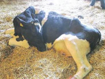
If the condition deteriorates significantly, the cow cannot rise to her feet. The position in which the animal lies is also specific: the legs are tucked under the stomach and the head is turned to the side. The neck is arched in a characteristic S-shaped curvature. The legs and horns are cold to the touch, the eyes are half-closed and watery, the pupils are dilated. In severe cases, the overall body temperature drops to 35 o -36 o.
The animal does not urinate or excrete feces. When the pharynx is paralyzed, the tongue falls out of the mouth and profuse salivation occurs. Some individuals are in an excited state for a short time and can sharply shake their head, throwing it back. They roll over, grind their teeth, hit the walls, and moo loudly. After short-term activity, the cow calms down and goes into a depressed state.
Treatment
Postpartum paresis in a cow should not be left to chance. Treatment started immediately reduces mortality from the disease by up to 4%. For comparison: if no measures are taken in the first hours, 70% of animals die.
Using a special Evers apparatus, air is blown into all four lobes of the udder through the nipples. To do this, they are pre-treated with a 70% ethanol solution. Before the procedure, the cow is milked in a dorsolateral position. Air is introduced using sterile mammary catheters. Injection is carried out until the folds of skin on the udder are straightened. Be sure to massage the udder to distribute air evenly.

To prevent air from escaping, the nipples are tied at the base with a bandage or strips of gauze. The animal is left in this position for 30-45 minutes. If after 8 hours there is no improvement, the procedure is repeated. After the cow gets up, after 1-2 hours she can be milked, without squeezing the air out of the udder.
Instead of air, you can introduce 200-500 ml of fresh milk into each lobe of the udder. It must come from a healthy cow.
To alleviate the animal’s condition, a 20% glucose solution up to 200 ml and a 10% calcium chloride solution up to 150 ml are administered intravenously. A 10% caffeine solution is injected subcutaneously. Active rubbing of the sides and limbs, wrapping the animal, and a hot enema (up to 45°C) are recommended.
Complications
With a rapid and severe course of the disease, the life of the animal may be threatened by tympany; this is one of the complications that causes postpartum paresis in the cow. Treatment boils down to piercing the scar with a thick needle or trocar, after which up to 400 ml of a 5% alcohol solution of ichthyol or up to 40 ml of a 40% formaldehyde solution is injected into the cavity.
Under no circumstances should liquid medicine be administered through the mouth; due to partial paralysis of the pharynx, it may enter the trachea.
The measures taken will completely cure the animal in 2-3 days without any consequences for its health. Perhaps this will never happen again, or perhaps the cow will experience postpartum paresis every time after calving.
Prevention of postpartum paresis
To avoid illness, adhere to the following rules:
- do not allow overfeeding of cows in and during the waning stage of lactation;
- carefully monitor the balance of the diet in micro- and macroelements;
- must be strictly observed percentage roughage and concentrated feed in the diet of animals;
- control the timely start of cows;
- provide the livestock with regular active exercise.
Farms for cows must be equipped with special birthing boxes where the animal is placed before calving. There should be no drafts in the maternity ward.
For highly productive cows that have previously suffered from postpartum paresis, the following are provided: preventive measures: twice, 7-10 days before calving, vitamin D 3 is administered intramuscularly at a dose of 3-4 million units. Sugar is introduced into the diet, 200-300 grams per day for several days before and after calving.
Postpartum paresis (maternity paresis, coma of dairy cows) is an acute, severe nervous disease of animals, accompanied by a paralyzed state of the pharynx, tongue, intestines and limbs with loss of consciousness.
Paresis occurs mainly in cows, goats and rarely in pigs.
Cause the occurrence of paresis is still remains unclear, but at the same time it was established that this disease observed mainly:
- In well-fed cows, whose diet is dominated by concentrated feed.
- In cows with high milk production(the disease in outbred cows is extremely rare).
- At 5-8 years of age those. period of highest milk production.
- In winter - stall period of maintenance.
- In the first three days after calving, rarely happens after a few weeks or months.
With this disease, blood sugar levels drop sharply (hypoglycemia) with simultaneous sharp drop calcium content (hypocalcemia), as a result of dysfunction thyroid, parathyroid and pancreas. Paresis is also associated with overstrain of the nervous system as a result of impulses coming from the baro- and chemoreceptors of the reproductive apparatus and other internal organs involved, one way or another, in the birth act (a good therapeutic effect is obtained from blowing air into the mammary gland).
Signs of illness. Initially, the animal stops chewing cud, frequently steps from limb to limb, trembling and twitching of muscles, and an unsteady gait appears. As the disease progresses, the animal falls and when trying to get up falls again. At mild form course of the disease - the cow is lying down, the neck is curved in an S-shape.
In severe forms of the disease— the cow lies on its side with its limbs extended, its head thrown back on its chest. If the head is forcefully pulled to the side, the animal returns it to its original place (chest). The eyes are half-closed, the cornea is cloudy, the pupils are dilated. The tongue falls out of the half-open mouth, and mucus accumulates in the mouth. Breathing is wheezing and harsh. Belching and chewing gum are absent, atony of the proventriculus develops, and sometimes rumen tympany. Intestinal motility stops, defecation and urination are absent. Body temperature drops to 36-35 degrees. The whole body, especially the horns and limbs, is cold. The animal does not react to needle pricks.
Forecast without treatment - unfavorable, the animal dies within 1-3 days from the onset of the disease.
With timely provision medical care, as a rule, is favorable, usually within 2-3 hours the cow gets to her feet, her body temperature rises and the animal begins to take food. IN in rare cases Relapses of the disease are possible 20-36 hours after treatment.
Treatment. A sick cow urgently needs to be given intravenously 200-400 ml of 10% calcium chloride solution and 200-250 ml of 40% glucose solution and also do subcutaneous injection 20% solution of caffeine - sodium benzoate in a dose of 15-20 ml. If available, it is also advisable to administer intramuscularly 40 ml of a 25% solution of magnesium sulfate and 2,500,000 units of vitamin D2. In most cases, the animal’s recovery occurs after the above assistance is provided. Practicing veterinarians simultaneously with medications pump air into the cow's udder. To do this, the cow is placed in a lateral position, the udder is milked a little, and the tops of the nipples are wiped with a swab moistened with 70% alcohol. Through a sterile milk catheter connected to the Evers apparatus, air is pumped into the udder, starting from the lower nipples. The air must be pumped not very quickly, but in sufficient quantity so that each quarter of the udder is tight (when clicking a finger, there is a sound like when clicking a finger on a tense, “inflated” cheek that is strongly stretched with air). We bandage the nipples for 15-30 minutes and massage gently for several minutes. If the cow does not get up, then after 6-8 hours we repeat the air pumping procedure. It is recommended to milk a cow in 12-24 hours after she gets up.
In all cases of postpartum paresis, the sick animal must warm up, for this (from the rump to the withers) the animal is rubbed on the sides with strands of straw or hay and covered with a warm blanket, under which heating pads or bottles of hot water (50-55 degrees) are placed.
In severe cases of the disease, it is recommended to periodically empty the rectum of feces and remove urine by massaging the bladder through the rectum. When tympany develops, gases are removed using a trocar. During the medical procedures cannot be set medicinal substances through the mouth, since due to paralysis of the pharynx they can enter the trachea.
Prevention. Avoid overfeeding cows in the stage of fading lactation and during the dry period, refuse the same type of highly concentrated feeding. In the diet of a dry cow hay must be at least 8kg., concentrates – no more than 2-3kg. One-time in 5-8 days intramuscular injection before calving vitamin D2 in a dose of 10 million. ED can to a certain extent prevent postpartum paresis. Due to the fact that most owners of private household plots and peasant farms are practically unable to balance their diet with vitamins and microelements, modern industry produces for them a large number of premixes containing macro, microelements and vitamins (). In order to prevent postpartum paresis, most practicing veterinary specialists recommend drinking energy drinks after calving, which contain a vitamin-mineral mixture, glucose, calcium propionate, electrolytes, probiotics and a sweetener. One of these energy drinks is produced by industry - Vitamas Energy. One kilogram of this energy drink is dissolved in 20-40 liters of warm water. Most cows drink this kind of swill themselves; some are given it through a bottle or tube. Propylene glycol is a popular liquid energy drink. It is given to a cow as in pure form, and with water. Dose from 300 to 500ml. Since cows rarely drink propylene glycle themselves, it has to be given to the cow using a bottle (rubber, plastic). One to two weeks before birth and within 7-10 days after birth, it is necessary from the diet remove concentrates and succulent feed, daily exercise, especially during the dry period. In cowsheds and maternity wards eliminate drafts.
In goats and sheep The disease proceeds in the same way as in cows. Treatment of paresis like in cows. In pigs, paresis occurs 2-5 days after birth and is manifested by severe depression. A sick pig is lying down. All reflexes are weakened, breathing is groaning. The mammary gland is very red and full. Body temperature drops to 37-37.5 degrees. The outcome of the disease is favorable.
Treatment: warm wrapping, massage of the breast with simultaneous rubbing camphor oil, sugar enemas and medium salt laxatives.
70 comments to “Postpartum paresis”
please help, the cow has not gotten up for 3 weeks, they called a veterinarian, she gave injections, gave intravenous injections, but nothing helps, her hind legs do not move, we massage her, turn her from one side to the other, she eats well, drinks water, we give crushed grain little by little with crusting, but nothing helps, the vet says we need to cut, but our hand won’t rise, we’re scared to think, what to do?
In recent years, due to gross violations committed by owners in feeding highly productive cows, postpartum diseases of cows have occurred in household plots, and the first thing a cow owner has to face is postpartum paresis. As a result of improper feeding, large calves are born; the owner of the cow often has to resort to the help of several people during calving in order to “pull out” the calf. At the same time, in such a cow, along with the development of paresis, damage to the ligaments and nerves in the pelvic area occurs.
To “put” a cow on its feet, veterinarians often have to inject large doses of a 10% solution intravenously up to three times. calcium chloride with 40% glucose solution, cardiac, etc. From your message it is clear that postpartum paresis has passed in your cow, and there is paresis or paralysis of the pelvic limbs due to childbirth (damage to ligaments, pelvic bones, nerves). Treatment of this pathology is long-term and mostly ineffective in LPH conditions. Therefore, unfortunately, from an economic point of view, you will most likely have to listen to the advice of the veterinarian who conducted the clinical examination and treatment of your cow.
Hello! Thanks for the answer, but we are not going to cut, we will continue to treat, our cow calved at the end of February, our mistake is that we did not kill the calf, he suckled her for a month, she weakened, the calf was removed, the next day we kicked the cow out into the street, she didn’t eat, mooed loudly, was eager to go into the barn to the calf, and the next day she didn’t get up, it seems to us that she suffered a lot of stress, if we had known in advance, we would have fought off the calf earlier, now we we slowly pulled her out into the street, covered her warmly for the night, did a massage, her hind legs barely started to move, please tell me what else can be done, but we won’t cut her, she’s cheerful, she eats everything, we make an infusion of different herbs, the goat doused herself, a liter at a time We give milk in the evening, if only we could give more. He drinks with pleasure, for lunch we give him 2 raw eggs with an infusion of herbs, we continue to give all the bait that the veterinarian said, please tell me, is there anything else you need?
From your new message it is clear that the cow did not have a birth canal injury, but was weakened as a result of improper feeding during the dry period (2 months before calving). Correct advice can only be given after you have conducted a full clinical examination of your cow and made the appropriate diagnosis.
Raw chicken eggs, which have great nutritional and biological value, are usually used for newborn calves instead of colostrum if the cow has mastitis.
Previously, we can recommend that you organize dietary feeding of your cow by introducing vitamin hay or good quality hay into the diet, giving oatmeal, a mixture of concentrates (wheat-barley), if possible, purchasing specialized feed for dairy cows, continuing to give vitamin and mineral supplements, such as "Burenki", BVMD, Minvit, "Vitasol". Carry out a course of vitamin therapy (tetravit, trivitamin intramuscularly 10-15 ml, three times with an interval of 10 days), administer intramuscular vitamin E - selenium in a dose of 10 ml.
Thank you very much, we will do as you wrote.
Hello. My mother-in-law's cow calved for the first time. She gave birth in 4 hours. They helped stretch out the calf a little. The udder was very full, tight, and just huge. They gave in to the last drop. After 3 days, she stopped eating and drinking. She began to pull her head out and wouldn’t lie down. The veterinarian said it was birth paresis. m Amoxicillin, glucose and chloride. Injected for 2 days. There are no improvements. Maybe something else needs to be done?
Intravenous administration of large doses of 40% glucose solution (200-300 ml) and 10% calcium chloride solution (150-200 ml) by a veterinarian is in any case justified.
It is necessary to additionally inject 10 ml of a 20% caffeine solution subcutaneously. For traumatic reticulitis - dietary feeding and symptomatic treatment. In recent years, with postpartum paresis, glucose and calcium chloride have to be administered intravenously repeatedly.
valentine
Hello. My cow calved 4 days ago. On the first day after calving she got up, but on the second she couldn’t get up. A veterinarian came, examined her and said that she had postpartum paresis. He did all the procedures. After 5 hours the cow got up on her feet, but she had no appetite never showed up. The veterinarian said that under no circumstances should she be given water now, but I see that the cow is very thirsty. I don’t even know what to do...
Water is not given to the cow or given from a bottle when pharyngeal paralysis occurs due to paresis. In your case, if the cow has stood up and has normal swallowing reflexes, you can, under your control, place an incomplete bucket of salted water and see how the cow drinks. If the cow accepts water without any difficulty, then water can be given under your control.
Hello, Leonid Stepanovich. Please help me with an answer. 3 weeks have passed since the cow calved. We noticed that she began to arch her back, stretch her hind legs a little, and somehow stepped over. In general, some of the symptoms coincide with “milk paresis.” We called a doctor, she examined her and, it seems, injected only calcium. The cow seemed to feel better. After 4 days, she began to produce less milk than all these days, we thought that everything had returned to normal. And today, after lunch milking, she perked up somehow strangely, then she drank the second bucket of water through her teeth and pulled her neck a little upward. And a couple of hours later, having arrived at the barn, we found her lying with her head wrapped up, her tongue fell out, her eyes were closed, blood was flowing from her nose and from all other holes, her last breath could be heard... There is nothing you can do to help, but the question is why all this blood? We think maybe she broke her neck when she fell? The doctor will arrive only tomorrow and will take a brain test; there will be no autopsy. Maybe you have any ideas why this happened?
The cause of the cow's death was most likely postpartum paresis, the treatment of which should be comprehensive ( intravenous administration large doses of calcium chloride, 40% glucose solution, subcutaneous injection of 20% caffeine solution. Moreover, in recent years this has had to be done more than once). A brain test won't tell you anything. There is simply a standard method - pathological material (spleen, pieces of lung, liver, kidney) is taken from each dead animal to the veterinary laboratory. to rule out an infectious disease. Insurance companies are also trumpeting mandatory research material in the laboratory, the conclusion of which is indicated in the case report, which must be written by a veterinary specialist, must be examined by a veterinary laboratory. A more accurate answer to the question about the cause of death is possible only if the animal is present at the autopsy and the treatment carried out by the veterinarian is correct.
Hello. Yesterday the goat lambed, after a while we tried to feed her, but she refused the food. He doesn’t get up, we tried to lift him up, but he gets up with difficulty and screams. Everything points to postpartum paresis (in goats it probably occurs like cows). There is no veterinarian in our village, we give injections ourselves, we even learned how to administer them into a vein. But we can’t pump air into the udder. Inject glucose and calcium chloride + cardamine. My question is: how many times a day can these drugs be administered and whether we have a chance for a favorable prognosis.
These medications can be administered to a sick goat up to twice a day. There are always chances for a favorable prognosis with complex treatment.
Hello dear Leonid Stepanovich. Our cow calved three weeks ago, but 4 days ago the cow fell on her feet. We called a veterinarian and he diagnosed postpartum paresis. They started giving an intravenous injection - catazal 10 cc, 200 grams of calcium chloride 10%, 200 grams of glucose 40% and caffeine sodium benzoate 20% 20 mg. We give injections for 4 days, once a day, but the cow does not get up on her feet. Tell me what to do?
In recent years, postpartum paresis in cows has been severe - large doses of calcium chloride and glucose have to be administered intravenously, sometimes up to 4 times. Try increasing the dose of calcium chloride. Call your veterinarian and have him conduct a thorough heart examination. If all else fails, then you probably need to think about taking the cow to a meat processing plant for forced slaughter.
Hello! The cow calved well, but the calf does not get up, its legs and head are stretched out, as if it has paralysis; its muscles are hardening. Then he tries to get up: he kicks all his legs, waves his tail, raises his head. rapid breathing. He sucks milk from the nipple himself, but gets tired quickly. When the cow licks him, he shudders, as if his spine hurts. When he was born, he didn’t knock, he started breathing right away on his own. We tried to lift it, but it was no use. What to do?
According to your description, the calf has white muscle disease. Look at the article on our website: "". The most effective drug is a 0.1-0.5% solution of sodium selenite administered subcutaneously or intramuscularly. Nowadays, drugs are being produced that can be used for the treatment and prevention of white muscle disease - E-selenium, Selemag, Tocoselen and others. Can accurately diagnose a calf veterinarian based on the clinical examination performed, as well as on the basis therapeutic effect from the use of these medications.
Svetlana
Good afternoon, Leonid Stepanovich.
Our cow suffered paresis after calving, a month later. Caffeine injections, calcium chloride, borgluconate, glucose, gemodets were infused, 2 times a day, all this for about 10 days. The milk did not disappear at all, chewing gum appeared with difficulty on the 5th day. The tips of the ears were cold and fell off. It was very difficult to get out of this state. Then the forelimb began to swell a little, they began to inject penicillin and streptomycin for 10 days. Then a tumor appeared on the ankle, it abscessed and festered; it started about a month ago. Now below the wrist the leg is cold, on the wrist itself the skin is dry and hard, it’s as if cut wound, there is still pus coming out, but not much anymore. There is ichor near the hoof. And today we also noticed that from the end of the tail, 5 cm higher, the tail is very hard and hard.
We treat the leg with furatsilin and apply levomekol. We took a course - 10 days of vitamins. It feels like the blood is not reaching the end of the limb. The cow's appetite is good, her milk is increasing. She walks with difficulty and does not step on her sore leg.
Please tell me how else we can help the cow, can we cure her?
As a result of a deep metabolic disorder in the cow's body, which resulted in postpartum paresis, various secondary diseases may appear, especially if the zoohygienic rules of keeping and feeding are violated. It is quite difficult to give an accurate forecast for the full restoration of the health and productivity of your cow. A thorough examination by an experienced veterinarian is necessary; it is possible that the cow has some additional disease.
Svetlana
Thank you. On will come in a few days veterinarian, but she already told us that most likely we will have to surrender the cow. Now we treat it with furatsilin, then apply it with levomekol, and massage the metacarpus. But it feels like we are marking time.
Postpartum paresis in a cow. The veterinarian arrived, administered a solution of calcium chloride and glucose, gave an injection of caffeine, and did everything as it should. Then he pumped air into the udder, it expanded to an unprecedented size, and ordered her to milk it every 2 hours and remove the air from there. The first time it worked, the air came out, but then during milking very little air came out, but meanwhile the milk was milked (compared to the air, there was a lot of milk). But the size of the udder remained the same. What to do? Should the air come out on its own? I couldn't find an answer to this question on the Internet. Maybe we went too far with air pumping or air pumping speed?
Anything is possible, the air should gradually come out of the udder, for this purpose it is necessary to massage the udder towards the nipples during the milking process.
Hello. The cow calved a month ago. because she suffered from cuts in previous calvings, her condition was constantly monitored. Everything was fine at the beginning, after 2 weeks the milk began to decrease, although it was good to eat mixed feed, but hay was worse. Now he practically doesn’t eat hay, chewing gum is rare but is available, the horns are cold, he has difficulty getting up, but his legs may hurt. We don't have a veterinarian. Started giving calcium chloride and
glucose in the drink, at least like this: there is no one to give it intravenously, I injected 10 mg of caffeine subcutaneously. Yesterday she became a little happier, chewing gum appeared, and her horns became warmer. This morning everything is bad again. How many days and times a day should you inject caffeine, and in what dose. the cow has lost a lot of weight now
The dose of caffeine subcutaneously is 15-20 ml per cow. per day. In your case, I would recommend a thorough examination of the cow by a veterinarian, since in addition to the main disease (Postpartum paresis), the cow may have other concomitant diseases (ketosis, etc.). Considering that postpartum paresis occurred before, in the future, in order to prevent it, add additional vitamins to the diet a month before calving. mineral supplements, carry out vitaminization vitamin preparations(tetravit, trivitamin, etc.) in a dose of 10-15 ml intramuscularly 3 times with an interval of 10 days, and also administer vitamin E-selenium intramuscularly in a dose of 10 ml. Additionally, see the article on our website -. If possible, then change your cow in the future, since repeated exposure to postpartum paresis for a cow does not go away without leaving a trace.
Hello. Tell me please. The cow's calving went well on the 4th day. The veterinarians gave her a vaccination. She immediately became weak. Her legs were shaking. The temperature was below normal. There was liquid feces. No urine coming out. They gave her a chloride system with glucose. They say the cut is postpartum. But she gets up. The man could barely contain the reaction to the puncture of the vein.
There's something wrong here. A cow is never vaccinated after calving until the genitals return to normal (14-21 days), i.e. their normal involution will not take place. Then there is the rule is vaccination is carried out only in clinically healthy animals (temperature is normal, respiratory organs, heart, kidneys, digestion, reproductive system etc). As with people, after any vaccination, a cow may occasionally have anaphylactic reaction to the introduction of a foreign protein or antigen. In your case, most likely, the onset of maternity paresis overlapped with vaccination.
Hello! A cow with postpartum paresis has stood up and her hind leg hurts. The next day she cannot get up. We lifted her but her back legs were shaking
It is necessary to re-inject the cow intravenously with a large dose of a 10% solution of calcium chloride with 40% glucose. If this does not help, then repeat the intravenous administration of calcium chloride with glucose the next day.
Hello. After calving on the ninth day, the cow began to feel unsteady, raised her hind legs high, and had cold horns. The veterinarian administered calcium and glucose intravenously. as well as caffeine. Almost immediately the cow began to eat. The horns became warm. On the 11th day everything happened again (they were treated again), on the 13th day the same symptoms. Although productivity does not drop. What could it be? Last year the postpartum cut was on the 19th day, the veterinarian said it was mild.
Postpartum paresis, as a result of unbalanced feeding, lack of easily digestible carbohydrates in the diet (sugar, fodder beets, molasses, etc.), lack minerals(calcium, phosphorus). Repeat intravenous administration of large doses of calcium chloride (200 ml), glucose (400 ml), and introduce premixes for cows into the feeding ration.
Hello. Last year, after calving, the cow suffered postpartum paresis on February 1, 2017. When she calved, they didn’t see her. They didn’t wait yet. In the morning they came, the calf was running, the cow was standing. She didn’t drink or eat well. the placenta did not separate on its own, so in the evening they called a veterinarian and she cleaned the cow by hand. At 8 o’clock in the evening they milked and fed the calf, at 10 o’clock they went to check, she was lying down and did not notice anything suspicious, and the next morning at 7 o’clock they came, she was lying on her side unconscious and wheezing. They urgently called a doctor, gave him glucose and calcium chloride. caffeine was introduced. After 2 hours the cow regained consciousness, and after another two hours she stood up. everything was fine afterwards. It didn’t affect the milk yield, it started milking again on May 6, and now it’s time for calving again, we’re waiting for February 14-15, we started it according to the rules 70 days before calving, we reduced the feed. Hay, water and once a day 1 kg of feed. I’m very worried about how calving will be and whether a repeat of last year is possible
Last year, the cow suffered postpartum paresis a day after calving, and did not free herself from the placenta, only with the help of a veterinarian. . everything was fine throughout the year, she became pregnant on May 6, we are waiting for calving again on February 14-15. Is it possible for the disease to recur?
Hello! In cows, especially highly productive ones, postpartum paresis may recur the following year. Advice - stock up on medications (10% calcium chloride solution, 40% glucose solution, caffeine). Strengthen control over the cow, measure the temperature in the rectum more often (it is observed to decrease below normal - 37.5 and below), and if the first symptoms appear, immediately contact your local veterinarian.
Tell me, a day after the separation, the cow fell, I was injected with Calfoset and Gamavit. Is it still possible to administer E-selenium?
The introduction of E-selenium to a cow can only be welcomed. E-selenium, being a powerful antioxidant, will help remove accumulated toxic substances from the cow’s body, plus it will additionally increase its immunity and overall body resistance.
Good afternoon, the cow fell a day after giving birth. They inject caffeine and calcium, they also give her an IV, she has chewing gum, she has a good appetite and goes to the toilet, she also gives milk, she also turns over on her sides, they picked her up, she stood for 30 minutes while she went to the toilet, and she walked a little and fell, what should I do?
It is necessary to repeat the therapeutic manipulations - the introduction of large doses of 10% calcium chloride solution, 40% glucose solution, caffeine. It is a good idea to additionally use E-selenium during treatment.
Additionally, the use of premixes for cattle in the feeding ration, which are especially effective for the prevention of postpartum paresis in the cow, retention of the placenta and subsequent increase in milk production. One of these premixes is - premix - Energotonic.
To my question about the recurrence of a cow with postpartum paresis dated 02/3/18, an answer was received. 02/16/18 the cow calved. According to your recommendations, it was possible to prevent the complete development of paresis. Signs began to appear on the 3rd day. We carried out treatment. Currently, the cow is recovering, she has an appetite, but she has lost a lot of weight after calving. How long can it be fed and with what feed, in what quantity. Thanks for the advice. Best regards to you
Hello! From the first day after illness, the cow must be given a “chatterbox” of oatmeal, or wheat bran. Over the course of eight to ten days, the fresh cow is gradually transferred to a normal diet. It is advisable to include “” in the feeding diet.
Hello! A day after calving, paresis occurred. On the first day, the veterinarian injected intramuscular vitamins and intravenous glucose boroglyukol.caffeine, then she got up, ate and lay down and did not get up. Today I did everything again + pumped up the udder, but the cow did not get up. She ate some hay and is now responding to acupuncture. Tell me what she has and what else to do? Thank you!
Hello! The cow most likely has a severe form of postpartum cut. It is necessary to administer intravenously a large dose of 10% calcium chloride (300 ml.) in 200 ml. 40% glucose solution, 20 ml subcutaneously. 20% caffeine solution. In the future (the next day), it would be a good idea to administer 10 ml intramuscularly. E-selena. Include Energotonic in the cow's diet.
Hello, the goat suffered paresis on the tenth day after lambing. In the morning everything was as usual, she ate and milked well, we didn’t notice anything. And at lunchtime she was breathing heavily, her legs and muscles were trembling. When picked up, she falls, her legs give way, and after another two hours it was impossible to open her mouth, her jaws were clenched tightly. By the evening it was clear that the goat would not live to see the morning. Based on the symptoms, we identified it as paresis. It’s impossible to call Veta; no one is going to see the goat. Since the goat was not very productive, we decided to at least slaughter it to avoid losing meat. The question is, can this meat be eaten or fed to a dog? It seems that this is not an infectious disease, but a neuroparalytic one. We can't find the answer anywhere on the internet. Thank you.
With postpartum paresis, the animal's body temperature drops below normal. According to the rules of animal slaughter, animals with a temperature below normal are not subject to slaughter, and sick animals are also not subject to slaughter. In your case, meat from a killed goat can be fed to a dog, preferably pre-cooked.
Hello. After suffering postpartum paresis, the cow suffered from hind paralysis. right leg. She gets up, walks unsteadily, her left leg is weak, it’s clear that she’s paralyzed. They gave her 250 ml of butofen intravenously. Ringer's solution 500 ml. Will the leg come off or can we say goodbye to the cow? He has gained an appetite, chewing gum, and drinks water well too. When I milk, he stands still.
Hello! The paresis of a limb is a long-term process and should go away over time. To speed up treatment, as well as normalize metabolism in the cow’s body, I recommend introducing it into the feeding diet. Inject vitamin E-selenium intramuscularly into the cow in a dose of 10 ml.
Leonid Stepanovich, thank you very much for your help and advice. Thanks to your recommendations, the cow begins to recover, and I have hope that everything will be fine. With respect to you.
Leonid Stepanovich, Hello. The cow suffered birth paresis for the second time. He is recovering. What recommendations can YOU give before the next calving to prevent the development of paresis 3? I’m already afraid that it might happen again. Thank you.
To prevent postpartum paresis in a cow, the efforts of animal owners should be aimed at preventing factors that contribute to the development of postpartum paresis, namely:
— Strive to balance the feeding diet during pregnancy, especially during the dry period (45-60 days before calving) nutrients, especially the sugar-protein ratio (should be 1:1).
— Due to the great difficulties for owners of private household plots and peasant farms to provide competent, balanced feeding of their cows with a large number of components, I recommend introducing them into the cow’s feeding diet.
— In order to prevent postpartum paresis due to the inability to balance the diet according to the sugar-protein ratio, most owners of private farms and peasant farms introduce 500 grams of food into the diet daily 7-14 days before the expected calving. granulated sugar dissolved in water.
— Three times before calving (30, 20 and 10 days) intramuscularly administer vitamins (trivitamin, tetravit) in a dose of 10 ml and twice after calving on days 10 and 20 the same vitamins in the same dose.
— 1-2 weeks before calving, especially for well-fed cows, exclude concentrated feed from the diet, replacing them with good quality hay ad libitum.
— Owners of private household plots and peasant farms must strictly observe the existing zoohygienic rules for keeping cows (warm, dry room, no drafts, avoid hypothermia). Very good action Walking has an impact on a cow, especially in winter.
Hello. Leonid Stepanovich, the cow calved from March 8 to 9, at 1 am I checked there was nothing, at 7 am I came in with a calf, good healthy, I immediately gave the cow water with sugar, on March 11 she fell ill, still hasn’t gotten up, the doctor makes vitamins, gave boluses, he says it’s his first time, he can’t get up for so long, we don’t know what to do anymore, please help with advice!!!
The cow most likely has a severe metabolic disorder caused by inadequate feeding during the dry period. Only a veterinary specialist can make an accurate diagnosis after an in-depth clinical examination of the animal and a biochemical blood test in a veterinary laboratory. I can advise the veterinary specialist treating your cow to first rule out osteomalacia in the cow (), see our article “”), introduce good quality hay and root vegetables into the feeding ration, preferring oatmeal from concentrates, and be sure to include minerals in the feeding ration fertilizing, or better yet premixes (). Treatment for a cow can be very lengthy.
The cow fell ill with postpartum paresis. The veterinarian put on a drip with glucose and calcium. I injected caffeine, as well as Catozal. Pumped up the udder. The cow got up an hour later and started eating and drinking. Now two days have passed, and one leg is paralyzed. Maybe I still need to inject something - the veterinarian said it will go away, but I’m afraid it won’t. Tell me, please, I will be very grateful.
The veterinary specialist is right, paresis-like condition of the limbs sometimes occurs in cows after postpartum paresis. The process is long and must be completed over time. As a piece of advice, I can recommend administering 10 ml of E-selenium intramuscularly to the cow; if the paresis is prolonged, then repeat the administration of E-selenium after 10 days. Be sure to include it in your diet.
