Extract from the gynecological department curettage. Diagnostic curettage of the cervical canal and uterine cavity: purpose of the procedure, preparation and rehabilitation
One of the most common gynecological procedures is curettage of the uterine cavity (cleaning). Another name for the procedure is uterine curettage - a derivative of the surgical instrument curette, which directly performs curettage.
The concepts of “RDV”, “LDV”, “scraping”
In medicine, to refer to the operation of curettage of the uterine cavity, the terms RDV (separate diagnostic curettage) and LDV (therapeutic and diagnostic curettage) are used, depending on the purpose. The upper layer of the endometrium lining the uterine cavity is scraped. If necessary, the resulting tissue is used for further examination to determine the presence or absence of pathology.
Anatomy of the uterus
Uterus - organ reproductive system the female body in which the fetus is born and develops. Located in the pelvic cavity between bladder and rectum. For this reason, the vesical (anterior) and intestinal (posterior) surfaces of the uterus are distinguished.
The uterus is conventionally divided into three components:
- The fundus is located in the upper part above the line of junction of the fallopian tubes.
- Body – located in the middle part and is the largest part of the organ.
- The neck is located in the lower part.
In turn, the cervix has two parts. The lower part of the cervix protrudes into the vaginal cavity and is called the vaginal cavity. The upper part is located above the vaginal cavity and is called supravaginal. The cervix has a canal inside, the upper opening (throat) opens into the uterine cavity, and the lower opening into the vagina.
In sexually mature nulliparous women the volume of the uterus does not exceed 6 cm3, and the weight is 40-60 g. The walls of the uterus have exceptional elasticity, which determines the ability of this organ to increase in size throughout the entire period of pregnancy. This occurs due to the growth and hypertrophy of muscle tissue cells.
The walls of the uterus have a complex structure:
- The serous membrane, or perimeter, is a continuation of the serous covering of the bladder. Over a larger surface area of the uterus, it is tightly connected to the muscular layer;
- The mucous membrane, or endometrium, is the inner layer of the walls of the uterus. It is represented by a layer of columnar epithelium, in the jejunum of which there are simple tubular glands. The endometrium consists of 2 layers: superficial (functional) and deep (basal).
- the muscular layer, or myometrium, is a dense layer uterine wall, located between the serous and mucous membranes. The myometrium consists of three layers of smooth muscle:
- subserosal, or outer, layer - longitudinally located muscle fibers, tightly connected to the serous layer;
- vascular, or middle circular, is the most developed layer, most strongly represented in the cervix area. A large number of vessels are concentrated in this layer;
- submucosal, or internal longitudinal, is a thin layer with longitudinally located muscle fibers.
Possessing a developed muscular structure, the uterus is directly involved in the expulsion of the fetus during childbirth. After childbirth, over time, uterine cells come into normal condition, the uterus itself decreases in size, only a slight change in weight is observed up to 80 g, which is also associated with hypertrophy of muscle tissue cells.
When is curettage performed?
Depending on the purpose of the procedure, the time for its implementation is selected. The first days of the cycle are the optimal time for RDV. During this period, changes in the uterine cavity are most clearly manifested. Last days cycle are the best time to study the functions of the uterine mucosa.
The operation is not performed during menstruation.
Diagnostic purpose
Diagnostic curettage carried out to confirm the results of a preliminary examination or to establish accurate diagnosis if there is a suspicion of an inflammatory process of the uterine mucosa (endometritis), pathological growth of the uterine mucosa (endometriosis), the presence of a benign tumor (fibroids) or malignant neoplasms; identifying the causes of irregular or heavy menstruation, untimely bleeding; diagnosis of infertility.
Therapeutic purpose
The therapeutic purpose is reduced to direct surgical intervention to dissect the intrauterine septa and adhesions, extract polyps, remnants of embryonic tissue and amniotic membranes, and select neoplasm cells for histological examination.
Abortion
The manipulation of curettage of the uterine cavity is a method of terminating pregnancy. It is practiced when terminating a pregnancy up to 16 weeks. This method is considered the most traumatic, often with unpredictable consequences, but is still used in medical practice.
Frozen pregnancy
When a woman is diagnosed with a frozen pregnancy, immediate medical intervention is necessary, since in fact this diagnosis indicates the death of the fetus. Decomposition products, entering the mother's blood, can lead to adverse consequences for the body, including death. Therefore, the primary task for doctors is to remove the embryo and amniotic membranes from the uterine cavity. For these purposes, vacuum aspiration and curettage of the uterine cavity are used.
Preparing for surgery
Before surgery you need to take tests:
- general blood analysis;
- blood chemistry;
- determination of blood group and Rh factor;
- coagulogram;
- general urine analysis;
- smear on the flora of the vaginal mucosa;
- tests for HIV, syphilis, hepatitis.
Except laboratory tests, the woman is prescribed an ECG and ultrasound examination of the pelvic organs.
Before the operation, you need to avoid eating, take a shower, shave your hair, and give a cleansing enema.
Procedure technique

The operation is performed in stages and, given the painfulness of the procedure, under general anesthesia:
- using special expanders, the diameter of the cervical canal is gradually increased so that the curette can pass into it;
- curettage is performed cervical canal, and then - the uterine cavity;
- the resulting scrapings are sent for histological analysis to the laboratory.
Curettage for endometrial hyperplasia
The diagnosis of “endometrial hyperplasia” is made when the inner layer of the uterus grows to 15 mm or more. Ultrasound examination can reveal a disease, but its nature can only be determined by directly studying the cells of the mucosa. The solution to the problem is to reduce the functional layer of the mucosa by scraping the endometrium with a curette. This helps stop the bleeding, but does not solve the problem. Hormonal drugs are used to treat hyperplasia. Surgery may be indicated.
Hysteroscopy and RDV
Currently, RDV is performed in combination with hysteroscopy.
Hysteroscopy of the uterus is a visual method for diagnosing the internal cavity of an organ using an optical device - a hysteroscope. The capabilities of a hysteroscope allow the doctor to visually determine the condition of the uterine cavity, perform certain manipulations with greater accuracy during curettage surgery, and evaluate the result of the operation.
Histological examination
To make a diagnosis of RDV, cells are taken from the cervical canal, endometrium, and neoplasms located in the uterine cavity. Histological examination of the uterine mucosa is the most effective way determining the causes of infertility and missed abortion. Some diseases are asymptomatic and can only be diagnosed using histology.
Contraindications for surgery
Like any other medical operation, curettage has contraindications:
- acute infectious and inflammatory diseases of the genital organs;
- acute diseases of the urinary system;
- organ diseases gastrointestinal tract in the acute stage;
- suspicion of a violation of the integrity of the uterine wall.
In emergency cases, contraindications can be neglected (for example, in case of severe postpartum bleeding).
How to quickly restore the uterine mucosa?
Restoration of the uterine mucosa after RDV occurs quickly and without any complications, provided that certain recommendations are followed:
- Accept medications as prescribed by a doctor.
- If possible, limit physical activity, temporarily avoid going to the gym, and do not lift heavy objects during the rehabilitation period.
- Avoid using tampons during menstruation during the recovery period, as it is possible that the natural microflora of the genital organs may be disrupted.
- Pay special attention intimate hygiene– use neutral detergents that do not disturb the acidic environment of the vagina.
- In the first 10-14 days after RDV, it is necessary to abstain from sexual intercourse.
- Taking baths or going to the sauna is strictly prohibited - the likelihood of uterine bleeding increases.
After performing RDV during the recovery period, you should listen to your body and if unusual sensations occur, immediately consult a doctor.
Is discharge after surgery normal or pathological?
In the first few hours after the procedure bloody issues are considered the norm. During the first 10 days after curettage, brown or brown spotting indicates the normal course of the uterine restoration process. If the discharge stops or is absent and if pain occurs, you should consult a doctor.
The cause of pain is uterine spasms and blood stagnation.
The healing process does not always go smoothly, and in some cases, changes in the color and odor of the discharge may indicate serious problems. A yellowish color and a strong unpleasant odor indicate an admixture of pus, i.e. about inflammation, and you can’t do without antibiotics.
How long should I stay in the hospital after curettage?
If there are no obvious complications after diagnostic curettage, the patient can be sent home on the same day. After curettage surgery for a frozen pregnancy, termination of pregnancy, removal of tumors, as well as in the presence of complications, the length of stay in the hospital can be 5-7 days.
Exercising after curettage
Small physical exercise to maintain body tone can be done the very next day after surgery, but you can start playing sports with the same loads no earlier than 10-12 days later, provided there are no complications.
When does your period start after curettage?
If the operation is carried out correctly, the first menstruation should begin at the scheduled time, but a slight delay cannot be ruled out.
Ovarian cyst after curettage
The appearance of an ovarian cyst after curettage during a missed pregnancy or abortion is a kind of hormonal reaction of the body. In most cases, cysts disappear on their own after the cycle normalizes and hormonal levels are restored.
Complications after RDV and their treatment
Curettage of the uterus, like any other surgery, may be accompanied by a number of complications:
- Uterine bleeding- continuous heavy blood loss. To stop bleeding and further determine the causes, drugs that contract the muscles of the uterus, as well as hemostatic agents, are used. Oxytocin and Pituitrin, Desaminooxytocin are most often used.
- Endometritis– inflammatory process of the uterine mucosa. The cause of infection is poor quality sterilization of instruments used in RDV; genital tract infections; non-compliance with the recommendations of the gynecologist during the rehabilitation period. Signs include pain and fever. Antibiotics are used for treatment.
- Perforation of the uterine walls– damage to an organ by medical instruments during surgery. The consequence of this can be massive bleeding. Antibiotics and drugs that contract the uterus are used for treatment. Sometimes surgery is necessary to stitch the resulting wound.
- Asherman's syndrome– the occurrence of adhesions in the uterine cavity due to poorly performed curettage procedures with the subsequent development of bacterial diseases. The consequences are violations monthly cycle and decreased reproductive capacity. Treatment consists of surgical removal adhesions.
- Hematometer– accumulation of blood inside the uterus due to impaired outflow (blood clots clog the cervical canal). The likelihood of developing infectious diseases increases. The condition is accompanied by dizziness, nausea, high temperature. The problem is solved by simple probing of the uterine cavity.
After surgery to curettage the uterine cavity, symptoms of cystitis may appear. The reason for this may be infectious lesion urinary tract during surgery or the reaction of blood vessels to surgery. Diagnosis and treatment should be carried out under the supervision of a specialist.
Pregnancy after surgery
It is possible to get pregnant after RDV within a month, but it is worth considering that curettage depletes the mucous layer of the uterus and injures the walls of the organ, which can interfere with pregnancy. When planning a pregnancy after RDV, you should consult your attending gynecologist.
It is important to know
To diagnose and treat many diseases, surgery to curettage the uterine cavity is simply necessary. In any case, this procedure is a surgical intervention and is associated with certain risks, especially when terminating a pregnancy, and the results of the operation depend entirely on the professionalism of the doctor.
After examination by a gynecologist, the patient may be referred for curettage of the uterine cavity, but what is this? Among themselves, women call this process cleansing. Curettage is a surgical procedure to clean the uterus and cervical canal.
The uterus is the middle section of the reproductive system, a pear-shaped organ that contains a small cavity connected to the cervix. In this way the body controls external environment vagina. Main function uterus - bearing offspring.
The inside of the organ is covered with a special layer called the endometrium. During menstruation, it is rejected if pregnancy does not occur. Cleaning the uterus involves partial removal uterine mucosa, almost the same as during menstruation, only with the use of special tools. After the procedure, the endometrium grows again.
What is cervical canal curettage and why is it needed? Cleaning is carried out precisely from curettage of the cervical canal to the functional layer. A small part of the material is sent for a separate study. This procedure is necessary for severe uterine bleeding, to remove cervical polyps, or if any abnormalities are suspected.
Gynecological cleansing can be carried out in two ways:
- Separate diagnostic curettage (RDC). During this procedure, the surface is first removed from the cervical canal, and only then the endometrium is dealt with. After surgical intervention the patient is considered healthy, since the doctor removes all formations for which curettage was prescribed.
- Separate diagnostic cleaning under hysteroscopy control is one of the new techniques in gynecology. Conventional RDV is carried out without observing that instruments are removed, and RDV+GS is carried out with the help of direct control of the operation. First, an endoscopic device is inserted into the uterus, then the uterus is scraped out and then checked. This technology allows you to check the degree of purification, as well as find out whether any anomalies remain.
Indications
Gynecological cleansing is needed to obtain biological material, its research, as well as for the removal of pathological processes that have formed in the cervical canal or endometrium.
Curettage is carried out for the most part precisely to determine the diagnosis. Ultrasound diagnostics do not always accurately differentiate or help establish the name of the disease. Sometimes ultrasound is performed twice: before and after menstruation. All kinds of formations are rejected along with the endometrium; if this does not happen, then this indicates pathological processes. In this case, the woman is sent for cleaning of the cervical canal and cervix.
Curettage of the uterine cavity is carried out in case of heavy menstrual bleeding, a woman’s inability to become pregnant for six months, and also for an unclear reason feeling unwell. Other causes include abnormalities in the cervix and routine cleaning due to fibroids, while the uterus is preserved.
First of all, the indications for curettage are benign formations on the mucous membranes, in other words - polyps. Their peculiarity is the lack of response to treatment with pharmacological drugs. Polypoid growths can only be removed through surgery.
In addition, the procedure is prescribed for endometrial hyperplasia - this is an abnormal condition of the uterine mucosa, in which the structure and shape of the endometrium changes by enlarging cells.
Women during menopause are characterized by uterine hemorrhages and cleaning in such cases is carried out only to stop the bleeding. Cleaning also takes place after a miscarriage or abortion occurs.
Preparation
Preparation for curettage of the uterine cavity is an important and responsible task, which is carried out as intended (planned) or in emergency cases. If the procedure was planned, then it begins to be done at the same time when the endometrium itself should have begun to be rejected. In case of removal of polypoid growths, cleaning is carried out after menstruation, so that the exact location of the formation is known.
Gynecological curettage is rarely performed in the middle of the menstrual cycle, as this leads to prolonged bleeding. This process is explained by the fact that the growth of the endometrium and follicles are connected. And as a result, when the uterine mucosa is removed prematurely, the functioning of the ovaries and hormonal levels.
Preparation for cleaning the uterus includes the following tests:
- complex analysis of blood clotting indicators;
- general blood analysis;
- electrocardiogram;
- HIV test;
- analysis for hepatitis B and C;
- smear on flora.
At the appointed time before the procedure, you cannot eat and you need to remove pubic hair. Bring a robe, pads, and slippers with you.
Procedure
Patients are interested in how uterine curettage is done. In the office there is a chair similar to a gynecological one, where gynecological cleaning will take place. Before the procedure, you need to take a survey to find out if there is any allergy to the active ingredients of the drugs. Anesthesia is administered intravenously, and the duration of the entire operation is a maximum of 30 minutes. After curettage, the patient is transported to the ward, where she wakes up.
As instruments for the operation, the doctor uses a speculum, Schroeder forceps, an iron stick, dilators, and a curette. When separate diagnostic cleaning is used under hysteroscopy control, a tube with a camera is inserted after the uterus is dilated.
How is the uterus cleansed? The doctor treats the external genitalia with an antiseptic, then inserts a speculum to open access to the cervix. The next step is to open the cervix using dilators to gain access to the endometrium.
Using a curette, the doctor performs curettage, while he carefully inserts instruments along the inner layer of the uterus, removing it. When curettage using an endoscope, the doctor sees the uterus on a computer monitor, which allows you to remove only the affected areas of the endometrium without affecting healthy ones.
Many patients are interested in how long they need to stay in the hospital after curettage. Curettage is a serious intervention that can entail many severe consequences. Therefore, gynecologists recommend spending at least a week in a hospital setting.
At this time, the woman is prescribed to take antibiotics and vitamins. The doctor monitors the woman’s condition throughout the recovery period. Before discharge, it is mandatory to carry out ultrasonography.
Consequences
Currently, gynecological curettage is considered not the most the best method therapy, especially if it is carried out blindly, without the use of endoscopic equipment. The fact is that any careless movement by the doctor can lead to the formation of scars or fistulas. In addition, such a rough impact on the internal cavity of the uterus increases the risk of developing serious complications:
- Trauma to the cervix, its tear.
- Perforation of the uterus.
- Infection of the uterus and cervix with subsequent inflammation.
- Damage to the growth layer of the endometrium, which subsequently disrupts the process of growth of the uterine mucosa and causes infertility.
- The risk of bleeding increases.
Gynecological curettage will most likely not cause harm if the woman prepares for it correctly and does not refuse hospitalization after surgery. It is especially important to follow the advice of a doctor for women who have undergone a cleanse to terminate a pregnancy. The fact is that the pregnant body becomes many times more vulnerable to infection and the occurrence of inflammatory diseases.
Diagnostic uterine curettage- a form of biopsy during which the doctor takes samples of the mucous membrane from the uterine cavity for cytological examination.
Curettage is considered a minor gynecological operation and is widespread in the practice of gynecologists. It allows you to accurately diagnose and carry out effective treatment for many diseases of the female reproductive system.
The procedure is carried out under intravenous anesthesia, so the woman does not experience pain at the time of curettage. The operation is not considered highly traumatic; in fact, curettage is mechanical removal that part of the mucous membrane that itself is rejected during menstruation. After curettage, a germ layer of the endometrium remains, from which a new mucous membrane grows after 2-3 weeks.
Synonyms. You may come across different names for this procedure: endometrial biopsy, diagnostic cleaning of the uterine cavity.
Types of uterine curettage
- diagnostic curettage of the uterus- an operation that is performed to diagnose the condition of the endometrium. The inner layer of cells lining the uterine cavity is removed, followed by an examination of their structure;
- separate diagnostic curettage– removal of the inner layer of the cervical canal and the uterine cavity. At the first stage, the top layer of the mucous membrane of the cervical canal is removed, and at the next stage, the top layer of the mucosa lining the uterine cavity.
Purposes of curettage
- diagnostic– allow you to take material to study the characteristics of cells. The main task is to confirm or refute the presence of cancer cells in the thickness of the endometrium;
- medicinal-diagnostic– when curettage of the endometrium, polyps, pathological foci and growths of the endometrium are removed, which became the reason for prescribing curettage. Subsequently, the resulting material is sent for research.
Anatomy of the uterus
 The uterus is a hollow muscular organ located in the pelvic cavity between the bladder and intestines.
 The uterus performs two main functions:
- reproductive– a fertilized egg is attached here, from which the fetus subsequently develops;
- menstrual– if fertilization does not occur, the inner lining of the uterus peels off at the end of the cycle, which is manifested by menstrual bleeding.
- Bottom– the upper part lying above the entry point fallopian tubes, through which the egg enters the uterus;
- Body– the lateral walls of the uterus, which taper towards the cervix. In the body of the uterus is cavity, in which fetal development occurs during pregnancy. Due to the significant thickness of the walls, the size of the cavity does not exceed several cubic centimeters;
- Neck– the lower part of the uterus, which is a 2-3 cm long tube connecting the uterine cavity to the vagina. The cervical canal, or cervical canal, runs inside the cervix.
- Outer– perimeter is the peritoneum, the connective membrane covering the outside of the uterus.
- Average– myometrium – muscle layer. It is represented by non-striated smooth muscle fibers that intertwine in various directions to form a dense muscle wall.
- Interior– endometrium is a mucous membrane richly supplied with blood vessels. In the body of the uterus, it is smooth and represented by ciliated epithelium. In the cervical canal, the mucous membrane is folded and lined with columnar epithelium.
Endometrium or mucous layer - the inner mucous membrane of the uterine cavity. It has a smooth surface and contains uterine glands which open into the uterine cavity. The endometrium is a hormonally sensitive tissue and therefore undergoes changes depending on the phase of the menstrual cycle. So after menstruation its thickness is 2 mm, and in the second half of the cycle it can exceed 2 cm.
In the endometrium there are:
- Functional layer– the outer layer of the endometrium, which lines the uterine cavity and is shed with each menstrual cycle. Its thickness and structure largely depend on the phase of the cycle and the hormonal state of the woman, which must be taken into account when analyzing the results of curettage. Ciliated cells with numerous cilia make up the majority of epithelial cells. Their function is to promote the fertilized egg to the site of attachment.
- Basal layer the lower layer of the endometrium adjacent to the muscular layer. Its function is to restore the mucous membrane after menstruation, childbirth, and curettage. Contains vesicle cells, from which ciliated cells of the functional layer are subsequently formed. The bases of the glands and blood capillaries are also located here. Weakly responds to cyclical fluctuations of hormones.
- Stroma– the basis of the endometrium, which is a mesh of cells connective tissue. It is dense and rich in connective fibers. In the basal layer lie the uterine glands. Meet light cells– immature ciliated epithelial cells. True lymphatic follicles– accumulations of lymphocytes without signs of inflammation.
- Uterine glands simple tubular glands that secrete a mucous secretion that provides normal functioning uterus. They have a convoluted but not branched structure. The glands are lined in a single row with columnar epithelium. They are subject to changes under the influence of hormones.
Indications for separate diagnostic curettage
Diagnostic uterine curettage is indicated for the following conditions:
- menstrual irregularities;
- intermenstrual (acyclic) bleeding;
- spotting after menopause (menopause);
- suspicion of endometrial tuberculosis;
- suspected endometrial cancer;
- Ultrasound of the uterus during 2 cycles revealed changes that require clarification;
- suspicious changes on the cervix;
- after spontaneous abortions;
- to establish the causes of infertility;
- preparation for planned gynecological surgery for fibroids.
- inflammatory processes in the uterus or other genital organs;
- general infectious diseases;
- suspicion of pregnancy.
Methodology for separate diagnostic curettage of the uterus
 Timing for curettage
- 2-3 days before menstruation– in most cases with infertility, with suspected malignancy. The procedure is carried out within these periods so that the removal of the mucous membrane approximately coincides with the physiological process of its rejection.
- On the 7-10th day after the start menses and with menorrhagia - prolonged heavy menstrual bleeding;
- Immediately after bleeding starts with acyclic bleeding in the middle of the cycle;
- Between the 17th and 24th day of the cycle– to assess the response of the endometrium to hormones;
- Immediately after the end of menstruation– for uterine polyps. In this case, the polyp is clearly visible against the background of the thin endometrium.
Not recommended carry out the procedure in the middle of the cycle, since the hormones secreted by the ovaries will interfere with the growth of the mucous membrane, which will lead to prolonged bleeding.
Pain relief during uterine curettage
- Intravenous anesthesia– short-term general anesthesia – the patient is injected with sodium thiopental or propofol. She falls asleep for 20-30 minutes. Pain sensations are completely absent;
- Local paracervical anesthesia– variety local anesthesia. The tissue around the uterus and cervix is soaked in an anesthetic. Painful sensations are significantly dulled, but do not disappear.
Where and how is uterine curettage performed?
 The procedure for separate diagnostic curettage of the uterus is carried out in a small operating room on a table equipped with the same leg holders as the gynecological chair. The whole process takes no more than 20 minutes.
 The gynecologist sequentially performs several stages.
- Two-handed examination of the uterus to determine its size and position.
- Treatment of the external genitalia with alcohol and iodine solution.
- Dilatation of the vagina using gynecological speculum.
- Fixation of the cervix using bullet forceps.
- Studying the depth and direction of the uterine cavity using a probe - a metal rod with a rounded end.
- Expansion of the cervical canal using Hegar dilators - metal cylinders of small diameter. The width of the channel must correspond to the size of the curette (surgical spoon).
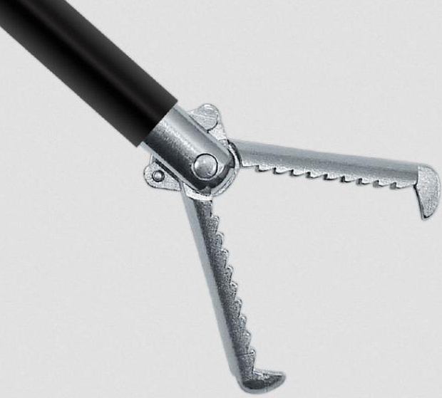
- Curettage of the mucous membrane of the cervical canal. A curette (a metal spoon with a long handle) is carefully inserted to a depth of 2 cm until internal pharynx. The curette is pressed against the wall of the cervical canal and brought out with an energetic movement. In this case, the curette scrapes out the epithelium. The action is repeated until all the mucous membrane from the walls of the cervical canal has been collected.
- Collecting material from the cervical canal into a container filled with a 10% formaldehyde solution.
- Curettage of the mucous membrane of the uterine cavity. Using a curette largest size scrape out the mucous membrane, vigorously pressing on the wall of the uterus. Start from the front wall, then move to the back and side walls. The gynecologist successively uses smaller and smaller curettes until the uterine wall feels smooth.
- Collect material from the uterine cavity into a container with formaldehyde solution.
- Treatment of the cervix and vagina with an antiseptic solution.
- Stop bleeding. Ice is placed on the abdomen for 30 minutes to stop bleeding.
- Postoperative rest. The woman is transferred to a ward, where she rests for several hours. The first 6 hours check the pressure, the nature of vaginal discharge on the pad, and the possibility of emptying the bladder.
- Extract. At the day hospital, discharge is carried out on the same day. The hospital discharges the woman the next day.
Modern version of the procedure – separate diagnostic curettage under hysteroscopy control(RDV+GS). If ordinary curettage is performed “by touch,” then in this case a hysteroscope is inserted into the uterine cavity - a miniature device that allows you to see everything that happens in the uterine cavity. This makes it possible to reduce trauma and check whether there are any areas of mucous membrane or formations that have not been removed.
In the laboratory, the resulting material is treated with paraffin and thin sections are made from it, which are then examined under a microscope.
How to prepare for the procedure?
 Curettage of the uterus is considered a minor gynecological operation and therefore requires preliminary preparation. The examination allows you to identify diseases that can cause complications after performing diagnostic cleaning. At the preliminary consultation, it is necessary to inform the doctor about the medications you are taking, especially those that affect the blood clotting process (aspirin, heparin).
 Required research:
- gynecological examination;
- Ultrasound of the uterus and pelvic organs.
- clinical blood test ;
- blood test for coagulation - coagulogram;
- blood test for HIV;
- blood test for syphilis - RW;
- blood test for hepatitis B and C;
- bacteriological examination of the contents of the genital tract;
In the evening before surgery, it is advisable to do a cleansing enema. This will avoid post-operative flatulence - painful bloating due to the accumulation of gases.
Before the procedure, you must take a shower and remove hair around the genitals.
What are the possible histology results?
 After examining the samples in the laboratory, a written conclusion is made. You will have to wait 10-20 days. You can find out the results from the doctor who performed the curettage or from your local gynecologist.
 The conclusion contains two parts:
- Macro description– description of tissues and discovered fragments. The color of the fabric, its consistency, and the weight of the sample are indicated. Presence of blood, mucus, blood clots, polyps. For example, material from the uterine cavity in large quantities may indicate the growth of the mucous membrane - endometrial hyperplasia.
- Microdescription– description of the detected cells and deviations in their structure. The detection of atypical cells indicates a precancerous condition (the risk of developing a cancerous tumor); the appearance of malignant cells indicates endometrial cancer.
| Phase of the menstrual cycle | Cycle days | Normal results | Pathologies with similar symptoms |
| Endometrium in the proliferation phase | Early stage of the proliferation phase 5-7th day of the cycle | Cuboidal epithelium on the surface of the mucosa. Glands are in the form of straight tubes with a narrow lumen. In cross section they have rounded contours. The glands are lined with low prismatic epithelium with oval nuclei. The nuclei are intensely colored and located at the base of the cells. The stromal cells are spindle-shaped with large nuclei. The spiral arteries are weakly tortuous. | |
| Middle stage of proliferation phase 8-10th day of the cycle | Prismatic epithelium lines the surface of the mucosa. The glands are slightly convoluted. A border of mucus along the edge of some cells. In the nuclei of cells, numerous mitoses (indirect cell division) are detected - the distribution of chromosomes between two daughter cells. The stroma is loosened and swollen. | ||
| Late stage of the proliferation phase 11-14th day of the cycle | Ciliated and secretory cells on the surface of the mucosa. The glands are tortuous, their lumen is widened. Cores in prismatic epithelium on at different levels. Some gland cells contain small vacuoles with glycogen. The vessels are tortuous. The stroma is juicy and loose. The cells enlarge and are stained less intensely than at the early stage. | a) Anovulatory cycle - a menstrual cycle during which there was no ovulation and no phase of development of the corpus luteum. The anovulatory cycle is evidenced by these cytological results that persisted during the second half of the menstrual cycle. b) Dysfunctional uterine bleeding against the background of anovulatory processes - bleeding not associated with menstruation. If curettage was performed during bleeding. V) Glandular hyperplasia– proliferation of endometrial glandular tissue. This pathology is indicated by the detection of tangles of spiral vessels against the background of changes characteristic of the proliferation stage. This is possible if during the previous menstruation the functional layer of the endometrium was not rejected, but it underwent reverse development. |
|
| Endometrium in the secretion phase | Early stage of the secretion phase 15-18th day | In the epithelium of the glands, large vacuoles containing glycogen are found, which push the nuclei into the center of the cell. The cores are located on the same level. The lumens of the glands are dilated, sometimes with traces of secretion. The endometrial stroma is juicy and loose. The vessels are tortuous. | Pathologies that are accompanied by such changes: a) Endocrine infertility associated with an inferior corpus luteum. In this case, these cytological signs are detected at the end of the menstrual cycle. b) Acyclic bleeding caused by the early death of an inferior corpus luteum. |
| Middle stage of secretion phase 19-23rd day | The lumens of the glands are expanded. The walls are folded. The epithelium of the glands is low. The cells are filled with secretion released into the lumen of the gland. The kernels are round in shape, pale in color. The vessels are sharply tortuous and form tangles. A decidua-like reaction occurs in the stroma - swelling, the formation of new blood capillaries. | During other periods of the cycle, this endometrial structure may be associated with: a) with increased function corpus luteum - an excess of its hormones; b) taking large doses of progesterone; c) with an ectopic pregnancy. |
|
| Late stage of the secretion phase 24-27th day | The glands have a stellate appearance in cross section. A secret can be seen in the lumen of the glands. The vessels form balls that are closely adjacent to each other. By the end of the cycle, the vessels are filled with blood. The height of the functional layer decreases. Infiltration (impregnation) of the stroma with leukocytes. Perivascular decidua-like reaction of the stroma - swelling, accumulation of nutrients and the formation of new vessels. Focal hemorrhages in the superficial layer of the mucosa. | A similar picture is observed with endometritis. However, in the case of disease, a cellular infiltrate (infiltration of leukocytes) is found around the vessels and glands. | |
| Endometrium in the bleeding phase | Stage of desquamation (separation of the functional layer of the endometrium) 28-2nd day | Accumulations of lymphocytes and leukocytes in the stroma. Endometrial necrosis. Collapsed glands with star-shaped outlines in necrotic tissue. | |
| Regeneration (recovery) 3-4th day | Diagnostic cleaning is not carried out so as not to damage the basal layer, which is responsible for the restoration of the endometrium. |
Terms that may appear in a cytological report:
- Endometrial atrophy– thinning of the endometrium of the uterus associated with age or hormonal changes in organism.
- Endometrial hyperplasia without signs of atypia– thickening of the uterine mucosa. An increase in the size and number of cells of the uterine mucosa without disrupting the structure of these cells.
- Endometrial hyperplasia with atypia– in the thickened endometrial mucosa, atypical cells are found that differ from normal ones, which indicates a precancerous condition. In 2-3% of women, it may develop cancer tumor.
- Remains of fertilized egg(the membranes surrounding the embryo at early stages) – detection of residues indicates termination of pregnancy.
- Cystic dilated glands– glands with an expanded lumen. They may be a normal variant at the late stage of proliferation (days 11-14 of the cycle) or indicate endometrial hyperplasia.
- Multinuclear epithelium- may be a sign of hyperplasia, as well as endometrial cancer.
- Lymphoid accumulations- accumulations of lymphocytes that can appear in healthy women before menstruation, and in other phases of the cycle indicate inflammation - chronic endometritis.
- Endometritis– inflammation of the uterine mucosa.
- Focal inflammation– foci of lymphocytes and leukocytes are found in the endometrium, which may indicate chronic inflammation.
- Endometrial metaplasia- degeneration of the epithelium. Unusual cells appear in the endometrium. In the presence of atypical cells there may be precancerous condition. In some cases it may indicate cancer.
- Endometrial adenocarcinoma – malignant tumor endometrium.
What diseases can be detected by this study?
| Disease | Signs revealed by microscopy of the endometrium |
| Hyperplastic conditions | |
| Glandular hyperplasia of the endometrium– thickening of the uterine mucosa. | The epithelium of the glands is multinucleated, located in several rows. The lumen (mouth) of the glands is expanded. There are no cysts of dilated glands. |
| Glandular cystic endometrial hyperplasia– proliferation and thickening of the endometrium, accompanied by blockage of the glands. | Large cells of cubic or columnar epithelium with large, sometimes polymorphic ( irregular shape) core. Cystic dilated glands. The cells are arranged in groups in a glandular substance. There are no cells in a state of mitosis. It is possible that the basal (lower) layer of the mucosa may thicken due to the proliferation of glands. |
| Atypical endometrial hyperplasia(synonyms: adenomatosis, adenomatous endometrial hyperplasia) is a condition in which active restructuring of the glands located in the mucous membrane of the uterus occurs. It is considered a precancerous condition - without treatment, after a few months or years, atypical cells can turn into cancer. | Glands of different sizes are separated from each other narrow stripes stroma. The epithelium of the glands is multinucleated. Individual nuclei are enlarged and of different shapes. Columnar epithelium forms growths into the lumen of the glands. |
| Endometrial polyps– local growths of the uterine mucosa. | Tangles of thick-walled vessels. The epithelium is tubular or villous. Atypical epithelial cells are rare. |
| Hypoplastic conditions | |
| Endometrial atrophy– thinning of the endometrium of the uterus. | The epithelium is single layer. Cells with signs of atrophy - decreased cell height, small nuclei. Small single glands or scraps of glands. There are no clear cells in the basal layer of the endometrium. |
| Hypoplastic endometriosis– a disease manifested by underdevelopment of endometrial cells. | Underdevelopment of cells of the functional layer. Indifferent type glands in the functional layer of the uterus. In some areas there are signs of mitosis. |
| Non-functioning endometrium– there are no signs of the influence of estrogenic hormones. | The structure of the epithelium does not correspond to the phase of the menstrual cycle. In some glands the cells are arranged in one row, in others the arrangement is multirowed. Uneven stroma density in different areas. |
| Inflammatory processes of the endometrium | |
| Endometritis– inflammation of the mucous membrane of the cervix | After staining, leukocytes are detected in the preparations. Diffuse-focal lymphocytic infiltration– accumulation of lymphocytes and plasma cells in limited areas mucous membrane. |
| Endometrial cancer | |
| Adenocarcinoma | Highly differentiated adenocarcinoma– increase in the size of endometrial cells.
|
| Squamous cell carcinoma– a cancerous tumor, the basis of which is squamous epithelium. | Large cells of different shapes and sizes, which can be located separately or in groups. The kernels are large, richly colored. Chromatin in the nuclei is unevenly distributed. The cytoplasm is dense and may contain various inclusions. |
| Undifferentiated cancer – a high degree of cell atypia does not allow us to determine which tissue became the basis of the tumor. | Violation of cell reproduction – signs of mitosis. Cells of all shapes and sizes. Enlarged multiple nuclei of irregular shape. |
What to do after curettage
 After curettage, pain is felt in the vagina, lower abdomen and lower back for several days. For the first 1-2 days, you can apply cold to reduce pain. Use a heating pad filled with cold water- every 2 hours for 30 minutes.
 Bloody discharge, as during menstruation, can last up to 10 days. During this period, pads are used. Tampons are prohibited.
It is necessary to carefully observe genital hygiene. Water procedures are recommended in the morning and evening, as well as after each bowel movement.
It is advisable to observe the first days after surgery bed rest. The sitting position is limited to reduce pressure on the uterus.
Medications after curettage:
- Analgesics(baralgin, renalgan, diclofenac) - eliminate pain and slightly reduce bleeding. For the first 1-2 days, take 1 tablet 3 times a day after meals. On the 3rd day, analgesics are taken once a day - at night.
- Antispasmodics(no-shpa) - to prevent uterine spasms and accumulation of blood in its cavity. Use 1 tablet 2-3 times a day for 3 days.
- Antibiotics a short course of up to 5 days (cefixime, cedex) to prevent the development of infection in the uterus. Take 400 mg orally 1 time per day, regardless of meals.
- Suppositories with iodine(iodoxide, betadine) prevent the development of infection in the vagina. 7 days, 1 suppository at night.
- Antiflex drugs(fucis, fluconazole). Prevention of the development of fungal infections - thrush. Orally 150 mg after meals once.
Healing after uterine curettage takes about 4 weeks. The place where the endometrium was removed is an open wound, so there is a high risk of bacteria entering there. To prevent the development of infections and bleeding For 4 weeks it is recommended to refrain from:
- sexual intercourse;
- physical activity– lifting weights more than 3 kg, visiting the gym;
- swimming in the pool and open water;
- taking a bath, only showers are allowed;
- visits to the bathhouse, sauna, solarium;
- use of vaginal medications without the consent of a doctor.
- The absence of bloody discharge during the first 2 days with severe abdominal pain indicates spasm of the uterus and accumulation of blood in its cavity;
- An increase in temperature above 37.5 may indicate inflammation;
- Severe pain in the abdomen and lower back - inflammation or infection;
- Deterioration in general condition may indicate an infection. It must be taken into account that on the first day, weakness and dizziness are the result of intravenous anesthesia;
- Heavy bleeding after scanty discharge may indicate new bleeding.
Curettage of the uterine cavity is carried out in order to identify diseases of the genitourinary system and their treatment. This may be blind cleaning using a curette or hysteroscopy with RDV. If all medical recommendations are followed, the risk of complications after such an operation is minimized.
Diagnostic curettage is a manipulation during which tissue is taken and sent to a histology laboratory. In this way, it is possible to accurately determine the type of pathology and select the appropriate treatment regimen.
During the operation, the uterine mucous membranes are removed. To do this, a special instrument (curette) is inserted into the vagina, penetrating the reproductive organ and excising areas of the endometrium.
Therapeutic and diagnostic curettage involves not only identifying the disease, but also eliminating pathological foci. These may be polyps, adhesions, or abnormally overgrown endometrial cells.
A more advanced method is hysteroscopy with curettage. Used for this special device– hysteroscope. It is equipped with a video camera, thanks to which the doctor has the opportunity to visually control the process, rather than perform it blindly.
Curettage of the uterus for diseases
The operation is indicated for. It is difficult to identify this pathology using other methods. Endometrial curettage is also indicated for uterine fibroids. In this case, first of all, a hysteroscopic examination is performed; if hyperplasia is detected, the functional layer is excised.
Curettage is also performed for various diseases of the cervix. Using this procedure, it is possible to determine the exact location and extent of the pathological process.
The uterus is cleaned during uterine bleeding. It is performed on an emergency basis without preparatory stage. Blood loss after excision of the endometrium in the uterus stops. The cause can be determined through microscopic examination.
Cleaning helps to identify and eliminate many pathologies, but after it is carried out, complications may develop. The risk of infection increases significantly. For this reason, endometritis sometimes develops after curettage. Undesirable consequences can be avoided only by strictly following medical recommendations. postoperative period.
Contraindications to manipulation
Curettage of the uterine cavity with fibroids or dysplasia is contraindicated if there is infectious pathologies genital organs, as well as inflammatory processes in the genitourinary system. In this case, planned cleaning is canceled until the associated diseases are eliminated.
If a woman begins to have uterine bleeding, then uterine curettage is performed, regardless of the presence of inflammation or infections in the genitourinary system. In this case, we are talking about saving lives.
Also, the procedure is not performed if the integrity of the uterus is damaged or the blood clotting process is disrupted.

Types of procedure
Therapeutic and diagnostic curettage involves excision of tissues exclusively in the cavity of the reproductive organ. In this way, it is possible to obtain material that is sent to the histology laboratory.
In this case, separate diagnostic curettage under the control of hysteroscopy and blindly is carried out somewhat differently. Initially, curettage of the cervical canal is performed and after that the uterus is cleaned.
Diagnostic curettage
Diagnostic curettage of the cervical canal and uterine cavity is performed in the event of the development of a number of diseases. Among the main ones are:
- . With this pathology, an abnormal growth and increase in the thickness of the cells of the uterine mucosa occurs, and neoplasms appear, the nature of which needs to be clarified. Ultrasound in this case turns out to be a low-informative method;
- . The functional uterine layer grows far beyond the reproductive organ;
- cervical dysplasia. Curettage of the uterine cavity is not performed; tissue for examination is taken directly from the cervical canal itself;
- polyps;
- myoma;
- disruption of the menstrual cycle.
It is worth noting that cleaning can lead to the development of infection in the uterus. For this reason, after it is carried out, you must strictly adhere to the doctor’s recommendations.
Medicinal
IN medicinal purposes Curettage is performed for uterine fibroids and a number of other pathologies. It is used in the following cases:
- uterine polyp. To remove it, you need to excise all the mucous membranes of the organ. In most cases, relapses are not observed;
- profuse, prolonged menstruation and acyclic discharge. The procedure is performed on an emergency basis without taking into account the menstrual cycle;
- infertility;
- the appearance of uterine bleeding during menopause and menopause;
- adhesions;
- myoma.
Carrying out the procedure
Hysteroscopy with RDV, as well as blind curettage of the cervix and uterine cavity, is performed in a strictly defined order. First of all, a woman needs to get tested. After receiving their results, a date for the operation is set.

Preparation
In preparation for hysteroscopy of the uterus and cleaning carried out for the purpose of diagnosing or treating diseases, several studies are done:
- examination on a chair;
- blood tests to determine the rate of coagulation, the presence of viruses and infections;
- cardiogram;
- taking a smear from the vagina.
Due to the fact that anesthesia is used during uterine curettage, it is extremely important to inform the doctor about all the medications taken before performing the operation. medications. As a rule, they are canceled several days before the procedure.
In the presence of diseases such as arrhythmia, epilepsy and diabetes, you must definitely consult with a doctor of the appropriate profile. A couple of days before the appointed date, intimate intimacy, douching and the use of vaginal suppositories should be avoided.
You cannot eat before surgery. The last meal should be at least 12 hours before. The intimate area should be shaved and the genitals thoroughly washed.
How is the operation performed?
Therapeutic and diagnostic curettage is carried out several days before the arrival of the regulator. When performing emergency surgery, the day of the cycle does not matter.
First of all, anesthesia is administered. After pain relief, the main manipulations begin. The intimate area is treated antiseptic, the cervix is exposed through the use of speculums. The organ is fixed in this position with forceps, and an expander is inserted into the canal.
When performing endoscopic control, a hysteroscope is inserted. After this, the cervix is scraped. The excised tissue is placed in a container. Then the reproductive organ itself is cleaned. In this case, uterine polyps and fibroids can be immediately removed and adhesions can be cut.
Finally, the neck is treated with an antiseptic and all instruments are removed. The woman remains under observation for a short time medical workers. If no complications are observed, then she is sent home in the evening.
Histology
After the hysteroscopy is completed and a cervical biopsy has been performed, the excised tissue is sent to histological examination. Its results make it possible to find out whether oncology has developed or whether the cells are benign.

Having received the results, you should definitely visit a gynecologist. At the same time, he will conduct an examination and find out what the consequences of curettage are.
Recovery period
For a couple of hours after the end of the anesthesia, intense pain in the lower abdomen is noted. Then it subsides and for another ten days it appears, but in a weak form. Also, after curettage of the uterus has been performed, the bleeding wound makes itself felt in the form of bloody discharge with blood clots. After a few hours their intensity decreases. During the week there is slight spotting.
An excessively rapid cessation of bleeding, accompanied by an increase in temperature, indicates blood stagnation and the onset of an inflammatory process. In this case, drugs are prescribed that stimulate contraction of the muscles of the organ.
The pain syndrome can be relieved by taking antispasmodics, which accelerate the process of release of residual blood, and painkillers. Also appointed antibiotic drugs, preventing the occurrence of inflammation.
A couple of weeks after the operation, a control ultrasound is performed to help determine the results of the procedure. If it is determined that the endometrium has not been completely removed, then the cleaning is repeated. During the same period, results come from the histological laboratory.
Menstruation will appear four or even five weeks after the operation. The cycle will be fully restored only after a few months.
If spotting after cleansing persists for more than ten days, and the pain becomes pronounced, then you should definitely seek help from a doctor. An alarming symptom An increase in temperature a few days after the operation is also considered. The reason for contacting a gynecologist is a change in the nature and volume of the regulation, and excessive pain in its course.
Complications and consequences
RVD in gynecology is considered minor operation, which can provoke the following complications:
- heavy bleeding;
- rupture of organ tissue and cervical canal;
- perforation of the uterus;
- infection of wounded endometrial cells;
- intrauterine synechiae. When they appear, repeated curettage will be required;
- complications from anesthesia.
Injuries and ruptures of the cervix
Rupture of the cervical canal most often occurs due to expansion of the cervix. Damage can also occur when the mucous membrane of the uterine layer is removed. Such complications can be avoided by preparing the organ for opening with the help of special preparations.

Perforation of the uterus
Gynecological cleansing can lead to perforation. This consequence of curettage is observed most often. The risk of occurrence increases when performing abortive measures, removing a placental polyp in postpartum period, abnormalities in the development of the reproductive organ. IN in rare cases similar disorders are observed during menopause and menopause.
If an organ is perforated with a blunt object, the woman is observed by a doctor for several hours, and no treatment is required. If the walls of the uterus were damaged by a curette or other sharp instrument, then laparoscopy is performed, especially in cases where heavy bleeding is observed. Sutures are also possible.
Signs of inflammation during perforation are observed only if curettage was performed in the presence of an inflammatory process in the cervix. IN for preventive purposes All women after surgery are prescribed antibiotic medications.
Intrauterine synechiae
Diagnostic curettage of the uterine layer can lead to the formation of adhesions inside the organ. They significantly complicate the operation in the future. The risk of perforation against their background increases significantly.
Usually, pathological process is asymptomatic. Women only experience scanty, irregular periods. In some cases, it may appear painful sensations lower abdomen during critical days. The epithelium becomes pale during this period.
You can suspect the proliferation of intrauterine synechiae with frequent spontaneous miscarriages and impossibility of conception. Often the pathological process becomes the cause of infertility.
Sexual life after curettage
Endometrial curettage, like laparoscopy, is a serious operation, after which the body needs time to recover. Curettage is performed in the process of diagnosing and treating pathologies in women reproductive age and after menopause. As a result of its implementation, bloody discharge is observed; intimate intimacy is impossible for this reason.
Doctors strongly recommend not to start sex life within two weeks after surgery. This is primarily due to the fact that during this period the risk of infection and the onset of an inflammatory process in the reproductive organ increases significantly. Damaged tissue much more susceptible to pathogenic microorganisms and cannot resist them.
In the postoperative period, cycle disruption is often observed, as well as pain, itching and discomfort during intimacy. This phenomenon is temporary.

Pregnancy after curettage
If diagnostic curettage passed without complications, then this procedure will not affect future pregnancy and childbirth.
It can happen as early as a few weeks after curettage, but doctors strongly recommend starting pregnancy planning no earlier than six months later.
Therapeutic cleansing also has a positive effect on the reproductive system. Depending on what pathology of the genital organs was eliminated, the recovery time will be different. As a rule, hormonal drugs are used during treatment. Conception becomes possible immediately after completing the course of therapy.
Curettage is one of the most common gynecological procedures. Allows you to identify diseases of the genitourinary system and eliminate pathological foci. This operation can be performed blindly and under the control of a hysteroscope. The body's recovery process proceeds quite quickly. After only a week and a half, the spotting disappears, and soon after it normal menstruation begins. Fully reproductive function however, it will recover only after three months.
Gynecology uses a lot of different methods to diagnose a patient’s condition. Some studies are carried out quickly and painlessly. For example, ultrasound. Others require the use of anesthesia and a hospital stay (laparoscopy). Today’s article will tell you about what RDV is in gynecology. You will learn about the features of this manipulation and indications for its implementation.
General information
What is hysteroscopy and RDV in gynecology? These are two diagnostic procedures that are combined with each other. Let's look at them in detail. The interpretation of the RDV in gynecology is as follows: “Separate diagnostic curettage.” This procedure necessary to confirm or refute the existing diagnosis. The doctor prescribes it when he himself is not sure or cannot confirm his verdict in other ways. It is worth noting that RDV in gynecology allows you to give a 100% reliable result. Whereas other diagnostic methods cannot provide such accuracy.
Hysteroscopy is an examination performed using a special magnifying device. It is called a hysteroscope. Diagnostics allows you to examine the uterine cavity and, if necessary, carry out therapeutic manipulations: remove polyps, take a biopsy, and so on. The study is carried out exclusively in a hospital. The combination of hysteroscopy and RDV in gynecology has given specialists greater opportunities to examine the patient and further prescribe the correct therapy.
When research is needed: indications
Separate diagnostic curettage is provided in the following situations:
- Neoplasms in the uterus or suspicions of them: fibroids, polyps, cysts, septums.
- Structural changes in the endometrium: hyperplasia or dysplasia.
- Menstrual irregularities of unknown origin. We are talking about long delays or heavy bleeding.
- Cancer of the cervix or genital organ at any stage. Including if there is a suspected pathology.
If the doctor suspects you have specified diseases, then he will give you directions to the Russian Far East. Gynecology provides free diagnostics for women according to indications. Manipulation is also carried out in private clinics. But such medical institutions provide a fee for the provision of their services.
Contraindications for manipulation
Some women are not allowed similar diagnostics. Let's consider the conditions under which you need to abandon the procedure:
- Inflammatory process. If during preparation it turns out that a woman has infectious diseases of the genital organs, then they must first be eliminated. Carrying out manipulation during the inflammatory process can increase the likelihood of complications.
- Stenosis of the cervix or cervical canal. With this pathology, narrowing of blood vessels occurs. The doctor simply cannot dilate the cervix without damaging it. Therefore, before manipulation it is necessary to relieve the spasm and undergo treatment.
- Pregnancy. If the patient is in an interesting position and wants to save the fetus, then such actions are strictly contraindicated. Any intervention in the reproductive organ and manipulation of the cervix can lead to termination of pregnancy.
- Viral and bacterial diseases. The separate curettage procedure is postponed if the patient is sick. Even a common cold, fever or flu become a contraindication.
- Use (spirals). Before diagnosis, such a device must be removed from the cavity of the reproductive organ.
Some sources indicate that RDV is not appropriate for advanced cervical cancer. However, this condition is doubtful. After all, the indication for surgery is cancer of the cervix and cervical canal. Therefore, in each individual case, the possibility of the procedure should be determined by the doctor.

Preparation for the Russian Far East
Before the procedure, the patient must undergo examination. A woman needs to donate blood for clotting. The presence of antibodies to HIV, syphilis, and STDs is determined. The gynecologist will also take a smear from the vagina, the examination of which will show the state of the microflora. Before the RDV, a woman needs to have a cardiogram, fluorography, and also visit a therapist. If you are allergic to any medicines, then be sure to inform your doctor about this. The manipulation involves preliminary hygiene procedures. The patient needs to wash and shave. When going to the hospital, take a change of underwear with you, sanitary napkin and documents.

Progress of the operation
Reviews about the RDV procedure (in gynecology) say that the manipulation is always carried out under anesthesia. Experts prefer intravenous. In this case, the patient sleeps and does not feel anything. Therefore, it cannot interfere with the work of doctors. If such anesthesia is not possible (for example, if there is an allergy), then the woman is simply injected into the cervix with painkillers. Next, the following actions are carried out:
- the vulva and cervix are treated with an alcohol antiseptic or iodine solution;
- the cervical canal is expanded with a probe;
- A hysteroscope is inserted into the cavity of the reproductive organ, which allows you to monitor the progress of the operation;
- Alternate curettage is done using a curette.
Separate diagnostic curettage got its name because material is first collected from and then from the uterine cavity. The procedure is carried out 2-3 days before menstruation or immediately after it.

During menstruation: the opinion of some doctors
There are gynecologists who prefer to perform manipulation during bleeding. They talk about the RDV procedure (in gynecology) that these are the same menstruation, only artificial. Surgery performed during this part of the cycle reduces the risk heavy bleeding and complications. When scraping, only the surface of the mucous membrane is separated, which grows over the course of a month. The basal layer, responsible for new cells, is not affected. However, performing RDV during menstruation has its risks.
Condition after the procedure
The manipulation lasts no more than 20 minutes. After this, the woman is transported to the ward, where she recovers from anesthesia. During this time, the patient is closely monitored. Usually during the day the woman remains within the walls of the hospital, where she receives antimicrobial therapy. In the absence of contraindications and complications, discharge is carried out the next day. However, after 7-10 days the woman must return to the clinic and undergo additional examination. It includes a gynecological examination, ultrasound diagnostics. The doctor assesses the condition of the mucous membrane and finds out how its healing is progressing.

Consequences of manipulation
Has RDV (in gynecology) consequences. But they occur quite rarely. Much depends on the qualifications of the doctor, the capabilities of the clinic and the modernity of the equipment. Among the complications are the following conditions:
- Perforation of the walls of the reproductive organ. Small lesions heal on their own, and large areas are sutured during additional surgery.
- Tear in the cervical region. It is fraught with the appearance of scars and difficulties during natural delivery.
- Formation of hematomas and hematometra. Blood accumulates in the uterine cavity, which cannot be released due to cervical spasm.
- Damage to the basal layer. There is no cure for this condition.
- Inflammatory process. It begins due to failure to observe asepsis and requires the use of antibiotics.
Almost all of the described complications have their own symptoms. This is an increase in body temperature, pain in the abdominal cavity, discharge from the genital tract with unpleasant smell. Contact your doctor if you find any.

Material research and result
After separate scraping, the resulting material is placed in sterile containers. In this state, it is sent to the laboratory. Specialists stain the cells in different colors, after which their reaction is determined. The diagnostic result is ready 10-14 days after the RDV. You can get a conclusion from the doctor who performed the procedure or your gynecologist. After this, you should definitely go to the next appointment with the doctor. The specialist will tell you about the values entered in the form.
The rest of the tactics are determined in accordance with the data received. Therapy depends entirely on the result of the operation. If polyps, fibroids, cysts are found, then it is prescribed hormonal correction. Sometimes surgery is required. Endometritis and inflammatory processes require antibacterial therapy with the use of immunomodulators and vitamin complexes. Treatment tactics are chosen in accordance with the patient’s age and desire to have children in the future.

Instead of a conclusion
You learned about the RDV procedure in gynecology. operations, indications for its implementation - everything is described in the article. Such manipulation is always planned; it has its limitations. If you are prescribed separate curettage, then you should not refuse and be scared. After all, this is the only way you can accurately know about your health. The results obtained reflect the condition of the cervical canal mucosa and the health of the reproductive organ. The manipulation allows you to evaluate the functioning of the reproductive system and hormonal levels in general. Good luck and good results!
