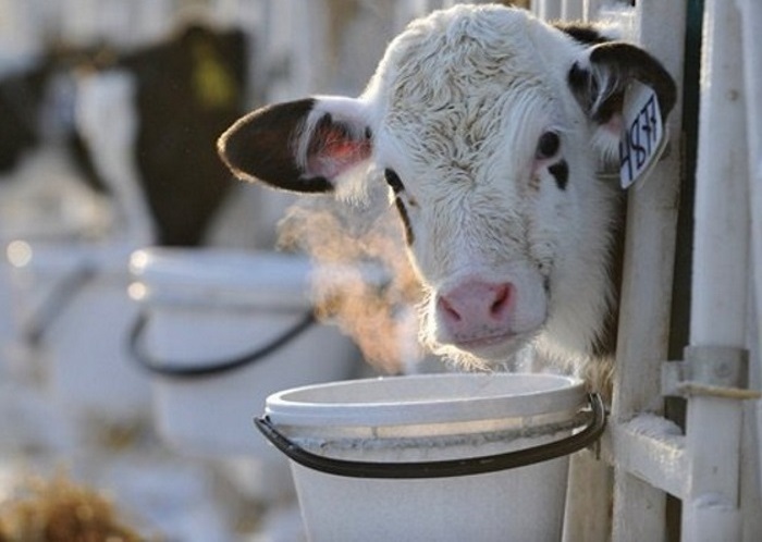
Catarrhal bronchopneumonia. A disease often found in calves is bronchopneumonia.
Bronchopneumonia in calves is a disease of non-contagious etiology that can cause large economic losses to a farm. Even though animals rarely die from pneumonia, the farmer still suffers losses. This article will discuss the causes of the disease, its manifestations in different forms of the disease, as well as methods of treatment and prevention.
What is bronchopneumonia?
This disease is characterized by the development of an inflammatory process in the bronchi and alveoli, accompanied by the accumulation of serous exudate in the bronchial tree. Inflammation quickly spreads, affecting all parts of the respiratory system. The disease always occurs against the background of a general weakening of the immune system, which is why it most often occurs in calves, whose resistance to pathogenic microorganisms is extremely low. The disease is accompanied by general intoxication of the body and can lead to irreversible pathological processes in the organs of the respiratory system, for example, necrosis of lung tissue.
Causes
The etiology of the disease is quite diverse. Let's consider the main reasons for the development of bronchopneumonia in young animals:
- Crowded housing of animals.
- Contaminated air.
- Poor quality of nutrition (lack of vitamins and amino acids in the diet).
- Unsuitable living conditions for animals (unheated room, dampness, lack of bedding).
- Stressful situations.
- Hypothermia.
- Congenital pathologies – short trachea, narrow lumens of the bronchi, rich mucous membrane respiratory tract capillaries, etc.
Under influence unfavorable factors a decrease in immunity occurs and pathogenic bacteria begin to rapidly multiply in the bronchi of the calf - streptococci, staphylococci, E. coli, Pseudomonas aeruginosa, pneumococci, fungi, etc. This leads to general intoxication of the body and destructive changes in the organs of the respiratory system.
Symptoms
Since bronchopneumonia can occur in three various forms, then the symptoms of the disease differ. Let's look at the general manifestations of the disease:
- Lethargy, depression.
- Cough (dry or wet).
- Nasal discharge is mucous or purulent
- Loss of appetite.
- Increased temperature (in the subacute form it increases only in the evening).
- Dyspnea.
- Intestinal upset due to intoxication.
- Most animals have pallor of the mucous membranes of the mouth and swelling of the conjunctiva.
Characteristic
To better understand exactly how bronchopneumonia occurs and how it manifests itself, let’s look at its three forms, because each of them has its own symptoms. There are the following forms of bronchopneumonia in cattle:
- Spicy.
- I'll sharpen it up.
- Chronic.
Acute form
Acute bronchopneumonia develops and progresses rapidly (up to 12 days) and is accompanied by pronounced symptoms:

- On the second or third day, body temperature rises to 40-42 degrees.
- Shortness of breath occurs.
- There is a dry, sharp cough.
- The conjunctiva is swollen, as is the nasal mucosa.
- Mucus is released from the nose, which contains purulent inclusions.
In the acute form of the disease, the cough gradually turns into a wet one, but at the same time it becomes more frequent. When listening to the calf, harsh breathing was noted, and moist rales were present. A blood test shows leukocytosis, with neutrophils shifting to the left.
Subacute form of bronchopneumonia
The subacute form is characterized by a longer course of the disease (from 15 to 30 days). Let's consider the clinical manifestations of the disease:
- Weakness, general depression, loss of appetite.
- The animal loses weight.
- The temperature is normal during the day, with a slight increase in the evening.
- The cough is wet and frequent.
- Dyspnea.
- Due to intoxication, intestinal upset occurs.
- When listening, a hard sound is noted bronchial breathing.
Reference. Under acute form Bronchopneumonia is often accompanied by periodic exacerbations, when the animal’s condition sharply worsens - the temperature rises, diarrhea occurs.
Chronic form
In the chronic course, the calf constantly has a cough, serous exudate is released from the nose, and the animal lags behind in weight. Appetite periodically returns to normal. When listening, dry wheezing is noted in the lungs.
Reference. The chronic form of pneumonia develops as a result of lack of treatment or improper treatment of the acute and subacute forms of the disease.
Diagnostics
To make a diagnosis, an external examination of the calf is carried out, and the conditions of its detention are taken into account. The veterinarian listens to heart sounds and breathing patterns. Great importance have blood indicators - as already mentioned, with inflammation in the lungs and bronchi, there is a shift of neutrophils to the left side and general leukocytosis. X-ray helps to identify multiple foci of inflammation in the lungs.

X-ray allows you to make a more accurate diagnosis and determine the form of the disease:
- On initial stage in acute and subacute forms of bronchopneumonia, blurred shadows are visible on the apical lobes of the lungs, while the anterior border of the heart is veiled, but still visible.
- In a chronic course, the foci of inflammation have clearer contours, and the border of the heart is practically not visible.
In large farms Where cases of animal death from bronchopneumonia have been recorded, it is recommended to perform an autopsy of animal corpses to identify the causative agent of the disease. This diagnosis makes it possible to exclude diplococcal infection, mycosis, salmonellosis, mycoplasmosis, viral infections, ascariasis and other diseases that have similar symptoms to bronchopneumonia.
Treatment
Very important in treatment A complex approach. Observations by veterinarians have made it possible to establish that the use of medications alone does not give the desired effect. First you need to create normal living conditions for sick individuals, which will contribute to their recovery:
- Move the calves to a warm room where there is no draft.
- Provide them with good bedding.
- Create optimal humidity conditions.
- Provide animals nutritious food, including vitamins and amino acids, to increase the body's resistance.
Antibacterial therapy
The main role in the treatment of bronchopneumonia is given to antibacterial agents. It is important to choose the right antibiotics; this will determine how effective the treatment will be. In the acute form of the disease and its subacute course, when gram-positive microflora predominates in the respiratory organs, high effectiveness of therapy is achieved after the use of penicillin or streptomycin. Streptomycin, oxytetracycline are prescribed for intramuscular administration with a solution of novocaine. Injections are given 3 times a day for 7-10 days.
Reference. Antibiotics are also used for injection into the bronchial tree using a special spray.
Sick calves are also administered sodium salts of sulfadimezine subcutaneously at 0.5-1 ml per kilogram of body weight. In total, no more than three such injections are required for the entire course of treatment. The frequency of administration is once every 4-5 days.

Antiallergic drugs
Treatment of calf bronchopneumonia always includes the use of antihistamines and drugs that reduce vascular permeability. This is necessary to avoid the development of allergies to waste products of the pathogen and to antibiotics. What remedies are recommended for sick calves:
- Suprastin.
- Pipolfen.
- Calcium gluconate.
- Sodium thiosulfate.
Attention! With catarrhal pneumonia, in rare cases, pulmonary edema develops. In this case, it is necessary to use a solution of calcium chloride at a concentration of 10%. The calf is injected into a vein.
Immunostimulants
Complex treatment of bronchopneumonia should include the use of immunoglobulins (gamma, beta) or polyglobulins to stimulate the immune system of sick calves. Instead of these drugs, the veterinarian may recommend an infusion of blood serum taken from healthy animals or hydrolysins.
Reference. Treatment is not advisable if large necrotic foci of inflammation are found in the lungs. Such animals are eliminated. Those whose disease has become chronic cannot be used as breeding animals.
Prevention
You can prevent the development of bronchopneumonia in calves in the following ways:
- Equipping premises for keeping cattle in accordance with all the rules recommended by the veterinary service (organizing good ventilation, using high-quality insulating materials during the construction of farms).
- Providing animals with good living conditions (clean, on warm bedding).
- Animals must receive adequate nutrition.

It is important to pay attention to strengthening the immunity of young animals, accustoming them to changes in temperature, ensuring regular walking. In the summer, all animals must be kept outdoors, equipped with canopies and protective fences from the wind. The premises where livestock are kept must be cleaned and disinfected. Particular attention is paid to the control of dust in the paddocks and in general on the farm territory.
Prevention of bronchopneumonia includes routine veterinary examinations and laboratory diagnostics infections occurring in a latent form.
Bronchopneumonia, although not a contagious disease, still poses a danger to livestock because the disease often becomes chronic. Chronicle animals are of no interest to the farmer, since they are unproductive and cannot be used as breeding stock. It is important to detect manifestations of bronchopneumonia in time in order to begin treatment as early as possible. It should be remembered that the best effect can be achieved only with complex therapy.
Bronchopneumonia (bronchopneumonia)– an animal disease characterized by the development of an inflammatory process in the bronchi and alveoli with effusion of serous-mucosal exudate into the latter. There are acute, subacute and chronic bronchopneumonia, and depending on the origin - primary and secondary. All types of animals of all ages are affected, but more often young animals (see Diseases of young animals).
Etiology . Bronchopneumonia is a disease of a polyetiological nature. All etiological factors of the disease can be divided into 2 groups: 1. reducing the natural resistance of the body and 2. opportunistic bacteria and viruses.
Factors that reduce natural resistance include disturbances in keeping and feeding animals (unsatisfactory microclimate, insufficient content of macro- and microelements and vitamins in the diet, especially vitamin A).
Against the background of reduced resistance, the evolutionarily developed balance between the macroorganism and opportunistic microorganisms, viruses, mycoplasmas, the total number of species of which can exceed 60, is disrupted. Violation of this balance causes the occurrence of the disease.
Secondary bronchopneumonia can be a symptom or complicate the course of bronchopneumonia, PCVD, gastroenteritis in horses, diseases of the uterus, udder, PCH, dictyocaulosis in cattle, dictyocaulosis, mulleriosis, necrobacteriosis in sheep, plague, metastrongylosis, vitamin deficiency in pigs.
Symptoms and course . At the onset of the disease, body temperature rises by 1-1.5 0, however, later it may drop to normal. Remitting fever. Breathing is rapid, shallow, mixed shortness of breath. The cough is short, muffled, and painful in acute cases.
Nasal discharge is serous-mucosal, and in subacute and chronic forms it is mucopurulent and purulent. With percussion, tympanic sounds are identified in the lesions, which later turns dull and dull, and with auscultation, bronchial breathing and small- and medium-bubble rales are heard. In healthy areas, upon auscultation, a rigid vesicular respiration. The functioning of the cardiac and digestive systems is disrupted.
The subacute and chronic form of the disease occurs with the same symptoms as the acute one, but less pronounced.
In acute and subacute forms, the number of red blood cells and hemoglobin in the blood decreases, and leukocytosis. The leukogram shows neutrophilia. In chronic bronchopneumonia, blood thickening is observed, accompanied by a relative increase in red blood cells and hemoglobin, leukocytosis with an increase in the number of lymphocytes.
With properly organized treatment, the acute form of the disease lasts 8-12 days, subacute 3-4 weeks, chronic - months and even years.
Diagnosis diagnosed on the basis of anamnesis, clinical manifestations, fluoroscopy, and laboratory tests.
Differential diagnosis . One should keep in mind lobar and symptomatic pneumonia in infectious and invasive diseases. (see Diseases of young animals).
Treatment . Greatest effect therapeutic measures are provided in the initial stages of disease development. An indispensable condition for a favorable outcome in the treatment of patients is the elimination of the causes of the disease and the creation of optimal feeding and maintenance conditions.
Antibiotics and sulfonamide drugs are used as antimicrobial agents after titrating them to the secreted microflora. Among antibiotics, penicillin, novocillin, ampicillin, ampiox, gentamicin, and tetracycline, morphocycline, olimorphocycline, etc. can be used according to the recommendations for use. Among other antimicrobial agents, it is effective to use -50 or 200, intramuscularly, 5 mg once a day for pigs, tilan - 10 mg, orally, 2 times a day. Of the sulfonamide preparations used, sulfadimizine, sulfadimethoxine, sulfamonomethoxine. The combined use of antibiotics and sulfonamides is effective.
In subacute and chronic cases, antibiotics and soluble sulfonamides are indicated to be administered intratracheally, and in case of mass disease - by aerosol method (see Bronchopneumonia of young animals).
Good results in acute and subacute forms are obtained using novocaine blockades of the stellate ganglion and splanchnic nerves and sympathetic trunks according to Shakurov. The effectiveness of treatment increases with the use of physiotherapy (ultraviolet irradiation, air ionization). Inclusion in complex therapy is mandatory symptomatic remedies(heart and digestive drugs).
To increase the natural resistance of the body, stimulants are used.
Prevention . Prevention of bronchopneumonia includes a complex of organizational, economic and special veterinary measures aimed at complying with zoohygienic standards for keeping and feeding animals, increasing the natural resistance of the body (see Bronchopneumonia of young animals).Bronchopneumonia is a disease manifested by inflammation of the bronchi and lobules of the lung with the accumulation of exudate and epithelial cells in the alveoli. The pathological process begins with the appearance of serous exudate in the bronchi and pulmonary parenchyma, which corresponds to the picture of catarrhal pneumonia in adult animals, but since the bronchi are primarily affected and the process quickly spreads along the bronchial tree to the parenchyma of the organ, such a disease occurs mainly in at a young age, called “bronchopneumonia”.
In addition to this disease, young animals may also have rhinitis, laryngitis, atelectatic, abscess and non-abscess pneumonia, but they are much less common and manifest themselves almost in the same way as in adult animals.
Calves, piglets, lambs, murrelets (reindeer calves), young fur-bearing animals and, less commonly, foals, become ill with bronchopneumonia.
The disease usually appears in calves at the age of 30-45 days, in piglets - 30-60 days, in lambs - 3-6 months.
Bronchopneumonia is more common in young animals that have been ill in early age acute digestive disorders and having, in connection with this, reduced resistance of the body.
Etiology. The disease occurs most often when the body’s resistance to adverse effects of factors decreases. external environment.
The occurrence of bronchopneumonia in lambs and piglets is often preceded by the presence of hypopneumatosis and small-focal atelectasis in the lungs that occur in hypotrophics, as well as due to blockage of the bronchi with mucus, which cannot be removed by sluggish coughing in weakened animals.
In young animals, in the first weeks and months of life, special anatomical and physiological prerequisites are created for the occurrence of bronchopneumonia. The short trachea and narrow bronchi, the richness of blood vessels in the mucous membrane lining the respiratory tract, its tenderness and slight vulnerability, the weakness of the elastic tissue of the walls of the alveoli and their saturation with lymphatic vessels favor the rapid transition of the inflammatory process from the upper parts of the respiratory tract to the deeper ones. The bronchi and alveoli of newborns and young animals are easily clogged with mucus.
The lack of retinol in mother food contributes to a decrease in the body's resistance and the appearance of bronchopneumonia. Due to the development of A-hypovitaminosis, the retinol content in the milk fed to calves, piglets, lambs and foals decreases sharply. Hypovitaminosis A disrupts the function of epithelial barriers, and their permeability to microorganisms increases.
Hypothermia and overheating of a young body lead to circulatory disorders, thermoregulation disorders, and the appearance of congestion in the lungs, which creates conditions for the occurrence of bronchopneumonia.
Keeping young animals in poorly equipped premises with poor ventilation, when dust, carbon dioxide, ammonia, hydrogen sulfide, methane, and water vapor accumulate in the air, has an adverse effect on the condition of the respiratory system.
Pathogenesis. Primary changes in the bronchi, and then bronchioles, infundibulae and alveoli create conditions for the development of opportunistic and saprophytic microflora entering the large quantities with inhaled air. This is also facilitated by changes in the epithelium under the influence of a lack of retinol. The resulting toxic waste products of microorganisms are absorbed and cause intoxication. As a result, the capillary walls become more permeable, effusions accumulate in the lung parenchyma, and catarrh. Blood and lymph circulation in the lungs is disrupted.
All listed pathological changes lead to a decrease in gas exchange, which leads to oxygen starvation fabrics. Under-oxidized metabolic products accumulate in tissues and blood and acidosis develops. The accumulation of acidic products causes further metabolic disorder, shortness of breath, nervous phenomena, weakening of cardiac activity, excretion large quantities basic salts in the form of alkaline phosphate and ammonium compounds, which are formed during the neutralization of acidic products. The tone of blood vessels, mainly arteries, arterioles and capillaries, decreases. There is an “equalization” of arterial and venous pressure. The speed of blood flow changes, and congestion develops. Dystrophic changes appear in the heart muscle. The excitability, conductivity and contractility of the heart are impaired, which results in shifts and changes in the electrocardiogram. The ECG shows a decrease in voltage in all leads, the disappearance of the P wave, a 2-fold decrease in the PQ interval, roundedness of the R wave, a decrease and stretching of the T wave, a sharp decrease in the TP interval, acceleration of the full cardiac cycle(the segment R-R is significantly shortened).
Liver function is also impaired. Changes in water-salt metabolism are primarily manifested by a decrease in the content of chlorides in the blood and their accumulation in tissues. The state of achlorosis sharply disrupts the formation and secretion of hydrochloric acid in the stomach (abomasum) and leads to disruption of the functions of the digestive organs, which in some cases gives reason to talk about diseases of young animals with pneumoenteritis.
In patients, kidney function is impaired: their filtration capacity changes, and protein appears in the urine.
In the bronchi, bronchioles, infundibulae and alveoli, desquamation of the epithelium occurs, which is mixed with a serous effusion containing leukocytes and erythrocytes. The presence of serous effusion in the lobules of the lung leads to increased vesicular and manifestation of bronchial breathing, the occurrence of wet and dry rales. Microbial toxins acting on the central nervous system disrupt thermoregulation processes, and patients develop fever.
Pathological changes. In most animals, during the acute course of bronchopneumonia, pallor of the mucous membranes, usually compaction of the lung tissue, especially in the area of the anterior lobes, sometimes atelectasis, hyperemia of the upper respiratory tract are found; in the bronchi and more often in the bronchioles - a mucous, easily squeezed out mass. Sometimes there is a catarrhal state of the stomach and intestines.
In the subacute course of bronchopneumonia, changes are detected in the upper respiratory tract (rhinitis) and bronchi (bronchitis). The lungs are variegated in color. The lesions are dense. The middle and anterior areas of the diaphragmatic lobes are most often affected. On a section of the lungs, viscous mucus or curdled whitish masses are squeezed out of the bronchi; the mucous membrane of the bronchi is hyperemic and edematous. The mediastinal and bronchial lymph nodes are enlarged and edematous; There are pinpoint hemorrhages on the section.
In some cases, there are signs of pleurisy in the form of fibrinous deposits on the pleura and the presence of straw-yellow or cloudy yellowish fluid in the pleural cavity.
The cardiac muscle is matte. The liver is enlarged, the gallbladder is filled with thick bile.
In the case of chronic bronchopneumonia in calves, areas of the lungs have a variegated color (reddish, yellowish, brown). On the cut, you can see an uneven surface with whitish partitions between the lobules. In piglets and very often in lambs, purulent encapsulated foci, indurative changes, pneumosclerosis and even petrified foci are found in the lungs. Foals may have separate areas of the lungs. Adhesive pleurisy, fusion of the pleura (costal with pulmonary) is often found in animals. The mediastinal and bronchial lymph nodes are enlarged, dark in color, without signs of pinpoint hemorrhages.
The heart sac is filled with a turbid fluid or is fused with the heart muscle. The heart is expanded. Changes characteristic of chronic gastroenteritis are possible.
Symptoms. There are acute, subacute and chronic course of bronchopneumonia. The acute course of bronchopneumonia occurs at a very young age and, as a rule, in hypotrophic patients. A subacute course is observed in young animals under unsatisfactory conditions of feeding, keeping and caring for them; it can also be a continuation of an acute disease.
The chronic course of bronchopneumonia is typical for young animals of the post-weaning period.
The acute course of bronchopneumonia, especially in animals (usually piglets and lambs) with a very low birth weight, can occur in an areactive form with a fatal outcome after 2-3 days of illness. Sick animals develop adynamia (stagnation), and some, in addition, have a decrease in appetite. Then they appear hard breathing, dry cough, dry wheezing can be heard. Later, nasal discharge, rapid breathing, moist wheezing and coughing are noticeable.
Visible mucous membranes become pale and cyanotic. Heart sounds are muffled and the pulse wave is weak.
The activity of the digestive organs is disrupted, peristalsis increases and diarrhea appears.
The subacute course of the disease is characterized by decreased appetite, stunted growth and poor nutrition of patients. They begin to experience shortness of breath, often of a mixed type, moist cough. It manifests itself especially clearly when pressing on the trachea in the upper part. On auscultation chest wheezing and bronchial breathing are heard. When the pleura is involved in the pathological process, friction noises appear. Body temperature periodically rises.
In lambs, coughing is noticeable after watering and rapid movements. Visible mucous membranes are hyperemic. Subsequently, depression intensifies, stagnation appears, sometimes fever (remitting), pulse rate and breathing movements increases. The cough becomes loud and comes into attacks; in piglets and gilts with symptoms of suffocation.
Percussion of the chest in calves reveals foci of dullness in the apical and diaphragmatic lobes of the lung. In sick young animals with this course of bronchopneumonia, the pulse becomes faster and weaker, the maximum arterial pressure decreases and the minimum arterial and venous pressure increases. The blood flow slows down, the mucous membranes become bluish, and blood stagnation occurs in the liver. Profuse diarrhea develops. Animals suffering from chronic bronchopneumonia are stunted in growth. Appetite is variable. In damp and warm weather, cough and mixed shortness of breath intensify. Body temperature either periodically rises to 40.5 ° C, or is constantly raised by several tenths of a degree.
There is periodic discharge from the nasal openings. On auscultation, wheezing is heard, and percussion reveals significant areas of dullness.
Diagnosis. When making a diagnosis, general data on the sanitary and zoohygienic conditions of raising young animals and the maintenance and feeding of mothers are taken into account. Pay attention to the animal’s behavior indoors, on walks and its general condition, taking into account clinical signs and pathological changes. At X-ray examination in sick piglets and lambs, varying degrees of shading of the pulmonary field are found, mainly in the apical and cardiac lobes, increased bronchial pattern, loss of visibility of the cardiophrenic triangle and the contours of the ribs in the affected areas. Great help Thoracofluorographic studies using the method of R. G. Mustakimov can help identify sick young animals, especially in the early stages of the disease.
Differential diagnosis. It is necessary to exclude streptococcal infection (presence of specific pathogen, temperature, appearance, in addition to pneumonia, damage to the joints, digestive organs, etc.), salmonellosis (initial dysfunction of the digestive organs, detection of the pathogen during laboratory testing, characteristic pathological changes). In case of diseases of young animals with pasteurellosis, rapid coverage of a large number of animals is established; During laboratory testing, the pathogen is isolated.
Viral pneumonia in calves and piglets can be distinguished from bronchopneumonia only by the results of a biological test (artificial reproduction of the disease) and histological examination of the affected lung tissue, as well as using serological and immunofluorescent reactions.
DISEASE HISTORY
DIAGNOSIS: CATARHAL BRONCHOPNEUMONIA
CASE HISTORY №______
Initial diagnosis bronchopneumonia.
__________________________________________________________________________
The diagnosis is final unilateral catarrhal bronchopneumonia.
__________________________________________________________________________
study of arteries and veins (rhythm, filling, pulse rate, venous pulsation) arterial pulse rhythmic, soft, medium filling, frequency – 71 beats per minute; venous pulse is negative, venous undulation is moderate, fullness is moderate;
Digestive organs:
appetite slightly reduced oral cavity no visible damage;
pharynx, esophagus there is no pain or swelling, patency is not impaired;
state of the proventriculus, stomach, intestines (frequency, activity of contractions: palpation, percussion, auscultation) Scar: 3 contractions in 2 minutes, percussion sound - tympanic, painless on palpation; the abomasum and intestines are painless on palpation, peristaltic sounds are of moderate strength, the percussion sound is dull;
Liver border does not extend beyond the edge of the last rib;
The act of defecation (regularity, frequency, consistency, color) not obstructed, feces of soft consistency, dark brown;
Respiratory system:
examination of the upper respiratory tract (inhalation and exhalation, nasal discharge, larynx, cough, sputum)shortness of breath and cough are observed. The nasal discharge is serous in nature, respiratory sounds are moderate in strength;
examination of the chest (shape, symmetry, type of breathing; palpation, percussion, auscultation of the lungs) breathing is of mixed type, the chest is symmetrical, round, painless on palpation, there are no tangible noises, the integrity of the ribs is not broken, the percussion sound is atympanic, there are areas of dullness. Mixed breathing. The posterior border of the lungs along the macular line is at the level of the 12th rib, along the line of the scapulohumeral joint - at the level of the 9th rib. Breathing noises – bronchial and hard vesicular breathing, dry wheezing can be heard.
Genitourinary organs (frequency, pain of urination: presence of blood, color, smell, clarity of urine; percussion of the kidneys) urination is not difficult, painless. Urine is clear, straw-yellow in color, without blood;
Nervous system (type, temperament; depression, excitation; tactile and pain sensitivity; paresis, paralysis; tendon reflexes) mobile, pain and tactile sensitivity is not impaired. There are no paresis or paralysis.
Sense organs:
Organs of vision (pupillary reflex; movement of the eyeball; transparency of the ocular media)the movement of the eyeballs is voluntary, the pupil is not dilated, accommodation is not impaired. The cornea is moist, transparent, its integrity is not compromised.
Hearing organs (reaction to auditory stimuli; presence of discharge from ears) the animal responds adequately to sound stimuli, there is no discharge from the ears;
Study of the pathological process area
(detailed description clinical signs of the pathological process).
The chick is depressed, her appetite is somewhat reduced. Mixed shortness of breath, moderate discharge of serous fluid from the nose, and cough are observed.
On percussion, foci of dullness are noted (in the area of the apical lobes).
On auscultation, hard vesicular and bronchial breathing is noted, as well as dry wheezing.
| date | T | P | D | Course of the disease | Therapy, diet, regimen content |
| 24.07 | 39,9 | 83 | 33 | Slight depression, slightly decreased appetite, cough, small amount of serous fluid discharge. On percussion of the chest, areas of dullness are noted. On auscultation of the chest, hard vesicular and bronchial breathing, as well as dry rales, are heard. | Rp.: Tetraviti – 20 ml D.t.d No. 1 in flaconis S. Rp.: Bicillini 3 – 600.000 ED D. t. d. No. 2 in ampullis D.S. Give moistened hay, sprinkles in the form of mash |
| 25.07 | 39,9 | 82 | 34 | Slight depression, slightly decreased appetite, cough, discharge of serous-catarrhal fluid from the nasal cavity. On percussion of the chest, areas of dullness are noted. On auscultation of the chest, harsh vesicular and bronchial breathing is heard, as well as dry wheezing that is less pronounced than yesterday. | Rp.: Bicillini 3 – 1.800.000 ED M.f.Solutio D.S. Intramuscular per injection |
| 26.07 | 39,7 | 81 | 29 | Slight depression, slightly decreased appetite, cough, moderate discharge of serous-catarrhal fluid from the nasal cavity, the nasal mucosa is hyperemic. On percussion of the chest, small areas of dullness are noted. On auscultation of the chest, hard vesicular and bronchial breathing is heard, as well as moist fine rales. | Rp.: Bicillini 3 – 1.200.000 ED Sol.Novocaini sterilisatae 0.5% - 10.0 M.f.Solutio D.S. Intramuscular per injection Give moistened hay, sprinkles in the form of mash |
| 27.07 | 39,7 | 82 | 33 | Slight depression, appetite is somewhat reduced, cough is observed periodically, moderate discharge of serous-catarrhal fluid from the nasal cavity, the nasal mucosa is hyperemic. On percussion of the chest, small areas of dullness are noted. On auscultation of the chest, hard vesicular and bronchial breathing is heard, as well as moist fine rales. | Rp.: Bicillini 3 – 1.200.000 ED Sol.Novocaini sterilisatae 0.5% - 10.0 M.f.Solutio D.S. Intramuscular per injection |
| 28.07 | 39,5 | 79 | 30 | Cough is observed very rarely, serous discharge from the nose is insignificant. On percussion of the chest, isolated areas of dullness are noted. On auscultation of the chest, hard vesicular and bronchial breathing is heard, as well as weakly expressed moist fine bubbling rales. | Rp.: Tetraviti – 20 ml D.t.d No. 1 in flaconis S. Intramuscularly at a dose of 8 ml, per injection Rp.: Bicillini 3 – 1.200.000 ED Sol.Novocaini sterilisatae 0.5% - 10.0 M.f.Solutio D.S. Intramuscular per injection |
| 29.07 | 39,4 | 74 | 29 | The cough has stopped and there is no discharge from the nose. When percussing the chest - an atympanic sound. Auscultation of the chest - bronchial and vesicular breathing. The animal recovered clinically. | |
Epicrisis according to literature
Bronchopneumonia – this is inflammation of the bronchi and alveoli with effusion in the latter of catarrhal exudate. It is recorded mainly in late autumn and early spring due to unstable weather and sudden changes in air temperature during the day. The pathological process begins with the appearance of serous exudate in the lungs and pulmonary parenchyma, which corresponds to the picture of catarrhal pneumonia in adult animals, but since the bronchi are primarily affected and the process quickly spreads along the bronchial tree, such a disease, which occurs mainly in young animals, is usually called bronchopneumonia .
On large farms, in specialized farms and industrial complexes, if veterinary and sanitary rules for keeping animals are violated, bronchopneumonia can become widespread, covering up to 30-40% of the total livestock in certain periods.
Bronchopneumonia is registered in different zones of the country and by specific gravity
takes second place after gastrointestinal diseases. According to a number of
authors, every year in the country 20-30% of young animals suffer from bronchopneumonia. As a result of illness, the average daily increase in live weight, productive and breeding qualities of animals are reduced, therefore the prevention of bronchopneumonia is a matter of paramount importance, which requires a timely and competent solution.
Etiology . Bronchopneumonia of calves is a polyetiological disease. According to some authors, bronchopneumonia is a disease of non-infectious origin; the microbial factor in the development of nonspecific bronchopneumonia in calves is not leading and has no pathogenetic significance. Microorganisms isolated from the lungs of sick and dead animals are saprophytic; they become pathogenic only when the resistance of the animal’s body decreases. Most often, pneumonia is isolated from staphylococci, streptococci, diplococci, pneumococci, and Escherichia coli. Many authors believe that bronchopneumonia manifests itself as a consequence of unsatisfactory living and feeding conditions. It is customary to distinguish between endogenous and exogenous causes of bronchopneumonia in calves. TO endogenous reasons include: incorrect selection of pairs during mating, inbreeding, leading to the birth of unhealthy young animals with reduced resistance and susceptibility to many diseases. Endogenous causes also include the anatomical and physiological characteristics of young animals: short trachea, narrow bronchi, richness of blood vessels in the mucous membrane lining the respiratory tract, weakness of the elastic tissue of the walls of the alveoli and their saturation with lymphatic vessels. These reasons contribute to the rapid occurrence and spread of the inflammatory process.
TO exogenous reasons The occurrence of bronchopneumonia includes: violations of the feeding conditions of breeding stock, in particular, insufficiency of vitamin A in their diets. This causes them to develop A-hypovitaminosis, as a result of which the content of vitamin A in the milk fed to calves decreases. Hypovitaminosis A causes calves to develop the barrier function of the mucous membranes, in particular the respiratory tract, as a result of which their permeability to microorganisms increases. Also exogenous factors include violation of the rules of hygiene for keeping young animals (hypothermia, overheating), which leads to impaired circulation, the appearance of congestion in the lungs and, thus, creates favorable conditions for the development of bronchopneumonia, especially in the absence of exercise in animals. Excessive accumulation of dust, carbon dioxide, ammonia, hydrogen sulfide, methane, water vapor in the air, or vice versa, its excessive dryness, also contribute to the occurrence of of this disease. Microbial air pollution is also an exogenous cause of bronchopneumonia in calves.
As a complication, catarrhal bronchopneumonia develops with diffuse and capillary bronchitis, pulmonary atelectasis due to bronchitis or malnutrition of newborns (atelectatic pneumonia).
A predisposing factor to the appearance of this disease is a decrease in the resistance of the animal’s body, which can occur against the background of stress (transport, industrial); against the background of gastrointestinal diseases suffered at an earlier age (for example, dyspepsia); in the absence of exercise; with a lack of natural or artificial ultraviolet radiation; with inadequate feeding (lack of protein, certain amino acids, vitamins, mineral components).
Pathogenesis . The pathogenesis of bronchopneumonia is quite complex, since all organs and systems of the sick animal are involved in the process. Pathogenesis is determined by the state of all organs and tissues, primarily by the state of the nervous system. Adverse factors primarily cause changes in the nervous system, therefore, a violation of humoral and nervous factors occurs, the body's defenses decrease, the concentration of lysozyme and histamine in the blood decreases, and the globulin fractions of proteins increase. This contributes to stagnation of blood in the lungs and swelling of the mucous membranes of the bronchioles and bronchi. The phagocytic activity of leukocytes and the lysozyme activity of bronchial mucus sharply decreases, and the barrier function of the epithelium decreases. Initial changes are characterized by exudative processes, leukocyte reaction, accumulation of serous exudate in the bronchi and alveoli. Accordingly, favorable conditions develop for the development of microflora, which can be both pathogenic and saprophytic. Microflora multiplies quickly, microbial enzymes and toxins accumulate in high concentration and cause necrosis of the mucous membranes and the development of the inflammatory process. Lobular inflammation and microbronchitis occur.
Subsequently, the affected areas merge and foci form. At the site of inflammatory foci, the lung tissue is compacted and has smooth surface. arise defensive reactions- snorting, coughing. Microbial toxins are absorbed into the blood, intoxication occurs, and consequently, vascular porosity occurs. Effusion accumulates in the lung parenchyma and catarrhal inflammation occurs. Ventilation of the lungs becomes more difficult, and the functioning of healthy areas increases. As a result, breathing intensifies and becomes more frequent. A decrease in the level of gas exchange in the lungs causes a decrease in gas exchange in the tissues, accumulation of under-oxidized metabolic products occurs, and acidosis develops. As a result, shortness of breath, nervous phenomena, weakening of the cardiovascular system, decreased tone of blood vessels and, consequently, decreased blood pressure occur. As a result of a decrease in blood flow, stagnation occurs, dystrophic processes occur in the heart muscle, and the functioning of the liver and pancreas changes. A lack of chlorides in the blood causes a disruption in the formation of hydrochloric acid in the stomach, and diarrhea develops.
The filtration capacity of the kidneys changes (protein appears in the urine). Microbial toxins affect the central nervous system, causing disruption of thermoregulation, and fever develops accordingly. With a favorable course and elimination of etiological factors, as well as with the provision of medical care, after 7-10 days recovery occurs.
If the course is unfavorable, the process can take on a lobar character, purulent-necrotic changes, pleurisy, pericarditis occur, and secondary immune deficiencies appear.
Clinical signs . The acute form is characterized by a relapsing-remitting fever with daily temperature fluctuations of up to 2°C (the temperature rises to 41-42°C). Cough. Bilateral mucous or mucopurulent discharge from the nose. Mixed shortness of breath with increased breathing. On percussion, there are foci of dullness of varying sizes in the lower third of the chest. Hard vesicular breathing, fine bubbling or dry wheezing in the form of squeaking, whistling; and above the foci of dullness there is an absence of respiratory sounds, sometimes bronchial breathing. The pulse is rapid, weak filling. Depending on the degree of damage to the lungs, decreased appetite, emaciation, stunted growth, decreased productivity and performance, a tendency to constantly lie down, pallor and cyanosis of the mucous membranes, decreased elasticity of the skin, ruffled hair or fur, and other signs are noted.
Urination is reduced . In the blood - neutrophilia, moderate leukocytosis. IN severe cases– leukopenia. In contrast to lobar pneumonia, the area of dullness in catarrhal bronchopneumonia is relatively smaller, the fever is not constant, and there is no natural change of stages or crisis.
The subacute and chronic forms of the disease occur at normal body temperature and with the same clinical symptoms as the acute form, but they are less pronounced. With these forms, rapid emaciation of animals is noted.
Uncomplicated forms of acute catarrhal bronchopneumonia end with recovery within 1.5 - 2 weeks. The subacute form lasts 18-25 days and, in the absence of course treatment, becomes chronic.
Secondary bronchopneumonia depends on the course of the underlying disease. Possible complications: purulent pneumonia, transition to a subacute or chronic form with the development of pneumofibrosis, heart failure.
Pathological changes . In most animals with acute bronchopneumonia, pale mucous membranes are detected. The lung tissue is compacted, in the apical and middle lobes there are multiple pneumonic lesions on the surface and in the thickness of the organ with a diameter of 1 to 2-3 centimeters, blue-red or pale gray in color, dense, with a specific gravity heavier than water. When these lesions are cut, catarrhal exudate is released.
At autopsy, edema and hyperemia of the upper respiratory tract, exudate in the bronchi and bronchioles are also noted. Mediastinal and bronchial The lymph nodes increased. In subacute pneumonia, exhaustion, cyanosis of the mucous membranes, purulent exudate in the bronchi. The mucous membrane of the bronchi is edematous, hyperemic, with hemorrhages. The affected areas of the lungs have a doughy consistency, are mottled, and sink in water.
The heart muscle is dull, the liver is enlarged, the gallbladder is filled with thick bile.
In chronic bronchopneumonia, the areas of the lungs are motley, and the growth of connective tissue is noticeable. The lung has a dense consistency, the surface is lumpy, granular when cut, pieces of the lung sink in water. In the chronic form, bronchiectasis, bronchostenosis, obliteration of the bronchi, and carnification of the lungs can also be observed.
Diagnosis . The diagnosis is made on the basis of anamnesis (taking into account general data on the sanitary and zoohygienic conditions of raising young animals, the maintenance and feeding of the mother), clinical, radiological and laboratory studies.
Making an early and accurate diagnosis is especially important in large livestock complexes and specialized farms.
Hematological research methods for bronchopneumonia reveal neutrophilic leukocytosis with a shift to the left, lymphopenia, eosinopenia, monocytosis, accelerated ESR, decreased reserve alkalinity, decreased catalase activity of erythrocytes, a relative decrease in the albumin fraction of blood serum and an increase in globulin fractions of proteins, a decrease in the degree of hemoglobin saturation arterial blood oxygen.
The most objective and exact method diagnostics - selective x-ray examination.
In the initial stages of bronchopneumonia, homogeneous foci of shading, blurring of the pulmonary field in the cranial areas of the lungs, and veiling of the anterior border of the heart are detected radiographically in the apical and cardiac lobes of the lungs. In chronic bronchopneumonia with localized lesions, dense, well-contoured foci of shading are visible in the area of the apical and cardiac lobes of the lungs. In this case, the anterior border of the heart is not visible in most cases. In patients with chronic confluent forms of bronchopneumonia with diffuse lesions of the lungs, X-ray examination reveals diffuse, extensive, intense shading in the anterior and lower parts of the pulmonary field. The borders of the heart, the cardiophrenic triangle and the contours of the ribs in the affected areas are not distinguished.
For mass research on large livestock farms, a fluorographic method for the differential diagnosis of bronchopneumonia of various forms in calves was proposed (R.G. Mustakimov, 1970).
In some cases, to clarify the diagnosis, a biopsy from the affected areas of the lungs, bronchography, bronchophotography, examination of tracheal mucus, nasal discharge, and other research methods are used. In system diagnostic measures When carrying out clinical examination, it is recommended to carry out selective pathological autopsies with histological examination animals suspected of having the disease and killed for diagnostic purposes.
Differential diagnosis . The differential diagnosis excludes infectious and invasive diseases that manifest themselves as symptoms of damage to the respiratory tract and lungs.
Should be excluded streptococcal infection(presence of a specific pathogen, temperature, appearance of lesions of the joints, digestive organs), salmonellosis(impairment of the functions of the digestive organs at the beginning, detection of the pathogen during laboratory testing, characteristic pathological changes). In case of disease in young animals pasteurellosis rapid coverage of a large number of animals is noted, and during laboratory testing the pathogen is isolated.
Viral pneumonia calves can be distinguished from bronchopneumonia only by the results of a bioassay and histological examination of the affected lung tissue, as well as by serological and immunofluorescent reactions.
Also exclude catarrhal pleuropneumonia, dictyocaulosis, ascariasis. All of the above diseases are characterized by massive damage to animals, and along with damage to the respiratory organs, damage to other animal body systems is noted.
At bronchitis, unlike catarrhal bronchopneumonia, body temperature is absent or slightly elevated and percussion of the chest does not reveal foci of dullness in the apical lobes of the lung.
Unlike lobar pneumonia The area of dullness in catarrhal bronchopneumonia is relatively smaller, the fever is not constant, and there is no natural change in stages. Lobar pneumonia is characterized by a staged course, persistent fever, fibrinous or hemorrhagic discharge from the nasal openings. The percussion sound changes in accordance with the stages of the inflammatory process - from tympanic to dull and dull.
It is also necessary to exclude mycoses, mycoplasmosis, metastrongyllosis and some other diseases.
Treatment . Treatment of sick animals must be carried out comprehensively, dividing patients into separate groups depending on the course of the disease and its severity. The main condition for successful treatment of bronchopneumonia is the elimination of etiological factors, the creation of optimal living conditions and the provision of adequate feeding. The animal is given rest and kept in a warm, clean room with good ventilation, but without drafts. Feed for sick animals should be high-calorie, easily digestible, low-volume and complete in vitamin and mineral composition (for example, soft hay, root vegetables, silage in moderation).
Complex treatment in combination with properly organized housing and feeding conditions leads to the complete recovery of animals with acute and subacute bronchopneumonia. Treatment of animals suffering from chronic bronchopneumonia does not lead to complete recovery, but it helps to stop the process. Young animals that have recovered from chronic bronchopneumonia cannot be used for breeding purposes and are subject to culling.
Complex treatment includes the simultaneous use of various means:
antimicrobial therapy (antibiotics, sulfonamides, nitrofurans, drugs
arsenic); pathogenetic therapy(expectorants and absorbents); replacement therapy (vitamins, macro and microelements, oxygen therapy, bronchial dilators: ephedrine, adrenaline); symptomatic therapy (cardiac drugs).
Treatment for all forms of bronchopneumonia comes down primarily to the elimination of secondary microflora of the lungs and respiratory tract, for which purpose antimicrobial therapy . At individual treatment antimicrobial drugs are given orally, administered intramuscularly, intratracheally, intravenously. Many authors emphasize the effectiveness of intratracheal administration of antimicrobial drugs. Antibiotics are prescribed taking into account the sensitivity of the microflora of the respiratory tract and lungs to them. Pulmonary sputum for research is collected with special devices, as well as by sucking from the lower third of the trachea with a sterile syringe or by biopsy from pneumonic foci. In the laboratory, samples are sown on nutrient media and the sensitivity of the microflora to antibiotics is determined using serial dilutions or using antibiotic disks. Long-term uncontrolled use of the same antibiotics on the farm reduces their therapeutic effectiveness and leads to the emergence of antibiotic-resistant races of microbes.
When choosing an antibiotic for treatment, it should be taken into account that during the acute course of the disease in the first days, gram-positive microflora predominates in the foci of inflammation. During this period, the best therapeutic effect is achieved with the use of penicillin and streptomycin. The sodium or potassium salt of penicillin or benzylpenicillin in a 0.5% solution of novocaine is administered intramuscularly 3-4 times a day at a rate of 7000-10000 units/kg per injection, treatment duration is 5-8 days. Penicillin-3 is prescribed in the form of an aqueous suspension in distilled water intramuscularly every other day at the rate of 10,000 -12,000 units/kg, for the entire course of 3-5 injections.
In subacute and chronic forms, when purulent-necrotic processes develop in the lungs, it is advisable to use broad-spectrum antibiotics: streptomycin, tetracycline, oxytetracycline, chloramphenicol, terramycin, monomycin. These antibiotics are administered intramuscularly or subcutaneously in prolonging solvents for 5-6 days in a row at 3 thousand - 5 thousand units / kg of calf weight.
Sulfonamide drugs(norsulfazole, sulfadimezin, sulfantrol, phthalazole, etazol, sulfdimethoxine) are given orally 3-4 times a day for 5-7 days in a row at the rate of 0.02-0.05/kg body weight. Sodium salts of norsulfazole or sulfadimezine can be used in the form of a 30% oil suspension, injected twice with a break of 4-5 days at the rate of 0.5 ml/kg of body weight. The oil suspension is injected warm through a thick needle subcutaneously in the neck area.
Good results in the treatment of bronchopneumonia in calves were obtained from penicillin, biomycin, and norsulfazole administered intratracheally. In the lower third of the trachea, 5 ml of a 5% solution of novocaine is injected over 0.5-1 minutes and, without removing the needle, 50 thousand - 100 thousand units of crystalline penicillin salt are slowly injected in 5-10 ml of sterile saline solution or 15 -20 ml of 0.5% biomycin, or 15-20 ml of 10% sodium salt of norsulfazole.
Novarsenol for treatment is given to calves older than two weeks of age by instilling a 50% novarsenol solution onto the conjunctiva of both eyes - 5-10 drops twice a day for 2-3 days in a row.
Calcium gluconate 0.25-0.5 g, suprastin 0.025-0.05 g, or pipolfen 0.025 g are recommended as anti-allergic agents that reduce the permeability of vascular walls for the entire period of treatment.
Against the background of active antimicrobial therapy, it is effective to carry out novocaine blockade of the stellate ganglia or the inferior cervical sympathetic ganglion. It is most suitable for calves: 20-30 ml of a sterile 0.25% novocaine solution. The injection is made with a large needle, stepping back 1-1.5 cm from the posterior edge of the transverse process 6 cervical vertebra. The needle is carefully advanced in the medial-caudal direction to a depth of 3-5 cm until it stops at the base of the body of the 1st or 2nd thoracic vertebra and then pulled back 1-3 cm and novocaine is immediately injected. During the course of treatment, it is recommended to carry out 2-3 novocaine blockades, which are done alternately on the right and left sides.
An economical and effective method of group therapy for bronchopneumonia in young animals in industrial complexes and farms is aerosol therapy antibacterial agents in accordance with approved recommendations and instructions attached to each specific drug. Special hermetic compartments or chambers are equipped for inhalation facilities inside the livestock building, preferably closer to the insulator (you can use plastic films). The cells must have sewerage and supply and exhaust ventilation. The volume of inhalers is determined based on an average of 1.5-2 m 3 per calf. Small chambers (10-20 m3) are used more often for aerosol therapy with antibiotics and sulfonamides, and large chambers (50-100 m3) are used for other antibacterial agents and for preventive group treatment of animals. Medicines are sprayed from aerosol generators SAG-1, SAG-2, VAU-1, DAG-1, DAG-2 and others, which are installed according to the instructions. For group aerosol treatment, many drugs are used: antibiotics (on average 400,000 - 500,000 units per 1 m 3 of air), sulfonamides (0.5 g of soluble norsulfazole in 1 m 3), novarsenol (5 ml of 1% solution in 1 m 3 ), lactic acid (0.1 g per 1m3) and other antibacterial agents. Therapeutic effectiveness increases with the combined use of antibacterial agents with vitamins and microelements. Cooking medicinal solutions on distilled water or a 1% solution of novocaine and immediately before spraying. The duration of one aerosol therapy session is 50-60 minutes, 2-3 such sessions should be performed per day, the course of treatment is 7-15 days (depending on the degree of lung damage).
Symptomatic and pathogenetic treatment is determined by the form and severity of the manifestation of bronchopneumonia.
Pathogenetic therapy includes the use of expectorants and absorbents. Ammonium chloride is given orally to calves as an expectorant.
bicarbonate of soda, and also use inhalation of turpentine and chloride vapors
In order to increase natural immunobiological resistance, they use replacement therapy . Sick animals are injected intramuscularly with nonspecific gamma globulins, gamma beta globulins, polyglobulins at a dose of 1 ml per kilogram with an interval of 48 hours 2-3 times.
There is a high therapeutic effectiveness of citrated blood in the treatment and prevention of acute respiratory infections in young cattle.
Hematotherapy is used to stimulate the body's defenses. For this
use the blood of the animal itself or another animal of the same species. Autologous blood is injected subcutaneously or intramuscularly into the neck, inner thigh or croup. In case of increased coagulability, for every 100 ml of blood, add 5 ml of a 5% sodium citrate solution or 10 ml of a 10% sodium salicylate solution.
Blood is injected into healthy tissue, bordering the affected ones, because in place
injection creates a short-term barrier that has autoantiseptic properties
properties. The dose of blood is determined each time depending on the characteristics of the sick animal and the nature of the pathological process in the body. In acute limited inflammatory processes, the recommended dose of autologous blood for large animals is 125-150 ml, and for small animals 5-50 ml. At the beginning of the disease, it is advisable to administer blood in the evening.
For diffuse inflammatory processes accompanied by a long-term febrile state, small doses of blood are used (2-25 ml for small animals). Single injections of blood rarely give a positive result; it is better to do 4-5 injections, although the first two are most effective. The interval between injections is from 48 hours to 4 days. The more severe the disease, the lower the dose and the longer the interval between injections. With each subsequent injection, the body's reaction weakens. Therefore, with repeated injections, it is necessary to increase the blood dose, but without exceeding the maximum. If after 2-fold administration there is no improvement in the animal’s general condition, the course of the pathological process and the morphological composition of the blood, autohemotherapy should be abandoned.
To enhance the therapeutic effect, physiotherapeutic procedures are used:
heating young animals with Sollux and Infraruge lamps, diathermy, aeroionization, rubbing the chest with irritating substances, mustard plasters, cupping.
Vitamins in the treatment complex for bronchopneumonia acquire particular importance, as they normalize metabolism and reduce side effect antimicrobial agents and increase their therapeutic effectiveness.
Symptomatic therapy includes giving cordials: 20% camphor
oil, 3-5 ml intramuscularly; 10% caffeine solution 1-3 ml subcutaneously; cordiamine, 1.5-2 ml subcutaneously; valerian tincture, 2-3 ml per glass of water orally per calf.
When pulmonary edema develops, a 10% solution of calcium chloride is administered intravenously at a dose of 5-10 ml per calf.
The use of replacement and symptomatic therapy contributes to a more rapid restoration of the physiological functions of the body.
Prevention Bronchopneumonia consists of a complex of organizational, economic, zoo-hygienic and veterinary-sanitary measures aimed at obtaining and raising strong, disease-resistant young animals. Particular attention is paid to creating optimal conditions for keeping and feeding breeding stock and young animals. Livestock premises must meet the zoohygienic indicators approved by the standards. It is unacceptable to place livestock buildings and feedlots in swampy, low-lying and flooded areas. Industrial specialized farms and livestock complexes should be built only according to projects approved and agreed upon with the veterinary service, and they must provide for the division of the territory into production and economic zones. In calf barns, the amplitude of temperature fluctuations should not exceed 5°C, relative humidity - 70%, air speed 0.1-0.3 m/sec., concentration of hydrogen sulfide and carbon dioxide 5 mg/m 3 each. Among the measures to prevent colds, favorable conditions for keeping animals, as well as regular walks for young animals, are important. To avoid overheating of animals in the hot season, make shady canopies or increase ventilation in the premises. It is especially dangerous to give cold water to hot animals; the temperature of the drinking water should not be lower than 15-20°C. To avoid colds, young animals should not lie on unheated cement or asphalt floors without bedding. In animal resting areas, cement floors must be covered with wooden flooring or movable wooden boards.
Important In the system of measures to prevent the disease of animals with bronchopneumonia, they include the fight against dust in the air of livestock yards, walking areas, for which they carry out landscaping of the farm territory, create forest protection fences around livestock buildings. Avoid long drives of livestock along dusty tracts, especially in the hottest part of the day. Bulk feed (compound feed, grass meal, chaff and others) is stored closed in separate rooms, and when distributed, they are moistened. It is better to prepare herbal flour in granular or tablet forms. In the premises where young animals are kept, a sanitary regime must be observed, cleanliness must be systematically maintained, and disinfection must be carried out.
Drugs that increase resistance are widely used in animal feeding.
body (premixes containing vitamins and minerals; gamma globulin and other agents are administered to weakened animals).
The effectiveness of methods for the prevention of bronchopneumonia using aerosol treatment is also noted. For this purpose, it is recommended to use substances that disinfect the air in livestock buildings and sanitize the respiratory organs of animals. This is forest balsam A in its pure form in a concentration of 0.3-0.5 g/m 2 room for 1-2 hours, iodine triethylene glycol at the rate of 0.15-0.3 g iodine per 1 m 2 for 40 minutes, iodine triethylene glycol in combination with turpentine and lactic acid in an amount of 0.3 ml/m2 with an exposure of 40 minutes. For these purposes, use 3% hydrogen peroxide, 5% water solution chloramine B, sodium hypochloride containing 1.5-2% chlorine, 4% alkali solution.
An indispensable condition for ensuring the effectiveness of the prevention of respiratory diseases is routine medical examination and periodic veterinary examinations using modern methods and diagnostic tools.
Creation of optimal conditions for feeding and keeping young animals, compliance
proper veterinary and sanitary rules ensure a reduction in diseases
and high safety of young animals.
EPICRISIS ACCORDING TO CURATION DATA
Etiology . Hypothermia, inadequate feeding, inhalation of polluted air or irritating substances, decreased immune status of the body
Pathogenesis . In the affected areas, catarrhal inflammation develops, then exudate enters the lumen of the alveoli and bronchi, tissue decay products and microorganism toxins are absorbed into the blood, thereby causing intoxication of the body. When etiological factors are eliminated, the exudate is gradually released when coughing and partially resolves.
Diagnosis was diagnosed based on the history and clinical signs. The animal is depressed, its appetite is somewhat reduced. There is shortness of breath of a mixed type, moderate discharge of serous fluid from the nose, and a dry cough. On percussion, foci of dullness are noted (mainly in the area of the apical lobes). On auscultation, hard vesicular and bronchial breathing is noted, as well as dry wheezing.
Treatment . Tetravit used to increase the overall resistance of the body. The combination of vitamins in the preparation acts synergistically to increase resistance against infectious diseases and diseases of young animals, promotes the growth of young animals, which is especially important in case of imbalance nutrients in diet and respiratory diseases. An antibiotic was used as etiotropic therapy to suppress pathogenic microflora Bicillin 3. To reduce the amount of dust in the air, the sprinkles and hay were moistened with water.
Prevention . It is necessary to improve the living conditions of animals, adjust the diet of pregnant dry cows and young animals. It is necessary to make a diagnosis comprehensively and in a timely manner.
Conclusion . The diagnosis of catarrhal bronchopneumonia was made taking into account the history and clinical signs. As a result of timely complex treatment a pronounced therapeutic effect was obtained. The outcome of the disease was the clinical recovery of the animal.
Bibliography
1.) Danilevsky V.M. "Treatment of internal non-contagious animal diseases." – M.: Kolos, 1967 – 184 p.
2.) Chernukha V.K. "Handbook of Ruminant Diseases". – Kyiv: Harvest, 1987.
3.) Zharikov I.S. "Handbook of diseases of farm animals." – Minsk: Urajai, 1985 – 344 p.
4.) Danilevsky V.M. "Handbook of Veterinary Therapy." – M.: Kolos, 1983 – 192 p.
5.) Kolesov A.M. "Internal non-communicable diseases of animals." – St. Petersburg: Kolos, 1972 – 544 p.
6.) Shcherbakov G.G., Korobov A.V. "Workshop on internal non-communicable diseases." – M.: Lan, 2003 – 544 p.
7.) Ionov P.S. "Internal non-contagious diseases of cattle." – M.: Agropromizdat, 1985 – 383 p.
8.) Lochkarev V.A., “Treatment of calves with bronchopneumonia” // Veterinary Medicine, 1992, No. 12
9.) Belopolsky V.A., Golovzin Yu.V., “Immunological basis for the treatment of calves
for bronchopneumonia" // Veterinary Medicine, 1993, No. 11
10.) Gyuruji-Ogly S.Zh., “ Functional status cardiovascular system in calves with bronchopneumonia" // Veterinary Medicine, 1995, No. 9
Vologda State Dairy Academy named after. N.V. Vereshchagina
Faculty of Veterinary Medicine
Department of Internal Non-Contagious Diseases, Surgery and Obstetrics
COURSE WORK
in the discipline "Internal non-communicable diseases of farm animals"
BRONCHOPNEUMONIA IN CALVES
Prepared
5th year student 753gr.
Zorina I.E.
Vologda – Dairy
Introduction
1. Literature review
1.1 Definition of disease
1.2 Etiology of the disease
1.3 Pathogenesis of the disease
1.4 Symptoms of the disease
1.6 Diagnosis of the disease
1.8 Course and prognosis of the disease
1.9 Treatment of bronchopneumonia
1.10 Disease prevention
2. Own research
2.1 Farm characteristics
2.3 Room microclimate
2.5 Medical history
Conclusion
Application
Introduction
The desire to maximize productivity through the implementation of intensive industrial systems without sufficient consideration physiological needs animals leads to a decrease in their immune reactivity, against the background of which non-communicable diseases arise, accounting for about 90% of the main types of farm animals.
Among all the pathologies of farm animals caused by the technology of keeping, feeding and using them, the largest share is occupied by non-communicable diseases of young animals. At the same time, gastrointestinal, respiratory diseases, metabolic diseases and feed toxicoses take first place in terms of frequency, mass and magnitude of economic damage. Diseases of the immune system are also widespread. Due to changes in animal habitats, wide application chemical substances V agriculture, antimicrobial and biological drugs In animal husbandry and veterinary medicine, the course and clinical and morphological manifestation of many diseases have changed significantly, and new forms of pathology have appeared. Associated diseases of a polyetiological nature have become increasingly common.
Statistics show that animal diseases accompanied by damage to the respiratory system account for 20-30% of the total number of non-communicable diseases and rank second in prevalence.
The wide spread of respiratory diseases is due to a decrease in the natural resistance of animals as a result of violations of housing technology (long-term transportation, hypothermia, dampness and gas contamination of premises, high concentration on limited areas, promoting airborne droplet transmission of infection, insufficient natural lighting in the premises and other factors that weaken the body’s defenses.
For correct and timely diagnosis pathology of the respiratory system, organization of prevention and treatment, it is necessary to clearly understand the multifaceted physiological role of the respiratory tract and lungs. The respiratory organs are closely connected through the nervous system, blood and lymph with all body systems. When the respiratory organs are damaged in the body, the functions of the cardiovascular, digestive, urinary and other systems change, the flow of air into the lungs decreases, which leads to a deterioration in gas exchange in them and the occurrence of shortness of breath.
The economic damage from diseases of the respiratory system consists of the death of sick animals, which reaches 10%, a decrease in the productivity of sick and recovered animals, and the cost of treatment.
1. Literature review
1.1 Definition of disease
Pneumonia (pneumonia) is widespread compared to other respiratory diseases and accounts for 80% of all respiratory diseases. All pneumonias are divided into lobar and lobular.
Lobular pneumonia is characterized by the gradual spread of inflammation in the lobes of the lungs. Unlike lobar pneumonia, it clinically manifests itself with less clear signs. Most often it occurs chronically, sometimes asymptomatically. This type occurs atelectatic (occurs as a result of the formation of airless areas in the lung tissue - atelectasis, or collapsed areas - hypopneumatosis), aspiration (occurs when foreign bodies enter the respiratory tract), metastatic, or purulent (occurs as a result of the introduction of bacterial microflora into the lungs from other organs and tissues of the body), purulent-necrotic, or pulmonary gangrene (purulent-putrefactive melting of lung tissue), hypostatic (a disease resulting from stagnation of blood in the lungs - hypostasis and the subsequent development of catarrhal inflammation) pneumonia.
Lobar pneumonia is characterized by the rapid spread of inflammation in the lungs, covering in typical cases, already in the first hours of the disease, individual lobes of the lungs or even the entire lung. Lobar pneumonia always occurs quickly with severe clinical signs. The disease has a pronounced staged nature. Lobar pneumonia (an acute disease that occurs in stages) and some infectious diseases (infectious anemia, contagious pleuropneumonia, pasteurellosis) occur in this type.
Pneumonia, according to the nature of the exudate formed, can be catarrhal, purulent, fibrinous, according to its course - acute and chronic, and according to its etiology - primary and secondary.
Since the inflammatory process is rarely limited to the mucous membrane of the alveoli (pneumonia), but also affects the bronchi or, on the contrary, begins in the mucous membrane of the bronchi (bronchitis), and subsequently continues to the alveoli, the disease is called bronchopneumonia. Of all the named forms of pneumonia, the most common is catarrhal bronchopneumonia.
Bronchopneumonia is an inflammation of the bronchi and lungs, characterized by the accumulation of exudate in the bronchi and alveoli, consisting of a large amount of mucus rejected by the epithelial cells of the mucous membrane, leukocytes, exclusion of the affected areas from the respiratory function, circulatory disorders and gas exchange with increasing respiratory failure and intoxication of the body.
The disease is characterized by the spread of the pathological process, which initially occurs in the bronchi, along the bronchial tree to the lung tissue.
In young animals, according to their origin, they are divided into primary and secondary bronchopneumonia. Primary bronchopneumonia usually occurs due to exposure to unfavorable environmental factors and abnormal intrauterine development. Secondary bronchopneumonia is observed in a number of infectious diseases (paratyphoid, hemorrhagic septicemia, piglet influenza, viral bronchopneumonia of pigs, ascariasis, dictyocaulosis); primary (non-contagious) bronchopneumonia is most common. On some farms they affect up to 50-70% of the young stock.
Bronchopneumonia is recorded mainly among young animals. The disease occurs more often in the winter-spring and summer seasons of the year. The winter-spring outbreak usually begins in February with the maximum number of patients and their deaths in March - April.
Calves from 2 weeks to 2-3 months of age are mainly affected. During the summer outbreak, calves at the age of 2-3 months and even 4 months become ill. Piglets and lambs become ill at the age of 2 months and older.
1.2 Etiology of the disease
Bronchopneumonia is a polyetiological disease and usually occurs as a result of a combined effect on the body of unfavorable factors (stressors) that weaken resistance. The most common external (exogenous) factors of bronchopneumonia are colds and others associated with irritation of the respiratory tract. These are increased humidity in the room, damp floors and walls, maintenance without bedding on cement floors, drafts, excessive accumulation of ammonia, hydrogen sulfide, etc. In early spring and autumn, due to unstable weather and sudden changes in air temperature during the day, the incidence increases significantly.
Primary bronchopneumonia occurs when the sanitary and hygienic conditions of maintenance are violated (dampness, overcrowding, increased content ammonia indoors, hypothermia in the wind, in the rain, exposure to low air temperatures) and as a result of a decrease in the natural resistance of the young organism due to insufficient or inadequate feeding of the breeding stock. Both groups of factors act interconnectedly. This means that the poor resistance of the offspring increases its sensitivity to changes in the external environment, and a poor microclimate, in turn, aggravates the susceptibility of weak offspring to respiratory diseases.
A decrease in the resistance of the young organism as a result of poor feeding of mothers is especially often observed in lambs. It is known that lambs born in winter (early lambing) are fuller, develop better and have less bronchopneumonia than lambs born in late spring. Pregnancy of ewes during early lambing occurs in late autumn and early winter, when the ewes’ bodies retain reserves of nutrients, minerals and vitamins accumulated during the period of summer and autumn grazing. These factors ensure normal intrauterine development of the fetus and the birth of strong lambs that can withstand winter cold and summer heat. With late lambing, nutrient reserves in the body are consumed during the stall period (especially with poor feeding), which can adversely affect the intrauterine development of the fetus. In such cases, he is born with a lower body weight, weak and more often susceptible to respiratory diseases. The death of lambs occurs during the summer heat, which the sick body cannot tolerate. The same situation can be observed for other animal species.
The occurrence of bronchopneumonia is also facilitated by factors that reduce the natural resistance of the animal body: the birth of underdeveloped, hypotrophic young animals with reduced vitality, lack of protein, certain amino acids, vitamins, mineral components in the diet, lack of exercise, lack of natural or artificial ultraviolet radiation, illness at a young age (especially during the colostrum period) gastrointestinal diseases.
The occurrence of bronchopneumonia in young animals is facilitated by a lack of vitamin A, since as a result, the ciliated epithelium of the respiratory tract is replaced by a flat multilayered one, which leads to a violation rheological properties bronchial secretion.
Bacterial microflora plays a major role in the occurrence and development of bronchopneumonia. Microorganisms can be isolated from pneumonic foci, tracheal and bronchial mucus in the majority of animals sick and dead from bronchopneumonia various types: pneumococci, staphylococci, streptococci, sarcina, proteus, yeast-like fungi, mycoplasmas, sometimes Pseudomonas aeruginosa. In most cases, bacterial microflora plays a secondary, complicating role in etiology. However, under certain conditions it can become the root cause of the disease. This can occur when the virulent or toxigenic properties of microbes are enhanced, when microbes that the body has not previously encountered enter the lungs, which happens during various rearrangements of animals and the replenishment of farms with young animals from other farms.
The etiological role of respiratory infections in the occurrence, development and spread of bronchopneumonia in young farm animals has been proven. Inflammatory processes in the respiratory organs can be caused by many viruses, including influenza viruses, parainfluenza, rhinoviruses, reoviruses, adenoviruses, etc. In some cases, respiratory viral infections are mild, without severe clinical symptoms, limited to damage to the respiratory tract. However, these infections can also occur with the development of bronchopneumonia, which usually happens with complications of a bacterial infection.
Those. The main causes of bronchopneumonia in young animals are:
1. poor adaptability of the body to environmental conditions on the soil malnutrition and improper maintenance of mothers and young animals, as well as other stress factors;
2. weakening of growth, development and resistance of the body can occur after birth, even taking into account the fact that intrauterine development was normal.
So, for example, the disease develops in calves at the age of 2-3 months because, after satisfactory milk feeding, they are transferred to feeding with roughage without concentrates and mineral and vitamin supplements, which sharply reduces their resistance.
Poorly developed young animals do not always develop bronchopneumonia. The following conditions contribute to its appearance:
1. insufficient functioning of the respiratory system due to prolonged cellular maintenance or insufficient (absent) exercise. As a result, insufficient expansion of the alveoli develops;
2.cold (which is associated with exposure to cold and dampness) as a result of which the body’s heat transfer exceeds heat production;
3.overheating – at high air temperatures, thermal regulation is disrupted in underdeveloped calves exposed to the scorching rays of the sun for a long time. As a result, the temperature rises and the breathing and heart rate increases;
4. long-term keeping of young animals in premises with a high concentration of ammonia and hydrogen sulfide, which is possible with crowded housing, poor ventilation and sewerage;
5. hypovitaminosis A, D
6. long-term and recurring gastrointestinal diseases;
7. microflora inhabiting the airways and becoming more active in the weakened body of young animals - streptococci, staphylococci, diplococci, sarcina.
1.3 Pathogenesis of the disease
Bronchopneumonia is considered not only as a local process localized in the lungs, but as a general disease manifested by a violation of all systems and functions of the body.
Under the influence of an etiological factor, for example, sudden hypothermia, the body develops allergic condition, manifested by a disorder of neurohumoral reactions, which ultimately leads to perversion normal function bronchi and pulmonary alveoli. In the submucosal layer of the bronchial membrane, a spasm is first observed, and then paresis of the capillaries and venous stagnation of blood; hemorrhages and swelling occur in the lung tissue. In the blood, the concentration of lysozyme and histamine decreases and the content of globulin coarse fractions of proteins increases, which irritate the lung tissue and contribute to stagnation of blood in the lungs and the development of edema in the mucous membranes of the bronchioles and bronchi. The phagocytic activity of leukocytes and the lysozyme activity of bronchial mucus decreases.
In healthy animals, the ciliated epithelium of the bronchi serves as a barrier to the microflora entering with the inhaled air; part of the microflora is phagocytosed by leukocytes. In sick animals, as a result of a decrease in the barrier function of the epithelium, conditions are created for the rapid proliferation of microflora in the bronchial mucosa and in the lumens of the respiratory tract, and its toxicity increases.
Mycoplasmas and viruses penetrate the epithelium of the mucous membrane, where they multiply. Therefore, the initial changes in these cases are most noticeable in the mucous membranes of the respiratory tract, and exudate accumulates in the bronchi and alveoli several days after complications with bacterial flora.
With the predominant participation of bacteria in the development of bronchopneumonia, the initial changes are characterized mainly by an exudative process and a leukocyte reaction, which is manifested by the rapid accumulation of first serous and then catarrhal exudate in the lumen of the bronchioles and alveoli. This is due to the fact that bacteria usually do not penetrate the wall of the mucous membrane, but multiply almost exclusively in the lumens of the respiratory cavities of the respiratory sections of the lungs.
The inflammatory process during bronchopneumonia can develop further from large bronchi to small ones, then to bronchioles and alveoli, i.e. as a complication of bronchitis. However, the inflammatory process can occur initially in the bronchioles and alveoli, and then move to the bronchi. In all cases, bronchopneumonia is characterized by a lobular (lobular) type of spread of the process in the lungs. The cranial areas of the lungs (apical and cardiac lobes) are almost always affected first.
The inflammatory process spreads along the continuation of the bronchial branches or through the lymphatic tract.
In the acute course of the disease, the superficial lobules of the lungs are usually affected first. In the initial stages of the disease, interlobular connective tissue serves as a barrier to the transition of inflammation from the affected lobules to healthy ones, but later this barrier function is lost.
In the chronic course of the disease, especially if the etiological factors are not eliminated and treatment is not carried out, the process can become lobar as a result of the merging of individual foci of inflammation into large foci (confluent lobar pneumonia). In patients with a chronic course, more often in pigs, there may be complications in the form of adhesive pleurisy and pericarditis, and pulmonary emphysema.
The nature of the inflammatory process in bronchopneumonia varies depending on the etiological factor and the degree of body resistance. In the initial stages of the disease, serous, serous-catarrhal or catarrhal inflammation develops in the bronchi and alveoli. Non-coagulable exudate, consisting of mucin, leukocytes, erythrocytes and bronchial epithelial cells and microbes, sweats into the lumen of the bronchi and alveoli. In chronic processes, the organization of exudate, induration and calcification of pneumonic foci, purulent-necrotic decay of lung tissue and bronchi occur.
As a result of the absorption of toxins and decay products from foci of inflammation into the blood and lymph, intoxication of the body is observed, accompanied by varying degrees of increased body temperature, dysfunction of the cardiovascular, respiratory, digestive, nervous and other systems.
In patients with bronchopneumonia, gas exchange is disrupted due to a decrease in the respiratory surface of the lungs, accumulation of exudate in the lumen of the bronchi and intoxication. In the initial stages of the disease, gas exchange disturbances are compensated by increased respiratory movements and heart function. In a chronic course with damage to large areas of the lungs (confluent pneumonia), oxygen consumption per unit of animal weight is sharply reduced, the degree of saturation of arterial blood with oxygen decreases, and tissue gas exchange is disrupted. In pigs with diffuse chronic lobar pneumonia, oxygen consumption decreases by 2-3 times, and arterial blood oxygen saturation sometimes decreases to 70-80% instead of 97-98 in comparison with healthy animals.
With a favorable course of the disease, with timely and correct treatment, on average, after 7-10 days, the respiratory tract and alveolar tissue are restored to normal condition, are freed from catarrhal exudate, after which the animal recovers. In an unfavorable course, when the etiological factors are not eliminated and treatment is not carried out, the affected lobes merge into large foci (confluent, lobar pneumonia), the inflammation becomes purulent-necrotic in nature, there may be abscesses in the lungs, complications in the form of pleurisy and pericarditis are frequent. In such cases, intoxication increases, respiratory and cardiovascular vascular insufficiency, which leads to the need for forced slaughter.
1.4 Symptoms of the disease
The clinical manifestation of the disease largely depends on the etiological factor, species and age of animals.
In horses and sheep, in most cases, there is a rapid spread of the inflammatory process in the bronchi and lungs compared to pigs and cattle.
In pigs, compared to other animals, a sluggish course and erased forms of bronchopneumonia are more often recorded.
In young and old animals, bronchopneumonia manifests itself in a more severe form.
According to the course, bronchopneumonia is divided into acute and chronic, sometimes subacute.
In the acute course, already on the first day of the disease, when inflammation passes to the lungs, general weakness and apathy, weakening or loss of appetite, and an increase in body temperature by 1-2 0 C are noticed. In animals that are weakened or exhausted, body temperature in most cases does not increase. On the 2-3rd day of illness, symptoms of damage to the small bronchi and lungs are revealed: first dry, then wet deep cough, intense breathing, mixed shortness of breath, mixed shortness of breath, serous-catarrhal or catarrhal discharge from the nasal openings, discharge of catarrhal exudate during coughing. On auscultation, hard vesicular breathing and fine bubbling rales are detected in the lungs. Percussion establishes limited areas of dullness (mainly in the area of the apical and cardiac lobes). Most animals experience a moderate increase in heart rate and an increase in the second tone.
The subacute course of bronchopneumonia is characterized by a more protracted course and longer duration of fever.
Chronic bronchopneumonia is characterized by a long course, often with periods of exacerbation and attenuation. Depending on the degree of lung damage, decreased appetite, emaciation, stunted growth, decreased productivity and performance, a tendency to constantly lie down, pallor and cyanosis of the mucous membranes, decreased elasticity of the skin, tousled hair and other signs are noted.
Body temperature at upper limits normal and subfebrile. Breathing is rapid and intense, expiratory shortness of breath with a predominance of abdominal breathing is clearly visible. The cough is prolonged and usually occurs when standing up. In pigs, coughing attacks can be observed (sometimes 30-40 coughing bursts in a row). During auscultation, hard vesicular breathing, dry or moist rales are heard, and in areas of large pneumonic foci, bronchial breathing or breathing sounds are not audible at all. Percussion reveals limited areas of dullness in the apical, cardiac and lower sections diaphragmatic lobes of the lungs.
The chronic course of bronchopneumonia in young animals often occurs on farms with unsatisfactory feeding of the breeding stock, as well as with untimely and unsystematic treatment at the onset of the disease.
1.5 Pathological changes
The most characteristic changes are found in the lungs and bronchi. In the initial stages of bronchopneumonia and during its acute course, multiple lobular lesions are found in the apical and cardiac lobes in the form of pneumonic foci located superficially or in the thickness of the lung. Lesions ranging in size from one to several centimeters are blue-red or pale red in color, dense to the touch, sink in water, and when they are cut, catarrhal exudate is released from the bronchi.
Histological examination of the affected lobules reveals signs of catarrhal bronchopneumonia: in the alveoli and bronchi there is catarrhal exudate consisting of mucus, leukocytes, erythrocytes, bronchial epithelial cells, and microbes.
In chronic bronchopneumonia, depending on the duration of the process, the presence of extensive pneumonic foci formed as a result of the fusion of lobular lesions is characteristic; pleurisy and pericarditis are detected. Histologically, in these cases, induration, areas of purulent-necrotic decay of the lungs and bronchi, and petrification are found. Mediastinal lymph nodes are often enlarged. Nonspecific changes in chronic bronchopneumonia include exhaustion, degeneration of the myocardium, liver, kidneys, muscle atrophy, etc.
1.6 Diagnosis of the disease
The diagnosis is made on the basis of anamnesis, clinical symptoms and special laboratory diagnostic methods. Making an early and accurate diagnosis is of particular importance.
Hematological research methods for bronchopneumonia reveal neutrophilic leukocytosis with a shift to the left, lymphopenia, eosinopenia, monocytosis, accelerated ESR, a decrease in reserve alkalinity, a decrease in catalase activity of erythrocytes, a relative decrease in the albumin fraction of blood serum and an increase in globulin fractions of proteins, a decrease in the degree of oxygen saturation of hemoglobin in arterial blood.
The most objective and accurate diagnostic method is selective x-ray examination.
In the initial stages of bronchopneumonia, homogeneous foci of shading, blurring of the pulmonary field in the cranial areas of the lungs, and veiling of the anterior border of the heart are detected radiographically in the apical and cardiac lobes of the lungs. In chronic bronchopneumonia with localized lesions, dense, well-contoured foci of shading are visible in the area of the apical and cardiac lobes of the lungs. In this case, the anterior border of the heart is not visible in most cases. In patients with chronic confluent forms of bronchopneumonia with diffuse lung lesions, X-ray examination reveals diffuse, extensive, intense shading in the anterior and lower parts of the pulmonary field. The borders of the heart, cardio-diaphragmatic triangle and the contours of the ribs in the affected areas are not distinguished.
For mass research on large livestock farms, a fluorographic method for the differential diagnosis of bronchopneumonia of various forms in calves, sheep and pigs has been proposed.
In some cases, to clarify the diagnosis, a biopsy from the affected areas of the lungs, bronchography, bronchophotography, examination of tracheal mucus, nasal discharge, and other research methods are used. In the system of diagnostic measures during clinical examination, it is recommended to carry out selective pathological autopsies with histological examination of animals suspected of the disease and killed for diagnostic purposes.
1.7 Differential diagnosis
It should be taken into account symptomatic (pasteurellosis, salmonellosis, dictyocaulosis, metastrongylosis) and viral pneumonia (parainfluenza, adenovirus, mycoplasmosis), as well as viral diarrhea, infectious rhinotracheitis, chlamydia, etc. Differentiation is carried out taking into account epizootological data, clinical manifestations, bacteriological, virological and serological research methods.
In differential diagnosis, streptococcal infection is excluded based on the isolation of a specific pathogen during laboratory testing, changes in body temperature, the appearance of lesions of the joints, digestive organs and other characteristic symptoms, salmonellosis - at the beginning, dysfunction of the digestive organs, detection of the pathogen during laboratory testing, characteristic pathological changes. Catarrhal pleuropneumonia and ascariasis are also excluded. All of the above diseases are characterized by massive damage to animals and, along with damage to the respiratory organs, damage to other animal body systems is noted. Bronchitis and lobar pneumonia are excluded. With bronchitis, unlike catarrhal bronchopneumonia, body temperature is absent or slightly elevated and percussion of the chest does not reveal foci of dullness in the apical lobes of the lung. Lobar pneumonia is characterized by a staged course, constant fever and fibrinous or hemorrhagic discharge from the nasal openings. The percussion sound changes in accordance with the stages of the inflammatory process - from tympanic to dull and dull.
1.8 Course and prognosis of the disease
Catarrhal bronchopneumonia in the absence of medical assistance is characterized by a chronic (several weeks) course. Advanced cases of this disease result in abscess formation, pleurisy, gangrene, myocarditis, and endocarditis. Bronchopneumonia occurs more severely in emaciated and old animals. Timely provision of medical care prevents complications and allows us to hope for a favorable prognosis.
1.9 Treatment of bronchopneumonia
Pathological processes develop not only in the bronchi and alveoli of the lung, but also in other organs. In this regard, treatment of patients is carried out comprehensively using methods of etiotropic, pathogenetic, replacement and symptomatic therapy.
The effectiveness of treatment is largely based on the creation of favorable environmental conditions for animals. When sick animals appear and the first symptoms of the disease are established, it is necessary to take urgent measures to eliminate hypothermia, dampness, and the entry of cold air into the room, provide animals with bedding and create optimal temperature and humidity parameters for them. Sick animals are isolated in a separate room. Treating animals with medications only, without eliminating the etiological factors of the disease, gives a low therapeutic effect.
Antibiotics are widely used as nonspecific antimicrobial agents for bronchopneumonia; they are prescribed taking into account the sensitivity of the microflora of the respiratory tract and lungs to them. Pulmonary sputum is collected for examination. special device, as well as by sucking from the lower third of the trachea with a sterile syringe or by biopsy from pneumonic foci. In the laboratory, samples are sown on nutrient media to determine the sensitivity of the microflora to antibiotics. Long-term uncontrolled use of the same antibiotics on the farm reduces their therapeutic effectiveness and leads to the emergence of antibiotic-resistant races of microbes.
When choosing an antibiotic for treatment, it should be taken into account that during the acute course of the disease in the first days, gram-positive microflora, as a rule, prevails in the foci of inflammation. During this period, the best effect is obtained from penicillin and streptomycin. The sodium or potassium salt of penicillin in a 1% solution of novocaine is administered intramuscularly 3-4 times a day at a rate of 7000-10000 units/kg per injection. The duration of treatment is 5-8 days. Bicillin - 3 is prescribed in the form of an aqueous suspension in distilled water intramuscularly every other day at the rate of 10,000-15,000 units/kg, for the entire course of 3-5 injections.
For acute, subacute and chronic bronchopneumonia, streptomycin, ampicillin, kanamycin, neomycin, erythromycin, enroxil, gentamicin, baytril, tetracycline are prescribed. Streptomycin sulfate or oxytetracycline hydrochloride is administered intramuscularly in a 1-2% solution of novocaine 2-3 times a day for 5-7 days at the rate of 10,000-15,000 units/kg.
Sulfonamides are administered orally to young animals 3-4 times a day for 7-10 days in doses of 0.02-0.03 g/kg. Pigs, sheep and calves can be given subcutaneous sodium salts of sulfadimezine or norsulfazole in the form of a 10-15% suspension in fish oil. The suspension is administered at a dose of 0.5-1 ml/kg once every 4-5 days, for a total of 2-3 injections per course of treatment.
For purulent-catarrhal bronchopneumonia, intratracheal administration of sterile solutions of antibiotics or sulfonamides is indicated. First, 5-10 ml of a 5% solution of novocaine is injected into the lower third of the trachea with a syringe (slowly, over 0.5-1 min), and after the cough reflex subsides, without removing the needle, penicillin diluted in 5-7 ml of distilled water is injected into the dose of 0.05-0.1 g of dry matter per 1 kg of animal weight. Solutions of antibiotics or sulfonamides are prescribed 1-2 times a day for 3-5 days.
The use of novarsenol in the form of a 50% solution on the conjunctiva in the amount of 3-4 drops 1-2 times a day for 2-3 days in a row is justified.
Calcium gluconate 0.25-0.5 g, suprastin 0.025-0.05 g or pipolfen 0.025 g are recommended as anti-allergic agents and agents that reduce the permeability of vascular walls for the entire period of treatment (doses are indicated per calf) 2-3 times a day ). For the same purpose, a 5% aqueous solution of sodium thiosulfate can be used intravenously once a day at a dose of 1-1.5 ml of solution per 1 kg of animal weight, a total of 3-5 injections per course of treatment. When pulmonary edema develops, a 10% solution of calcium chloride is administered intravenously at a dose of 5-10 ml per animal.
To increase the nonspecific reactivity of the body, especially in the initial period of the disease, gamma - beta - globulins or nonspecific polyglobulins are administered in dosages according to the accompanying, methodological instructions or instructions on the package labels. Instead of globulins, you can use hydrolysins, blood serum of healthy animals, tissue preparations and other nonspecific stimulants.
The use of novocaine blockade of stellate (lower cervical) sympathetic nodes is indicated. Novocaine blockade is most appropriate for calves; they are injected into the area of the stellate node with 20-30 ml of a sterile 0.25% solution of novocaine. The injection is made with a large needle, stepping back 1-1.5 cm from the posterior edge of the transverse process of the 6th cervical vertebra. The needle is carefully advanced in the medial-caudal direction to a depth of 3-5 cm until it stops at the base of the body of the 1st or 2nd thoracic vertebra and then pulled back 1-3 cm and novocaine is immediately injected. Free entry of the solution indicates the correct position of the needle. For a course of treatment, 2-3 novocaine blockades are recommended, which are done alternately on the right and left sides.
It is recommended to heat sick animals with incandescent lamps, use diathermy, ultra-high-frequency therapy, ultraviolet artificial radiation, aeronization, rubbing the chest wall with irritating agents, mustard plasters, and cupping.
It is important to provide sick animals with vitamins, especially vitamin A.
It is useful for calves to administer 40 ml of glucose intravenously in the form of 20% solutions. Ammonium chloride, inhalation of water vapor of turpentine, tar, and ichthyol are used internally for patients.
An economical and effective method of therapy for bronchopneumonia is aerosol therapy with antibacterial agents. For aerosol treatment, many agents are used: antibiotics (on average 400,000-500,000 units per 1 m 3 of air), sulfonamides (0.5 g of soluble norsulfazole in 1 m 3 of air), novarsenol (5 ml of a 1% solution in 1 m 3), turpentine (5 ml of a 10% solution in 1 m 3), lactic acid (0.1 g in 1 m 3), iodinol (2 ml in 1 m 3) and other antibacterial agents.
1.10 Disease prevention
Prevention of bronchopneumonia includes a complex of organizational, economic and special veterinary measures aimed at complying with zoohygienic standards for keeping and feeding animals and increasing the body’s resistance. The most important element of properly organized prevention is maintaining an optimal microclimate. In a dispensary for calves, the temperature should be in the range of 16-20 0 C, relative humidity – 65-70%, CO 2 concentration – no higher than 0.15%, ammonia – 0.01 mg/l, the number of microbial bodies should not exceed 20 thousand/ m 3 air. In premises for calves from 20 days to 3 months of age, the temperature should be 15-17 0 C, relative humidity – 70%, CO 2 – 0.25%, ammonia content – 0.015 mg/l, microbial contamination – 40 thousand/m 3 air.
To avoid colds, young animals should not lie on unheated cement or asphalt floors without bedding. In animal resting areas, cement floors must be covered with wooden flooring or movable wooden boards. It is recommended to change the litter regularly. To avoid overheating during hot times of the day, animals are kept under shady canopies or indoor ventilation is increased.
A preventive measure for bronchopneumonia is also the fight against dust in premises and walking areas, for which they plant landscaping on the farm territory and create forest protection fences around livestock buildings. Avoid long drives of livestock along dusty tracts, especially in the hottest part of the day. Bulk feed is stored closed in separate rooms, and when distributed it is moistened.
In the complex of preventive measures, particular importance is attached to increasing the natural resistance and immunological stability of the animal body. Gradually accustoming young animals to fluctuations in external temperature and walking have a positive effect on the body's resistance to colds. Equally important is rational feeding of animals, especially pregnant women and young animals. They are provided with adequate feeding and include premixes containing vitamin and mineral components in their diet. Weakened animals are administered gamma globulin, antianemic and other stimulants.
Taking into account the possible etiological or complicating role of bacterial, fungal and viral microflora, a sanitary regime is maintained in animal premises, sanitization and disinfection are regularly carried out and the premises are used according to the principle “everything is occupied - everything is empty”.
An indispensable condition for ensuring the effectiveness of the prevention of respiratory diseases is routine medical examination and periodic veterinary examinations using modern methods and diagnostic tools.
2. Own research
2.1 Farm characteristics
The farm of SPK "Rus" is located in the Sheksninsky district of the Vologda region. At a distance of 85 km is the regional center - the city of Vologda, and at a distance of 47 km - the city of Cherepovets. The regional center, the village of Sheksna, is located 12 km from the farm. The village of Charomskoye is located 2 km away. At a distance of 1 km from the livestock facilities there is a local road Sheksna-Sizma. Livestock farms are located downwind of the settlement. They are surrounded by an iron fence and a green hedge consisting of trees and bushes. There is a disinfection barrier at the entrance to the farm. On the territory of the farm there are two barns for dairy herds with tethered housing and walking areas, a livestock building where heifers are kept loose, and a maternity ward. In addition, there are 2 calf barns - with tethered and loose housing.
The farm of SPK "Rus" is a dairy enterprise. In total, as of December 31, 2008, the farm contained 1,108 heads of Yaroslavl and black-and-white cattle:
Cows: main herd 480 head
fattening - heifers 43 heads.
heifers born 2005 - 2006 73 goals
2007 181 goals
2008 133 goals
bulls born in 2005 - 2006 - 2007 59goals
2008 138 goals
sire bulls 1 head.
The Yaroslavl breed is the main one in the agricultural production complex “Rus”, therefore the number of black and white breeds is significantly lower in relation to the first one.
The farm sells its products to the Federal State Unitary Enterprise “Educational and Experimental Dairy Plant” of the VSMU named after. N.V. Vereshchagina in the village of Molochnoe.
Milk is sold in high-quality varieties: “luxury”, “superior”, “extra”.
In addition, the farm cooperates with Vologda and Cherepovets meat processing plants.
Sales of livestock products
Milk, t 2700
Including: “luxury” 2106
"highest" 98
"extra" 477
1st grade - 2nd grade 7
off-grade 12
Cattle meat, t. 104.6
Including: highest fatness 82
average 8.6
below average 11
Many types of feed are prepared directly on the farm: silage, hay, forage, straw.
Land size and structure
Groups of crops:
Feeding area, total 1579 hectares
Incl. on arable land 1579 hectares
Of which: grains, fodder 522 hectares
annuals
silos 1330 ha
perennial grasses of previous years 2743 ha
incl. for hay 96 ha
improved hayfields 165 ha
improved pastures 134 ha
2.2 Zoo-hygienic conditions of detention
Veal barn No. 2 is located in the village of Alekseevo, s/s Charomskoye. The axis of the building is located in the direction from east to west. Livestock facility
is located on a slight hill. The calf barn is not typical. Previously, this building housed garages.
The walls of the room are made of concrete slabs. Metal doors without insulation. There are no vestibules.
The floor in all sections is made of concrete slabs, covered with rubber mats on top. Each section has two rows of places for calves to rest, which are slightly elevated in relation to the center of the section. There is no bedding used in the calf barn.
The windows are located on both sides of the room at a height of 2.5 meters from the floor. Double glazing, intermittent. The distance between windows is 1 meter. The glass in all windows is intact. Wooden frames. There are sources of artificial light on the ceiling - DRL lamps.
The ventilation system is a supply and exhaust system. Most often, doors or windows are opened because the ventilation system does not provide the required level of air in and out of the room. This in turn leads to strong drafts.
Feeding is carried out from a feeding table located in the center of the room. The width of the feeding table is 3 meters. Feeding is carried out 3 times a day from feed dispensers - mixers. The calf diet includes 2 kg of hay, 5 kg of silage, 1.5 kg of forage per feeding.
Calves are watered from group drinking bowls designed for two sections. Water is poured automatically as the liquid in the drinker runs out. The water is not heated. For watering, spring water is used, which passes through the water tower and enters the livestock buildings. If necessary, water is heated directly in the room in water heating devices: for washing cows, steaming feed in barns, holding a sanitary day, etc. The water is sent annually to a laboratory to determine its quality. According to the results of a water study in 2008, it meets the organoleptic and physicochemical requirements, but the bacteriological indicator is exceeded. The total number of bacteria in 1 ml of water is more than 100.
Manure removal is carried out using a scraper conveyor moving forward and backward. There are 2 manure removal lines installed in the calf barn, one conveyor on the right side, the other on the left. The manure is then moved along a vertical conveyor and placed in a cart, which is then carried away by a tractor.
Heating of the room occurs due to the release of heat by animals.
2.3 Room microclimate
There are certain microclimate parameters for premises where calves are kept from 4 to 12 months.
Microclimate parameters
In the calf barn, almost all microclimate parameters are disturbed.
Condensation accumulates on all enclosing structures and internal equipment of the room. This indicates that the air humidity in the room is high.
During feeding and manure removal, when the doors on both sides are open, a strong draft occurs in the room. At night, only the windows in the building remain open, through which the room cannot be well ventilated. A lot of humidity, heat and gases accumulate in the calf barn, which means microbial air pollution increases. In the morning, especially in winter, when the calf comes, she opens all the doors, thereby creating a rapid change in temperature in the room and increasing the speed of air movement, which leads to a sharp cooling of the animals.
In addition, the level of ammonia and hydrogen sulfide is always elevated in the room. This can be determined by applying the organoleptic method. When entering a room, we need to inhale the air and, if the level of gas pollution does not exceed the permissible level, then we will not notice any unpleasant odors or sensations in the nose. If gas pollution exceeds the normal level, an unpleasant odor and burning sensation in the nose are felt, and lacrimation may even begin. When entering the calf barn, watery eyes and irritation of the nasal mucosa occur. This indicates an overestimated level of gas pollution in the room.
2.4 Epizootic state of the farm
The farm of SPK "Rus" is free from contagious and infectious diseases.
The farm carries out all the necessary preventive anti-epizootic measures: vaccinations, deworming, stool and blood tests.
Calves are vaccinated against paratyphoid and trichophytosis. Serums against pasteurellosis and collibacteriosis are used for treatment.
Cows in the last month of pregnancy are vaccinated with the Koli-Vak vaccine against bovine escherichiosis.
After each transfer of a calf to a group cage, the individual cage is disinfected. Group cells are periodically disinfected. Every Thursday the farm holds a sanitary day. They wash all milking equipment, the milk pipeline, signs above the cows, and clean the cows whenever possible.
2.6 Calf treatment costs
Sv=Zo + Salary.
1. Salary per hour for a veterinary specialist
Monthly salary = 7000 rub.
Salary = 7000/(25.6*7) = 39.06 rub. in an hour
2. Time spent on treatment
Spent approximately 30 minutes per day
Treatment lasted 10 days
(30*1*10)/60min = 5 hours total spent
3. Salary for one calf
39.06 rubles * 5h = 195.30 rubles.
4. Medicine costs
Calcium borgluconate 20% - 14.47 rub.
Cyanocobalamin solution – 7.95 rubles.
Baytril – 5.00 rub.
Enroflon – 10 rub.
Liarsin – 8 rubles.
Penicillin – 3.70 rub.
Tetramag – 67.90 rub.
Novocain – 24.43 rubles.
Timpanol – 22.22 rubles.
Glucose – 28.07 rub.
Ringer-Locke solution – 7.69 rubles.
(14.47+10+28.07+7.69)*5+(5.00+3.70+67.90+24.43)*3+(22.22+8)*2 = 664, 68 rub.
5. Costs of treating one calf
Sv = 195.30+664.68=859.98 rub.
It is necessary to improve the living conditions of calves after they are transferred to the calf barn. It is necessary to ensure that a sufficient level of fresh air enters the room at an optimal air speed (0.5 m/s). I would recommend switching to a different ventilation system. For example, so that air comes from ventilation shafts in the ceiling and exits through the windows. This would ensure constant air movement, which is necessary to maintain the temperature at a certain level and clear the air of gases.
In addition, it is necessary to accustom young animals to temperature fluctuations. Set up a walking area for these purposes and let them go for walks for 20-50 minutes, depending on weather conditions.
I would recommend introducing mineral supplements and vitamin preparations into your diet. You can use chalk, table salt, calcium monophosphate and other trace elements (zinc) as a mineral supplement. As vitamin preparations liquid vitamin A that can be added to feed.
It is necessary to use bedding when keeping calves on rubber mats. I recommend chopped straw or sawdust as bedding. They will not clog the conveyor and are good to use, as they have absorbent properties, reduce odor and are poor heat conductors.
It is also necessary to begin treatment of sick animals on time. When the first signs appear, such as a decrease in appetite, even a slight one, the appearance of a rare cough, especially when eating food, it is necessary to examine all body systems and begin treatment, while it is necessary to create optimal conditions for keeping and feeding the animal.
We need to tell the calf about the most early signs arising from diseases of the respiratory system and when they are detected, so that she informs the veterinarian.
Conclusion
Bronchopneumonia is a polyetiological disease, that is, the occurrence and development of this disease is influenced by many factors. When preventing and treating this disease, they all need to be taken into account. Without removing the etiological factor, treatment of the animal will not be as productive as we would like.
In the case of bronchopneumonia in a calf that I supervised, I did not take into account all the causes of his illness, and as a result, I did not use all possible (and necessary) methods of treatment.
In addition, helping the calf became late. The animal had already developed an inflammatory process in the lungs.
I have seen in practice how important it is that the necessary veterinary care is provided in a timely manner and the need for preventive measures to prevent internal non-contagious diseases.
Bibliography
1. Anokhin B.M., Danilevsky V.M., Zamarin L.G. “Internal non-contagious diseases of farm animals” - M.: Agropromizdat, 1991
2. Balanin V.I., Davydov V.U. “Handbook of Veterinary Medicine” - L.: Kolos. Leningr. department, 1978
3. Danilevsky V.M. “Handbook of veterinary therapy” - M.: Kolos, 1983
4. Davydov V.U., Evdokimov P.D. “Textbook on non-communicable diseases for the operator of veterinary treatment of animals” - M: Kolos, 1982
5. Danilevsky V.M., Kondrakhin I.P. “Workshop on internal non-communicable diseases of animals” - M.: Kolos, 1992
6. Karput I.M., Porokhov F.F., Abramov S.S. “Non-contagious diseases of young animals” - Mn.: Harvest, 1989
7. Kolesov A.M., Tarasov I.I. “Internal non-contagious diseases of farm animals” - M.: Kolos, 1981
8. Lecture notes on internal non-communicable diseases.
9. Lemekhov P.A. “Basic techniques of therapeutic techniques and physiotherapeutic procedures in veterinary medicine” - Vologda, 2000
